†Digdem Ozer Etik, http://orcid.org/0000-0002-4724-0728 ‡Nuretdin Suna, http://orcid.org/0000-0001-6234-7788
Received: December 27, 2018. Revised: February 22, 2019. Accepted: April 3, 2019
© The Author(s) 2019. Published by Oxford University Press.
This is an Open Access article distributed under the terms of the Creative Commons Attribution Non-Commercial License (http://creativecommons.org/ licenses/by-nc/4.0/), which permits non-commercial re-use, distribution, and reproduction in any medium, provided the original work is properly cited. For commercial re-use, please contact [email protected]
225 Oxford Medical Case Reports, 2019;5, 225–227
doi: 10.1093/omcr/omz037
Case Report
C A S E R E P O R T
When abdominal pain knocks the door: an unusual
presentation of chronic lymphocytic leukemia
Digdem Ozer Etik
1,
*
,†
, Nuretdin Suna
1,‡
, Pelin Borcek
2
and
Fatih Hilmioglu
1
1
Department of Gastroenterology, Ankara Hospital, Ba¸skent University Faculty of Medicine, 06490 Ankara,
Turkey,
2Department of Pathology, Ankara Hospital, Ba¸skent University Faculty of Medicine, 06490 Ankara,
Turkey
*Correspondence address: CevherSok. No. 7/12 Kurtulus, Ankara 06511, Turkey. Tel:+0090 5332402368; Fax: 0090 312 223 73 33; E-mail: [email protected]
Abstract
A 67-year-old man presented non-specific abdominal pain. Polypoid mass at appendiceal orifice in the cecum was found on endoscopic investigation without appendicitis sign. Histopathology elucidated underlying mucosal infiltration that was chronic lymphocytic leukemia. This is an isolated and unusual gastrointestinal involvement of hematologic disorder in an older patient.
INTRODUCTION
Chronic lymphocytic leukemia (CLL), which accounts for∼25% of adult leukemias, is one of the commonest adult leukemia in the western world [1]. Clinically, this disease is character-ized by peripheral blood lymphocytosis, lymphadenopathy, hep-atosplenomegaly, autoimmune hemolytic anemia and autoim-mune thrombocytopenia [2]. CLL can exhibit extramedullary organ involvement such as the central nervous system and skin [1,2]. The reports of CLL cases involving infiltration of the gastrointestinal tract are relatively rare in the literature and its incidence, based on autopsy, ranges from 5.7% to 13% in patients with CLL [3]. Herein, we report a case where CLL was incidentally discovered in a 65-year-old man’s appendix orifice in the cecum following colonoscopy, who had no history of CLL prior to the endoscopic procedure.
CASE REPORT
A 67-year-old man, having mild intermittent pain in the right lower quadrant abdomen for 1 month, was admitted to the
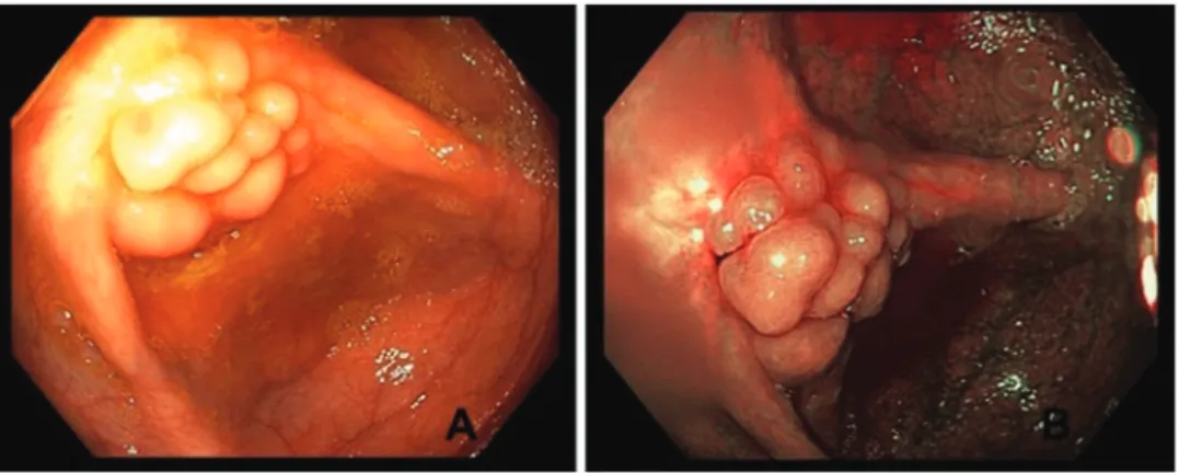
gastroenterology outpatient clinic. Upon enquiry, he denied any fever, weight loss, night sweat or nausea, vomiting and diarrhea, and his past medical history had only arterial hypertension controlled by calcium channel blocker. The physical examination of his abdomen showed no distention, tenderness, defense or palpable mass, and bowel sounds were observed to be present. Initial laboratory tests demonstrated the following findings: hemoglobin value of 14 gr/L, leukocyte count of 23 000/μL, lymphocyte count of 17 840/μL and platelet count of 256 000/μL, in addition to normal liver and kidney function test results. Mild erythematous gastritis was seen during gastroscopy. A polypoid lesion∼3 × 3 cm dimensions, mildly protruding into the lumen of cecum, completely covering appendix orifice, having normal mucosal surface without ulcer or erythema, was observed in white light and narrow band imaging (Fig.1A and B). Upon identification of the lesion, biopsy samples were obtained from its edges and center. Histopathologic analysis revealed the infiltration of a uniform population of lymphoid cells that were relevant to CLL. The immunohistochemistry of the specimen indicated that the cells were positive for CD5, CD19, CD20, CD23 and CD4 but negative for CD10, Bcl-6 and Cyclin D1
226 D. Ozer Etik et al.
Figure 1: Endoscopic view of polypoid lesion completely covers appendix orifice on white light (A) and narrow band imaging (B).
(Figs 2 and3). Additionally, there were high levels of lactate dehydrogenase [540 U/L (125–245 U/L)] alongside normal levels of beta-2 microglobulin [1.29 mg/L (0.9–2.6 mg/L)] and immunoglobulin G [1028 mg/dL (700–1600 mg/dL)]. The diagnosis of B-cell CLL was made by peripheral blood flow cytometric analysis. However, the circulating lymphocytes were found to be negative for CD38 antigen, which is a known prognostic indicator of CLL. The computed tomography scan of chest, abdomen and pelvis showed no contrast media passage into appendix due to the closure of its orifice but showed increased cecum wall thickness and paraaortic lymphadenopathy of 13 mm in diameter (Fig.4). The mediastinal, celiac, retroperitoneal and pelvic lymphadenopathies were not observed. Based on Rai and Binet CLL staging systems, the patient’s disease was classified as Stage 0 and Stage A, respectively. Considering the early stage of this disease, the strategy of ‘wait and see’ was proposed after hematology consultation.
DISCUSSION
CLL originates in the bone marrow and can spread to blood and organs of the hemopoietic system [2]. In intra-abdominal CLL, unifocal or multifocal lymphadenopathy or diffuse soft
Figure 2: Abdominal computerized tomography imaging revealed no contrast
media passage into the appendix.
tissue infiltration throughout the mesentery, retroperitoneum and solid organs might be seen [4]. Numerous enlarged lymph nodes or organ infiltration can lead to liver failure, cholecysti-tis, peritonicholecysti-tis, pancreatico-biliary obstruction or thrombosis of surrounding mesenteric arteries and veins [5]. In rare cases, the leukemic cells can possibly infiltrate the gastrointestinal tract and cause ulcer, polypoid lesions and clinical pictures mimicking malabsorption disorders or chronic inflammatory bowel disease [6–8]. The GI complications may occur after transformation of CLL to diffuse large B-cell lymphoma (Richter syndrome) [1]. Ratterman et al. [4] earlier complied a comprehensive systematic review of published extramedullary involvement of CLL case reports between the year 1975 and 2012. The involvement of the skin and central nervous system was the most commonly reported extramedullary manifestation that accounted for 33% and 27% of the cases, respectively [4]. There were 26 cases related to gastrointestinal involvement [4]; however, the involvement of appendiceal orifice in the colon, like in our patient, was never mentioned. Several studies have emphasized the unfavorable prognosis of extranodal localizations of lymphoproliferative dis-ease [1,2]; however, it may depend on the site and the extensity of involvement [1,2]. A follow-up for 18 months revealed that our patient was still asymptomatic and in the early stage without chemo- and/or immunotherapy.
Figure 3: Nodular mucosal lymphoid infiltrates in histopathological examination
(Hematoxylene and eosin,×20 original magnification).
An unusual presentation of CLL 227
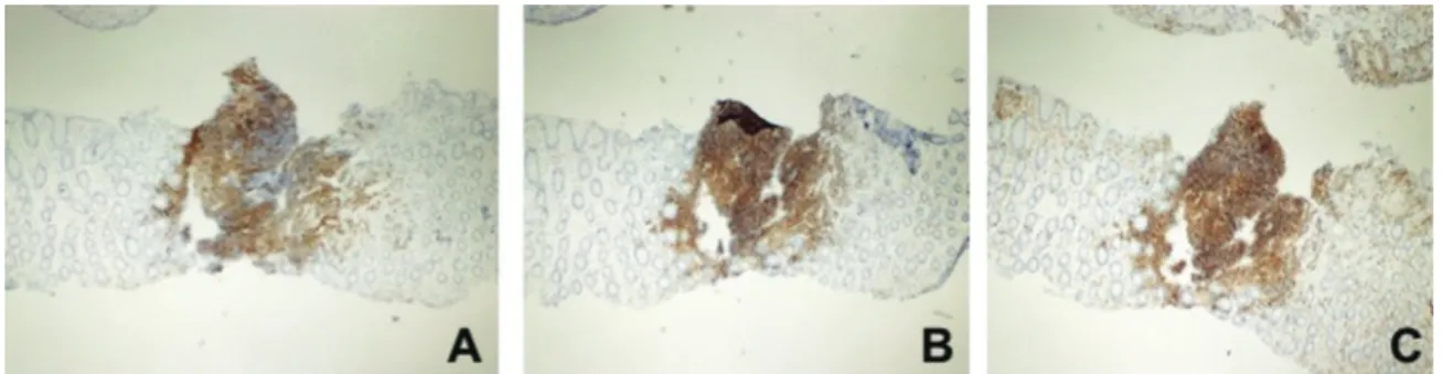
Figure 4: Immunohistochemical CD20 positivity (A), CD5 positivity (B) and CD43 positivity (C;×40 original magnification).
Our case is unique for several reasons; firstly, the abdom-inal pain that was an initial vague symptom yielded to the diagnosis of CLL in an older patient. Secondly, CLL presented with the involvement of the gastrointestinal tract despite the early stage of the disease. Thirdly, there existed mucosal inva-sion with malignant lymphocytes in the extranodal organ with-out transformation to lymphoma. Additionally, gastrointestinal involvement was occult, and it did not cause any ulceration, inflammation and obstruction even though the polypoid lesion closed the appendiceal orifice. The localization and presentation without signs of appendicitis make it a truly noteworthy case. Finally, the hematologists and gastroenterologists must keep in mind the possibility of extramedullary localizations of a lym-phoproliferative disease when a geriatric patient complains of abdominal pain.
ACKNOWLEDGEMENTS
We would like to thank the nurses in our Endoscopy Unit for their assistance and colleagues from the Department of Radiology for their scientific expertise.
CONFLICT OF INTEREST
None declared.
FUNDING
This study was not funded by any organization.
ETHICAL APPROVAL
The ethics committee approval was received for this case from the local ethics committee of our institute.
CONSENT
A written informed consent form from the patient and his wife was obtained before this report was prepared.
GUARANTOR
Professor Dr Mehmet Haberal confirms that all information pro-vided is true and accurate.
REFERENCES
1. Kipps TJ, Stevenson FK, Wu CJ, Croce MC, Packham G, Wlerda WG, et al. Chronic lymphocytic leukaemia. Nat Rev Dis
Primers 2017;3:16096.
2. Hallek M, Cheson BD, Catovsky D, Caligaris-Cappio F, Dighiero G, Döhner H, et al. iwCLL guidelines for diagnosis, indications for treatment, response assessment, and support-ive management of CLL. Blood 2018;131:2745–2760.
3. Barcos M, Lane W, Gomez GA, Han T, Freeman A, Preisler H,
et al. An autopsy study of 1,206 acute and chronic leukemias
(1958 to 1982). Cancer 1987;60:827–837.
4. Ratterman M, Kruczek K, Sulo S, Shanafelt TD, Kay NE, Nabhan C. Extramedullary chronic lymphocytic leukemia: systematic analysis of cases reported between 1975 and 2012.
Leuk Res 2014;38:299–303.
5. Kreiniz N, Beyar Katz O, Polliack A, Tadmor T. The clinical spectrum of hepatic manifestations in chronic lymphocytic leukemia. Clin Lymphoma Myeloma Leuk 2017;17:863–869. 6. Gonçalves R, Sousa R, Banhudo A, Pereira E, Pereira R,
Castel-Branco Silveira J Endoscopic findings in chronic lymphocytic leukemia. Am J Clin Oncol 2000;23:599–601.
7. Faigel DO, Vaughn DJ, Furth EE, Metz DC. Chronic lymphocytic leukemia: an unusual cause of upper gastrointestinal hemor-rhage. Am J Gastroenterol 1995;90:635–637.
8. Arkkila PE, Nuutinen H, Ebeling F, Elenon E, Kärkkäinen P, Karjalainen-Lindsberg ML. Colonic involvement in a patient with chronic lymphocytic leukaemia. Gastroenterol Res Pract 2008;2008:1–4.