A.Ü. Vet. Fak. Derg.
41 (3-4): 533 - 539,1994
GROSS ANATOMY OF DURAL SINUSES IN SHEEP
R. Merih Hazıroğlu! İsmet Takçız
Koyunda sinus durae matris'in makro anatomi<;i
Nejat ÇiftfP
Suınmary: The macro anatomy of dural sinuses in the head (n: 30) of whitekaraman native breed sheep obtained from an local abattoir was exam-ined. The dorsal system of dural sinuses was consisted of the dorsal sagittal si-nus, straight sisi-nus, transverse sisi-nus, temporal sinus and sigmoid sinus. The ven-tral system of dural sinuses composed of the eavernous sinus, intercavernous sinus, dorsal petrosal sinus, ventral petrosal sinus and basilar sinus. The lacu-nae laterales were present at the dorsal cerebral veins in which entered to the dorsal sagittal sinus. The straight sinus emptied in the dorsal sagittal sinus shortly before the formation of conjluence of sinuses (n: 21) or in the transverse sinus on the occasion of nine cases. There was intracranial connection between dorsal and ventral system of dural sinuses. The anterior eavernous sinus was present together with posterior eavernous sinus in six cases. The basilar sinus was eontinued as the ventral internal vertebral plexus inside the vertebral
ca-naL.
Özet: Ankara Et ve Balık Kurumu Mezbahasından temin edilen 30 adet Ak-karaman koyun başında sinus durae matris'in makro anatomisi incelendi. Dor-sal sinus sisteminin sinus sagittalis dorDor-salis, sinus reetus, sinus transversus, si-nus temporalis ve sinus sigmoideus'tan, ventral sinus sisteminin ise sinus cavernosus, sinus intercavernosus, sinus petrosus dorsalis, sinus petrosus ven-tralis ve sinus basilaris'ten oluştuğu görüldü. Vv. eerebri dorsales'in sinus sa-gittalis'e açıldığı yerde lacunae laterales'in varlığı dikkati çekti. Sinus rectus'un sinus sagittalis dorsalis'e (n: 21) ya da sinus transversus'a (n: 9) açıldığı görüldü. Dorsal ve ventral sinus sistemi arasında ilişkinin varlığı saptandı. Altı olguda sinus intercavernosus eaudalis ile birlikte sinus intercavernosus rostral-is'in bulunduğu gözlendi. Sin us basilarrostral-is'in eaudal ve ventral'de plexus venosus vertebralis internus şeklinde devam ettiği görüldü.
Introduction
The dural sinuses mostly loeated intraeranially between dura mater's peri-osteal and meningeal parts. Generally they laek tuniea media in their walls, and
1. Doç. Dr., A.Ü. Vet. fakültesi, Anatomi Anabilim Dalı, Ankara. 2. Araş. Gör. Dr., KA.U.Vet.fak., Anatomi Anabilim Dalı, Kars. 3. Araş.Gör.Dr., A.U.Vet.fak., Anatomi Anabilim Dalı, Ankara.
they do not have valves in their lumina. The dural sinuses are divided into dor-sal and basilar systems. The dordor-sal system eonsists of dordor-sal sagittal, straight si-nuses and transverse sisi-nuses. The basal system eonsists of the eavernous, inter eavernous, basilar, and dorsal and ventral petrosal sinuses (3,5,6). Anatomic deseriptions of these are available for domestieated animals in the standart textbooks (5,6,14). The horse has been used as the basis for anatomie informa-tion in these textbooks, but the informainforma-tion on the sheep dural sinuses is very seanty.
The purpose in the present report is to give a gross deseriptions of the dural sinuses in whitekaraman sheep.
Material and Methods
Thirty adult, loeal breed (whitekaraman) sheep heads were obtained from the loeal abattoir (The sheep body weights and sexes were not eonsidered). The heads were tlushed with normal saline solution. Of these heads, 18 were injeet-ed latex (blue) via the external jugular veins, six sheep heads were injeetinjeet-ed with methylmethaerylate, six sheep head were injeeted radioopaquae contrast media for venograpy via the external jugular vein.
Speeimens injeeted with latex were disseeted and stored in 4% formalin so-lution. The other heads were maeerated with 33% KOH solution for 2 to 3 weeks or 5% HCl solution for one week. Sehematie illustrations were drawn from the vaseular casts.
Results
DorsaL system of duraL sinuses
The dorsal sagittal sinus (=sinus sagittalis dorsalis) (Figs. la, 3b, 4A a) was formed by the eontluenee of right and left external ethmoid veins Ieaving oph-thalrnie plexus. From near the eribriform plate of the ethmoid bone, it run eaudo dorsally in the attaehed edge of the falx cerebri and terrninated into 2 transverse sinuses near the internal oecipital protuberance. The dorsal sagittal sinus was measured 6-6,5 cm in length and 2-3 mm in width. Six or 7 cerebral veins en-tered this sinus and lacunae laterales were present (Fig. 3a). It also received the diploic veins of the frontal and parietal bones.
The straight sinus (=sinus reetus) (Figs.ib, 4A b) was formed at the level of splenium eorpus callosi by the eontluence of the great eerebral veins ventral-Iy, and the vein of corpus eaııosum dorsaııy. it was about 2-2,5 mm in diameter and 8-9 mm in length. The straight sinus emptied in the dorsal sagittal sinus shürtly befüre the formation of the eontluence of sinuses (2icases). Sometimes (9 cases) it drained into transverse sinus.
The transverse sinus (=sinus transversus) (Figs. le, 4A c) was paired and loeated inside the membraneous tentorium of eerebeııum. it supposed ventrolat-eral continuation of the dorsal sagittal sinus. it was merged with its fellow to
GROSS ANATOMY OF DURAL SINUSES IN SHEEP 535
form the confluence of the sinuses. This triplc merging of sinuses was located within the occipital bone. The transverse sinus occasionally received the straight sinus (9 cases). The transverse sinus ran into transverse groove and ter-minated at the end of this groove. At this point transverse sinus divided into the temporal and sigmoid sinuses.
The temporal sinus (=sinus temporalis) (Fig. 4A d) was larger than sigmoid sinus, continued rostroventrally as the rostral branch of the tranSYerse sinus. It was located with in the temporal canal. it emerged as the emissary vein of the retroarticuler foramen and joined maxillary vein and pterygoid plexus. The tem-poral sinus was about 2,5 cm in length and 2,5 mm in diamater. it anastomosed with the cavemous sinus.
The sigmoid sinus (=sinus sigmoideus) (Fig 4A e) continued caudoventral-Iy as the caudal branch of the transverse sinus. it was "S" shaped and joined the emissary vein of the condylar foramen. Ifs branches finally emptİed into the oc-cipital vein and basilar sinus.
Ventral system of dural sinuses
The cavemous sinus (=sinus cavemosus) (Figs. 2c, 4B d) located on both sides of the hypophysis cerebri on the basisphenoidal bone. The carotid rete bathed in the cavemous sinus at the base of the brain. The cavemous sinus was
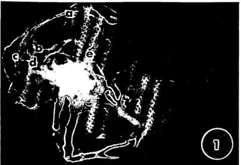
Fig. i.Lateral view of the venous drainage of the head in the sheep. a)Dorsal sagillal sinus. b) Straight sinus. c) Transverse sinus. d) Dorsal petrosal sinus. e) Ophthalmic plexus.
Fig. 2. The cavemous sinus of the shcep-vcntral view. a) Angularis oculi v. b) Ophthalmic plexus c)Cavcrnous sinus. d) Caudal intcrcavernous sinus.
Fig. 3. The dorsal cerebral veins and dorsal sagittal sinus. a) Lacuna lateralıs. b) Dorsal sagittal sinus.
GROSS ANATOMY OF DURAL SINUSES IN SHEEP 537
A
B
a
;w
f
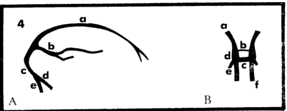
Fig. 4A. Diagram of the dorsal system of dural sinuses-lateral view.
a) Dorsal sagittal sinus. b) Straight sinus. c) Transverse sinus. d) Temporal sinus. e) Sigmoid sinus. 4B. Diagram of the ventral system of dural sinuses-dorsal view. a) Ophthalmic plexus.
b) Rostral intercavernous sinus (inconstantly). c) Caudal intcrcavernous sinus. d) Cavernous sinus. e) Dorsal petrosal sinus. f) Basilar sinus.
connected to the ophthalmic plexus (Figs. le, 2b, 48 a) via the emissary vein of the orbitorotundum foramen. There was a large venous pathway from the anteri-or panteri-ortion of the nasal mucosa to the cavernous sinus [danteri-orsal and lateral nasal veins-angular oculi vein (Fig. 2a), supraorbital vein-opthalmic vein-cavernous sinus]
The right and left cavernous sinuses were transversally connected to each other via a well-developed caudal intercavernous sinus (Figs. 2d 48 c) at the caudal aspect of the hypophysis cerebri in all speciemens. There were both ros-tral and caudal intercavernous sinuses in the six speciemens. The rosros-tral interca-vernous sinus (Fig. 48 b) was smailer than those of caudal ones.
Dorsocaudally, the cavernous sinus was connected with the rostral branch of the transverse sinus. This connection could be named as the dorsal petrosal sinus (es) (=sinus petrosus dorsalis) (Figs.ld, 48 e). Caudally the cavernous si-nus was continued the basilar sisi-nus.
Ventral petrosal sinus (=sinus petrosus ventralis) was ventrocaudal contin-uation of the cavernous sinus and connected with the emissary veins of the ju-gular foramen and the hypoglossal canal. Emissary vein of hypoglossal canal drained into the basilar sinus. The ventral petrosal sinus appeared as the extra-cranial continuation of the cavernous and basilar sinuses.
The basilar sinus (=sinus basilaris) (Fig. 48 f) was paired and located on the basilar part of the occipital bone. Rostrally, each basilar sinus anastomosed with the cavernous sinus. Caudally the basilar sinus continued as the ventral in-ternal vertebral plexus inside the vertebral cana\. There was no interbasilar si-nus. The basilar sinus was released to the occipital vein by the emissary vein of the hypoglossal cana\. Dorsolaterally, the basilar sinus connected the caudal branch of the transverse sinus (sinus sigmoideus).
Dİscussİon
In the whitekaraman native sheep, dorsal system of dural sinuses was com-posed of the dorsal sagittal, straight, tranverse, temporal and sigmoid sinuses. Dorsal sagittal sinus was formed by the confluence of the right and left external ethmoid veins. it was similar reported results in pigs (7,8). It has been recorded that dorsal sagittal sinus has constructed right and left rhinal veins draining 01-phactory bulb in dog (13) and camel (16). Dorsal sagittal sinus in dogs arises from the area of the ethmoidal sinuses as the ethmoidal vein entering the skull through one or two foramina (9). The lacunae laterales in horse (5) and dog (I, 5) have only been reported but they have not see n in Indian buffalo (12). In this study, it was encountered that lacunae laterales were observed at the dorsal cer-ebral veins in which entered to the dorsal sagittal sinus.
The straight sinus has been emptied in the dorsal sagittal sinus shortly be-fore the formation of the confluence of sinuses (3, 8, 14). In our cases, addition-ally, it was seen that straight sinus was occasionally (n: 9) drained to the trans-verse sinus. This type drainage was also recorded in camels (16).
Intracranial connection has not been reported between dorsal and ventral system in horse (5,6). However, this connection was present in whitekaraman sheep corresponding in camel (16), caule (15), dog (13) and pig (8). The caver-nous sinus was connected with the rostral branch of transverse sinus via the dor-sal petrodor-sal sinus and the basilar sinus connected the sigmoid sinus in whiteka-raman sheep.
The ventral system of the dural sinuses consisted of the cavernous sinus, intercavernous sinus, dorsal petrosal sintis, ventral sinus and basilar sinus in whitekaraman sheep.
The anterior intercavernous sinus is absent in sheep and go ats (5,14). The cavernous sinuses are communicated by means of the anterior and posterior in-tercavernous sinuses on the anterior and posterior of the hypophysis cerebri in cattle (4, i5). In this study, the anterior cavernous sinus was present together with posterior cavernous sinus in six cases. These findings were identical to the results of Khamas et ai. (10).
Carotid rete baths in the cavernous sinus, therefore, brain temperature is regulated by the cavernous sinuses (2, iO, 11, 17). We also believed that the role of these sinuses are counter current heat exchanger.
In whitekaraman sheep, it was seen that the basilar sinus continued as the ventral internal vertebral plexus inside the vertebral canal. In this point, there is agreement in the literature (16).
As a conclusion, we hope that these results could be beneficial to the con-ceming researchers with this subject.
GROSS ANATOMY OF DURAL SINUSES IN SHEEP
Kaynaklar
539
i. Armstrong, L.O., and Morowitz, A. (I 97 I). The brain venous system of the dog. Am. I. Anat., 132: 479-490.
2. Baker, M.A. and Hayward, J.N. (I 968). The injluence of the nasal mucosa and the caratid
rete upon hypothalamic temperature in sheep. I. Physiol., 198: 561-579.
3. Barone, R. and Payan, M. (1976). Drainage veineux de ['encephale et sinus veineux de la
dure-mere chez Bos taurus. Revue Med. vet., 127: 447-458.
4. Boyd, W.H. (1960). The relationship of the cranial dura to the cavemous sinuses in the cow.
Anat. Rec., 136: 413-415.
5. Elleberger, W. und Baum, H. (1974). "Handbuch der vergleichenden Anatomie der Haus-tiere" 18 ıh cd. Springer Verlag, Berlin.
6. Getty, R. (1975). "Sisson and Grassman's Anatomy of the Domestic Animals" 5th ed., Vol I., W.B. Saunders Company, Philadelphia.
7. Ghoshal, N.G. and Khamas, W.A. (1986). Blood supply of the nasal cavity of the normal
pig. Anat. Hislol. Embryol., 15: 14-22.
8. Ghoshal, N.G. and Zgui~al, H. (1986). Dural sinuses in the pig and their extracranial ve. nous connections. Am. I. Vet. Res., 47: ı165- i 169.
9. Hegedus, S.A. and Shackeırord, R.T. (1965). A comparative.anatomical study of the crani. ocervical venous systems in mammals, with spedal refaence to the dog: Relationship of anatomy to measurement of cerehral hloodjlow. Am.I. Anat., 116: 375-386.
10. Khama ••, W.A. and Ghosal, N.G. (1982). Blood supply to the na.wl cavity of sheep (ovis ar.
ies) and its significance to hrain temperature regulation. Anat. Anz., 151: 14-28.
iı. Khamas, W.A., Ghosal, N.G. and Bal, H.S. (1984). Histomorphologic structure of the ca. rotid rete.cavemous sinus complex and its functional importance in sheep (ovis aries). Am. I. Vet. Res., 45: 156- i58.
12. Lakshminarasimhan, A. (1974). Morphological variations of the sinus durae matris of the Indian huffalo (Buhalus huhalis). Anat.HistoI.Embryol., 3: 57-62.
13. Miller, M.E., Christensen, G.C. and Evans, H.E. (1965). "Anatomy of the Dog". 2nd ed., W.B. Saunders Company, Philadelphia.
14. Nickel, R., Schummer, A. und Seiferle, E.(ı975). "Lechhuch der Anatomie der Haustiere" Bd LV, Paul Barey. Berlin und Hamburg.
15. Uehara, M., Kudo, N. and Su~imura, M. (1978). Morphological studies on the rete mira. hle epidurale in the caif. Ipn. I. Vet. Res., 26: 11.18.
16. Zguigal, H. and Ghoshal, N.G. (199ı). Dural sinuses in the camel and their extracranial venous connections. Anat.HisıoI.Embryol., 20. 253-260.
17. Zguigal, H. and Ghoshal, N.G. (1991). Gmss and histologic study of the mstral epidural