E-mail: [email protected] doi:10.3906/sag-0902-12
Effects of long-term passive smoking on vascular
endothelial growth factor and apoptosis markers
expression in lens and corneal epithelial cells: An
experimental study*
Aim:To determine the effects of smoking on vascular endothelial growth factor (VEGF) expression and apoptosis in lens and corneal epithelial cells.
Materials and methods:This experimental study included 38 Sprague–Dawley rats. Rats were randomly assigned into 4 groups. Groups 1 (males) and 2 (females) were exposed to smoke beginning on the 21st day after birth, whereas groups 3 (males) and 4 (females) were not exposed to smoke. At the end of the fourth month, right eyes of all rats were enucleated. Immunohistochemical analysis was performed in enucleated eyes in terms of VEGF and apoptosis markers, namely caspase-3, Bcl-X, and p53. Comparisons between the groups were performed using the Mann-Whitney U test.
Results:There was no significant difference in the lens epithelial cells in terms of VEGF and apoptosis markers between the rats exposed to smoking and those not exposed. However, there was a significant increase in VEGF expression among female rats exposed to smoking while there was no significant difference in terms of apoptosis markers between the groups in corneal epithelial cells.
Conclusion:Our findings show that smoking does not affect the VEGF and apoptosis markers in lens epithelial cells, but it significantly increases the VEGF expression in corneal epithelial cells among female rats. In our opinion, the increase in the dry eye symptoms due to smoking may be caused by the differences in tear composition from change in permeability of the ocular surface vessels related to VEGF. Key words:Smoking, vascular endothelial growth factor, apoptosis, lens, cornea
Uzun dönem pasif sigara maruziyetinin lens ve kornea epitel
hücrelerinde vasküler endotelyal büyüme faktörü ve apoptozis
markerları üzerine etkisi: Deneysel çalışma
Amaç:Sigaranın lens ve kornea epitel hücrelerinde vasküler endotelyal büyüme faktörü (VEBF) ve apoptozis üzerine etkisini belirlemek.
Yöntem ve gereç:Çalışmaya 38 adet Sprague–Dawley rat alındı. Ratlar randomize olarak dört gruba ayrıldı. Grup 1 (erkek) ve 2 (dişi) doğumdan sonra 21. günden itibaren sigaraya maruz bırakılırken grup 3 (erkek) ve 4 (dişi) sigaraya maruz bırakılmadı. Dördüncü ayın sonunda tüm ratların sağ gözü enüklee edildi. Enüklee edilen gözlerde VEBF ve apoptozis markerları (kaspaz-3, Bcl-X, ve p53) yönünden immünohistokimyasal analiz yapıldı. Gruplar arası karşılaştırmalar Mann-Whitney U testi kullanılarak yapıldı.
Bulgular:Sigaraya maruz kalan ve kalmayan ratların lens epitel hücrelerinde VEBF ve apoptozis markerları yönünden anlamlı fark yoktu. Bununla birlikte, kornea epitel hücrelerinde sigaraya maruz kalan dişi ratlarda anlamlı VEBF artışı varken apoptozis markerları açısından gruplar arasında anlamlı fark yoktu.
Sonuç:Bulgularımız sigaranın lens epitel hücrelerinde VEBF ve apoptozis markerlarını etkilemediğini fakat dişi ratların kornea epitel hücrelerinde anlamlı VEBF artışı yaptığını gösterdi. Sigaraya bağlı kuru göz semptomlarındaki artışın VEBF’ye bağlı olarak oküler yüzeydeki damarların permeabilitesinin değişmesi sonucu gözyaşı bileşimde oluşacak farklılıktan kaynaklanabileceğini düşünmekteyiz. Anahtar sözcükler:Sigara, vasküler endotelyal büyüme faktörü, apoptozis, lens, kornea Fatih ÖZCURA1
Sema ORUÇ DÜNDAR2 İbrahim METEOĞLU3 Mehmet Erkut KARA4
1 Department of Ophthalmology, Faculty of Medicine, Dumlupınar University, Kütahya - TURKEY 2Department of Ophthalmology, Faculty of Medicine, Adnan Menderes University, Aydın - TURKEY
3
Department of Pathology, Faculty of Medicine, Adnan Menderes University, Aydın - TURKEY
4
Department of Anatomy, Faculty of Veterinary, Adnan Menderes University, Aydın - TURKEY Received: February 19, 2009 Accepted: August 03, 2009 Correspondence Fatih ÖZCURA Department of Ophthalmology, Faculty of Medicine, Dumlupınar University,
The Central Campus, 43270 Kütahya - TURKEY
Introduction
Smoking and exposure to smoking are two serious health problems around the world. It is known that cigarette contains more than 4000 toxic substances in the form of gas or particles (1). Smoking is a serious risk factor for life threatening diseases, such as cardiovascular and respiratory diseases as well as malign neoplasm (2). In addition, it was reported that smoking is also a risk factor for serious and common ocular disorders, such as cataract, glaucoma, age-related macular degeneration, Graves’ ophthalmopathy, and dry eye syndrome (DES) (2,3). Cataract is the leading cause of blindness in the world. The epidemiological relationship between smoking and cataract has been displayed by case-controlled, cross-sectional, and prospective studies (2). Hiller et al. reported as a result of their 30-year prospective study that smoking increased cataract incidence significantly (4). DES is another common ocular disorder; its prevalence is reported to be between 10.8% - 57.1% (5). Sahai et al. and Moss et al. reported that smoking increased DES prevalence (5,6). Although it is reported that smoking increases both cataract and DES prevalence, the mechanism of these effects is not clearly understood.
We are unaware of previous reports regarding the effect of smoking on vascular endothelial growth factor (VEGF) expression and apoptosis in lens and corneal epithelial cells. In our study, in order to analyze the effect of smoking on DES and development of cataract, we investigate the effect of smoking on the VEGF and apoptosis markers in the lens and corneal epithelial cells of the rats that were passively exposed to smoking.
Materials and methods
This study has been approved by the Animal Ethics Committee in Adnan Menderes University, Aydın, Turkey. Twenty female and 10 male Sprague-Dawley rats were used in the study. The rats were placed into 28 × 28 × 16 cm polycarbonate cages each of which has 2 females and 1 male. The rats were fed with special feed obtained from Gebze Best Yem Factory (İzmit, Turkey) and were provided water ad libitum. After mating, the male rats were taken out of the cages. The newborns were kept with their mothers until they were 3 weeks old.
Thirty eight healthy newborn rats (19 males and 19 females) were included in the study. Group 1 (9 males) and Group 2 (10 females) were subjected to passive smoking 21 days after their birth. Group 3 (10 males) and Group 4 (9 females) were control groups and were not subjected to smoking. For those rats exposed to passive smoking, their feed was taken out of their cages so that the smoke did not contaminate it and the rats would not receive nicotine orally; their feed was taken out of the cages for 2 h every day to air. After the smoke exposure period was over, the rats were fed. At weekends, they were given unlimited feed. The rats assigned to be exposed to passive smoking were exposed to smoke in a unit for 120 min a day for 5 days a week for 4 months. The smoke entered from one side of the unit and the air in the unit was circulated by an aspirator (the power of which was adjustable). The brande name of the cigarette used in the study was Birinci (85 mm, Tekel, Turkey), which has a high level of nicotine. The amount of smoke exposure was gradually increased during the study. The animals were exposed to smoke of 6 cigarettes in 120 min periods for two weeks, the smoke of 9 cigarettes for the next 2 weeks, and the smoke of 13 cigarettes for the following 12 weeks. The percentages of CO and CO2were measured at certain intervals to evaluate the CO and CO2levels created by the smoke and to test the reliability of the test environment (Sun Modular Gas Analyzer 1200; England, UK). At the end of the 4th month, right eyes of all rats were enucleated. Immunohistochemical analysis was performed in enucleated eyes.
Immunohistochemical evaluation
Four μm-thick sections from formalin-fixed, paraffin-embedded tissues were placed on coated slides. Immunostaining was performed using the avidin-biotin complex method. After deparaffinization and dehydration, sections were treated twice for 5 min in citrate buffer (0.01 mol/l, pH 6.0) in a microwave oven at 700 W. The slides were then cooled to room temperature for 1 h. Endogenous peroxidase activity was blocked by immersing the sections in 3% hydrogen peroxide in methanol for 30 min. Sections were then incubated with primary antibody for 1 h at room temperature. Biotinylated goat anti-rabbit secondary antibody was applied for 60 min at room temperature. Bound antibody was visualized with avidin–biotin– peroxidase complex (HISTOSTAIN – PLUS KITS,
Zymed, San Francisco, USA, code no: 85-9843) for 1 h at room temperature. The color was developed by 3,3’-diaminobenzidine tetrahydrochloride. Between steps, the slides were rinsed 3 times for 10 min in tris-buffered saline (pH 7.6). The slides were counterstained lightly in Harris’ hematoxylin, and then were dehydrated and mounted. The antibodies used were as follows: VEGF, (Neomarkers, CA, USA, cat no: RB-222-R7), Bcl-X, (Neomarkers, CA, USA, cat no: MS-715-R7), Caspase-3 (Neomarkers, CA, USA, cat no: RB-1197-P0), and p53 (Neomarkers, CA, USA, cat no: MS-104-R7).
In the immunohistochemical staining for positive control, various tissue samples were used. As negative control the primary antibody phase was skipped and staining process continued. The intensity of staining (p53 nuclear, Caspase-3, Bcl-X, and VEGF cytoplasmic) was scored by the investigator (IM) on a scale of 0 to 3 as follows: (0); absent, (1); weak and focal reaction, (2); moderate reaction, and (3); strong reaction.
Data were analyzed using SPSS for Windows (version 11.0, Chicago, IL, USA). Comparisons between groups were performed using the Mann-Whitney U test. P values below 0.05 were considered as statistically significant.
Results
Primarily, rats were evaluated in terms whether they were exposed to smoking. Then, to understand if smoking caused any different effect on different sexes, female and male rats were compared between each other.
Table 1 shows the average staining scores of VEGF and apoptosis markers of lens epithelial cells. As a result of the immunohistochemical analysis of the lens epithelial cells, there was no significantly difference between rats that were exposed to smoking and those not exposed in terms of both VEGF and apoptosis markers (Table 2).
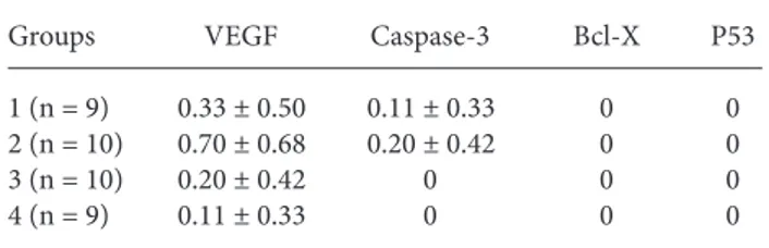
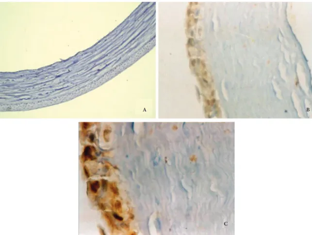
Table 3 shows the average staining scores of VEGF and apoptosis markers of corneal epithelial cells. As a result of the immunohistochemical analysis of the corneal epithelial cells, it was found that there was a significant difference between rats exposed to smoking and those not exposed in terms of VEGF while there was no difference in terms of apoptosis markers. There was a significant increase in VEGF expression among female rats exposed to smoking while no such increase was observed among male rats (Table 4). Figure 1 shows anti-VEGF staining pattern in corneal epithelium.
Table 1. The average staining scores of lens epithelial cells.
Groups VEGF Caspase-3 Bcl-X P53
1 (n = 9) 0.11 ± 0 33 0 0 0
2 (n = 10) 0 0 0 0
3 (n = 10) 0 0.20 ± 0.42 0 0
4 (n = 9) 0 0 0 0
VEGF: Vascular endothelial growth factor
Table 2. The statistical analysis results of the lens epithelial cells (P values).
Groups VEGF Caspase-3 Bcl-X P53
1+2 – 3+4 0.317 0.152 1.0 1.0
1 – 3 0.292 0.167 1.0 1.0
2 – 4 1.0 1.0 1.0 1.0
1 – 2 0.292 1.0 1.0 1.0
3 – 4 1.0 0.167 1.0 1.0
VEGF: Vascular endothelial growth factor
Table 3. The average staining scores of corneal epithelial cells.
Groups VEGF Caspase-3 Bcl-X P53
1 (n = 9) 0.33 ± 0.50 0.11 ± 0.33 0 0
2 (n = 10) 0.70 ± 0.68 0.20 ± 0.42 0 0
3 (n = 10) 0.20 ± 0.42 0 0 0
4 (n = 9) 0.11 ± 0.33 0 0 0
VEGF: Vascular endothelial growth factor
Table 4. The statistical analysis results of the corneal epithelial cells (P values).
Groups VEGF Caspase-3 Bcl-X P53
1+2 – 3+4 0.035* 0.075 1.0 1.0
1 – 3 0.521 0.292 1.0 1.0
2 – 4 0.030* 0.167 1.0 1.0
1 – 2 0.212 0.606 1.0 1.0
3 - 4 0.606 1.0 1.0 1.0
VEGF: Vascular endothelial growth factor, * statistically significant P < 0.05
Discussion
Cataract is a multifactorial disease and it has been shown in many studies that smoking and cataract progression are significantly related. Increase in the number of cigarettes consumed daily and long term smoking accelerate cataract progression (2,3). Nonetheless, how smoking accelerate cataract progression has not been clarified yet. The fact that smoking causes oxidative stress in the lens as it does in the whole body is the first theory. Animal, laboratory, clinical, and epidemiologic data support the relationship between cataract prevention and diets rich in nutritional factors with antioxidant properties, such as riboflavin, vitamins C and E, and the carotenoids (7,8). It is thought that oxidative stress in the lens, which is caused by the decrease in the body’s antioxidative capacity -related to smoking-, accelerate cataract progression. The second theory about this subject is that cigarette smoke contains
large amounts of heavy metals, such as cadmium, lead and copper, which appear to accumulate in the lens and exert further toxicity. Cekic found that copper, lead, and cadmium concentrations were significantly higher in cataractous lenses than normal human lenses and reported that cigarette smoking might be cataractogenic (9). Avunduk et al. found that there was a significant increase in iron and calcium concentrations in the lenses of the rats that were exposed to smoking compared to the control group; however, there was a decrease in the antioxidant zinc concentration. In addition, they found that lens epithelial cells had hypertrophy and hyperplasia (10). Therefore, in our study, in order to find out if there are different mechanisms in the development of cataract, we analyzed the changes of VEGF and apoptosis markers in the lens epithelial cells of the rats that were exposed to smoking passively.
A
C
B
Figure 1. Anti-VEGF staining pattern in corneal epithelium; A- Score 0 (Anti-VEGF, ×100), B-Score 2 (Anti-VEGF, ×200), C- Score 3 (Anti-VEGF, ×400).
VEGF plays an important role in physiological vascularization and contributes to pathologic neovascularization induced by local tissue ischemia (11). It was reported that there were VEGF and receptors in human and rat lens epithelial cells (12-14). We found no study in the literature reporting the effect of exposure to smoking on VEGF in the lens. It was shown that especially in the embryologic period, VEGF has a role in the formation of vascular structure around the lens. On the other hand, the role of VEGF in the mature lens is not known clearly (12,13). In Shui et al.’s study suturing the lids of adult mouse or rabbit eyes for 3 days was used to induce lens hypoxia and as a result of the study they found that the VEGF level in the lens epithelium and fibril cells increased significantly (12). In the study of Avunduk et al. (10), the increase of hypertrophy and hyperplasia in the lens epithelial cells as a result of exposure to smoking might be caused by growth factors. However, in our study, we did not find any significant VEGF difference in the lens epithelial cells of the rats that were exposed to smoking.
The studies of the relationship between apoptosis and cataractogenesis in different animal models provide strong support for the hypothesis that stress induced apoptosis is the common initiating event during cataractogenesis. It is thought that by the increase of apoptosis in the lens epithelial cells, the regulatory effect of the lens epithelial cells over lens fibril cells will decrease and therefore transparency in the lens will decrease accordingly. The hypothesis also predicts that the lens epithelium isolated from cataract patients should have apoptotic cells. Moreover, as the lens gets old, the number of lens epithelial cells is expected to decrease. Indeed, when the human lens epithelial cells from the surgically isolated capsular epithelium were examined by TUNEL, a varying level of apoptotic cells were detected in different types of cataracts. A decrease in the number of lens epithelial cells was found and correlated with aging. It has been shown that there was an apoptosis in the lens epithelial cells as a result of cataractogenic effect caused by hydrogen peroxide, abnormal calcium homeostasis, ultraviolet, selenite, and hypergalactosemia (15). We found no study in the literature questioning how smoking affects apoptosis in lens epithelial cells. In our study, we observed no
increase of apoptosis in the lens epithelial cells of the rats.
The most common symptoms of those who smoke and exposed to smoking are dry eye symptoms (3,16). Satici et al. reported that there was an increase in the eye irritation scores, Schirmer I-test values, and conjunctival squamous metaplasia scores while there was a decrease in tear break up time and in the tear lysozyme concentration in chronic smokers (16). Grus et al. reported that there was an increase in the protein peaks and there was a correlation between subjective dry eye symptoms and electrophoretic changes (17). In our study, we found that there was an increase of VEGF in the corneal epithelial cells with exposure to smoking. Grus et al. reported that the increased tear protein peaks of smokers might be resulted from a change in protein permeability because of a possible failure in ocular surface vessels (17). The increased VEGF level that we detected in our study is in accordance with Grus’s theory. Also, there was a significant VEGF increase in female rats but not in male rats in our study. Therefore gender might be a reason.
The clinical and experimental studies showed that apoptosis has an important role in the DES pathogenesis (18-20). Yeh et al. performed an experimental dry eye model in mice and 12 days later, they detected a significant apoptosis increase in central and peripheral corneal epithelium, bulbar and tarsal conjunctiva epithelium, tarsal conjunctival stroma, and eyelid margin (20). It is thought that with an increase in apoptosis, there was a rapid cell loss in ocular surface and therefore the protective effect of ocular surface decreased and resulted in punctate erosion. The role of apoptosis in DES pathophysiology is not clearly understood. In our study, we detected no apoptosis marker increase in the corneal epithelium related to smoking.
As a conclusion, we did not detect any significant VEGF and apoptosis difference in the lens epithelial cells. While there was a significant VEGF increase in the corneal epithelial cells, there was no difference in the apoptosis markers. We think that eye irritation and the increase in DES symptoms related to smoking may be caused by differences in tear composition as a result of a change in permeability of the ocular surface vessels in relation to VEGF.
1. Kjaergaard SK, Pedersen OF. Dust exposure, eye redness, eye cytology and mucous membrane irritation in a tobacco industry. Int Arch Occup Environ Health 1989; 61: 519-25. 2. Cheng AC, Pang CP, Leung AT, Chua JK, Fan DS, Lam DS. The
association between cigarette smoking and ocular diseases. Hong Kong Med J 2000; 6: 195-202.
3. Solberg Y, Rosner M, Belkin M. The association between cigarette smoking and ocular diseases. Surv Ophthalmol 1998; 42: 535-47.
4. Hiller R, Sperduto RD, Podgor MJ, Wilson PW, Ferris FL, Colton T, et al. Cigarette smoking and the risk of development of lens opacities. The Framingham studies. Arch Ophthalmol 1997; 115: 1113-8.
5. Sahai A, Malik P. Dry eye: prevalence and attributable risk factors in a hospital-based population. Indian J Ophthalmol 2005; 53: 87-91.
6. Moss SE, Klein R, Klein BE. Prevalence of and risk factors for dry eye syndrome. Arch Ophthalmol 2000; 118: 1264-8. 7. Heseker H. Antioxidative vitamins and cataracts in the elderly.
Z Ernahrungswiss 1995; 34: 167-76.
8. Mares-Perlman JA, Klein BE, Klein R, Ritter LL. Relation between lens opacities and vitamin and mineral supplement use. Ophthalmology 1994; 101: 315-25.
9. Cekic O. Effect of cigarette smoking on copper, lead, and cadmium accumulation in human lens. Br J Ophthalmol 1998; 82: 186-8.
10. Avunduk AM, Yardimci S, Avunduk MC, Kurnaz L. Cataractous changes in rat lens following cigarette smoke exposure is prevented by parenteral deferoxamine therapy. Arch Ophthalmol 1999; 117: 1368-72.
11. Ferrara N. Molecular and biological properties of vascular endothelial growth factor. J Mol Med 1999; 77: 527-43.
12. Shui YB, Wang X, Hu JS, Wang SP, Garcia CM, Potts JD, et al. Vascular endothelial growth factor expression and signaling in the lens. Invest Ophthalmol Vis Sci 2003; 44: 3911-9. 13. Mitchell CA, Risau W, Drexler HC. Regression of vessels in the
tunica vasculosa lentis is initiated by coordinated endothelial apoptosis: a role for vascular endothelial growth factor as a survival factor for endothelium. Dev Dyn 1998; 213: 322-33. 14. Gilbert RE, Vranes D, Berka JL, Kelly DJ, Cox A, Wu LL, et al.
Vascular endothelial growth factor and its receptors in control and diabetic rat eyes. Lab Invest 1998; 78: 1017-27.
15. Yan Q, Liu JP, Li DW. Apoptosis in lens development and pathology. Differentiation 2006; 74: 195-211.
16. Satici A, Bitiren M, Ozardali I, Vural H, Kilic A, Guzey M. The effects of chronic smoking on the ocular surface and tear characteristics: a clinical, histological and biochemical study. Acta Ophthalmol Scand 2003; 81: 583-7.
17. Grus FH, Sabuncuo P, Augustin A, Pfeiffer N. Effect of smoking on tear proteins. Graefes Arch Clin Exp Ophthalmol 2002; 240: 889-92.
18. Gao J, Schwalb TA, Addeo JV, Ghosn CR, Stern ME. The role of apoptosis in the pathogenesis of canine keratoconjunctivitis sicca: the effect of topical Cyclosporin A therapy. Cornea 1998; 17: 654-63.
19. Brignole F, Pisella PJ, Goldschild M, De Saint Jean M, Goguel A, Baudouin C. Flow cytometric analysis of inflammatory markers in conjunctival epithelial cells of patients with dry eyes. Invest Ophthalmol Vis Sci 2000; 41: 1356-63.
20. Yeh S, Song XJ, Farley W, Li DQ, Stern ME, Pflugfelder SC. Apoptosis of ocular surface cells in experimentally induced dry eye. Invest Ophthalmol Vis Sci 2003; 44: 124-9.