osacea is a chronic, inflammatory skin disorder involving the central part of the face.1The main subtypes are erythematotelangiectatic,
papulopustular, phymatous and ocular types, besides them, there is another form, which is called extrafacial rosacea (ER). ER is reported rarely and it tends to involve the scalp, neck, chest, axillary region, shoulders, arms and legs.2-4The diagnosis is generally made through the clinical
symptoms and the history of the patient, but it’s difficult to make a diag-nosis in atypical cases.4
Herein, a 66-year-old man with a severe chest and scalp involvement of ER and the clinical course of the patient will be presented.
CASE REPORT
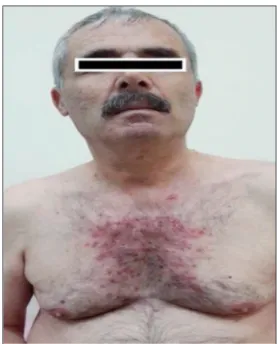
A 66 years-old man applied to our outpatient clinic with the complaints of some acneiform lesions on his chest and scalp for the last ten years. He had widespread erythematous papulopustular lesions on his scalp, chin and chest (Figure 1). He had a clear history in terms of other illnesses. The hemogram,
45
A Case with a Delayed Diagnosis of
Extrafacial Rosacea
AABBSS TTRRAACCTT Rosacea is a chronic, inflammatory skin disorder involving the central part of the face. There are four main subtypes of the disease which are most commonly seen; but also, when the dis-ease involves other parts of the body except the face, it’s called extrafacial rosacea (ER). ER is rarely reported in the literature and it may involve the scalp, chest and the back Herein, a man with ex-trafacially located rosacea who couldn’t have been diagnosed for long years will be presented be-cause of the rarity of the disease.
KKeeyywwoorrddss:: Extrafacial rosacea; atypical rosacea; rosacea Ö
ÖZZEETT Rozase sıklıkla yüzün santral kısmını etkileyen, kronik, inflamatuar bir deri hastalığıdır. Dört klasik formu sık görülürken; yüz dışındaki alanlar tutulduğunda ekstrafasiyal rozase (ER) ola-rak adlandırılmaktadır. ER, literatürde nadiren bildirilmiş olup; saçlı deri, göğüs, sırt gibi alanlarda görülebilmektedir. Burada uzun yıllar tanı koyulamamış atipik bir ekstrafasiyal yerleşimli rozase va-kası nadir görülmesi nedeni ile paylaşılacaktır.
AAnnaahh ttaarr KKee llii mmee lleerr:: Ekstrafasiyal rozase; atipik rozase; rozase
Hülya CENKa,
Gülbahar SARAÇb,
Yelda KAPICIOĞLUc aClinic of Dermatology,
Malatya Training and Reseach Hospital,
bDepartment of Dermatology,
İnönü University Faculty of Medicine, Malatya, TURKEY
cDepartment of Dermatology,
İstinye University Liv Hospital, İstanbul, TURKEY
Re ce i ved: 09.08.2018
Received in revised form: 02.11.2018 Ac cep ted: 05.11.2018
Available online: 25.04.2019 Cor res pon den ce:
Hülya CENK
Malatya Training and Reseach Hospital, Clinic of Dermatology, Malatya, TURKEY/TÜRKİYE
Cop yright © 2019 by Tür ki ye Kli nik le ri
biochemical parameters, microbiological and fun-gal culture values, which had been investigated in another medical care center before, were normal. He hadn’t responded well to 100 mg/day systemic doxycycline, topical mupirocin cream and keto-conazole shampoo treatments in 3 months, then, we thought that the patient had seborrheic der-matitis and our treatment with 400 mg/day sys-temic itraconazole for one week in a month didn’t relief the symptoms at all and the patient refused to continue to treatment. Afterwards, a standard-ized skin surface biopsy was performed to see any demodex spp, but the result was negative. Hence, a biopsy was performed from the lesion on the chest with the prediagnoses of ER and acne conglobata. Histopathological findings indicated subcorneal pustule formation, subepidermal edema, mixed in-flammatory cell infiltration, plasma cells, dilated vessels, and folliculitis. These findings were con-sistent with ER and 20 mg/day systemic isot retinoin and topical sodium sulfacetamide ments were started. After three months of treat-ment, the lesions regressed substantially and then the patient stopped the treatment by his own deci-sion because he thought that he was good enough (Figure 2). An informed consent has been obtained
to be able to share the data of the patient in any of the scientific journals or scientifical meetings.
DISCUSSION
Rosacea is a common, chronic, recurrent, and in-flammatory skin disorder.5 Mostly middle-aged
women with Fitzpatrick skin types 1 and 2 are af-fected.6 According to the classification system,
which has been composed by the National Rosacea Society Expert Committee in 2002; there are four main types (erythematotelangiectatic, papulopus-tular, phymatous and ocular) and one variant (granulomatous) of the disease.7
Generally, the convex parts of the face, except for the perioral and periocular region, are affected but the scalp, neck, chest, proximal back, shoul-ders, arms and proximal lower extremity involve-ments have been rarely reported.4 Extrafacial
region involvement is encountered mostly in men, as in our case. While ER has been usually reported in granulomatous or acneiform type, the erythe-matotelangiectatic type of ER can also be seen.3,4,8
Here, in this case, the lesions were consistent with the papulopustular type of ER.
The etiopathogenesis of rosacea hasn’t been fully understood yet. Naive immunity, neurovas-cular system disturbances, commensal microor-ganisms like demodex spp., topical steroids, systemic drugs like niacinamide, physical and psy-chological stress and environmental factors are considered to be among causes.1,4,6,8-10
Hülya CENK et al. Turkiye Klinikleri J Dermatol. 2019;29(1):45-8
46 FIGURE 1: Widespread papulopustular lesions in the chin and chest region.
Diagnosis of rosacea is easily made based on the clinical history of the patient, physical exami-nation and elimiexami-nation of the other differential diagnoses. But, in some cases which it is hard to diagnose, a biopsy may be required.10,11
Histopathology shows generally nonspecific find-ings. Here in our case, subcorneal pustule forma-tion, sub-epidermal edema, mixed inflammatory cell infiltration, plasma cells, dilated vessels, and folliculitis findings were consistent with papulo-pustular type rosacea.9,11
For the reason that the differential diagnosis depends on the type of ER, as for this case with papulopustular type of rosacea; acne conglobata, dermatophytosis, demodicosis, bacterial folliculi-tis, eosinophilic pustular folliculifolliculi-tis, halogeno-derma, seborrheic dermatitis were among differential diagnoses. There were no comedonal lesions and no neutrophilic infiltration extending beyond follicule in the histopathology for acne conglobata.11Microscobic examination of a pustule
smear with potassium hydroxide revealed no fun-gal elements for dermatophytosis. Histopathology and standardized skin surface biopsy were negative for demodex spp. as well. Microbiological culture was negative for bacterial folliculitis. There was no itching clinically and histopathology didn’t show eosinophil folliculotropism and this made it easier to exlude the eosinophilic pustular folliculitis.12
There was no drug history for halogenoderma. The patient didn’t respond well to seborrheic dermati-tis treatments also. After elimination of those dif-ferential diagnoses, with the support of clinical signs and histopathological findings, the patient was diagnosed as having ER.
Treatment options for ER are palliative rather than curative. Topical sodium sulfacetamide, aze-laic acid, metronidazole and brimonidine, and sys-temic sub-antimicrobial-dose doxycycline might be
used in the treatment. The treatment must be sup-ported with education of the patients about the dis-ease and by promoting the sunscreen and emollient usage.9,13,14 Systemic isotretinoin with
its sebum repressing and anti-inflammatory ef-fects has been also found to be effective on re-sistant papulopustular rosacea, rhinophyma and ER.4,13 Our case had systemic doxycycline
treat-ment for 1 month and systemic itraconazole treatment for 1-week course and there wasn’t any sign of regression. Afterwards, a low-dose systemic isotretinoin treatment provided a great relief in 3 months and there was just minimal erythema on the former location of the lesions after 3-months follow up period.
In conclusion, ER diagnosis may be very diffi-cult due to its atypical clinic presentation compared to rosacea, and that may be the reason of why the number of the reported ER cases on literature is very few.3Eventually, we think that the more
fre-quently it takes part in our differential diagnoses, the more cases will be reported.
S
Soouurrccee ooff FFiinnaannccee
During this study, no financial or spiritual support was received neither from any pharmaceutical company that has a direct connection with the research subject, nor from a company that provides or produces medical instruments and materials which may negatively affect the evaluation process of this study.
C
Coonnfflliicctt ooff IInntteerreesstt
No conflicts of interest between the authors and / or family members of the scientific and medical committee members or members of the potential conflicts of interest, counseling, ex-pertise, working conditions, share holding and similar situa-tions in any firm.
A
Auutthhoorrsshhiipp CCoonnttrriibbuuttiioonnss
All authors contributed equally while this study preparing.
Hülya CENK et al. Turkiye Klinikleri J Dermatol. 2019;29(1):45-8
Hülya CENK et al. Turkiye Klinikleri J Dermatol. 2019;29(1):45-8
48
1. Two AM, Wu W, Gallo RL, Hata TR. Rosacea: part II. Topical and systemic therapies in the treatment of rosacea. J Am Acad Dermatol 2015;72(5):761-70. [Crossref] [PubMed] 2. Vemuri RC, Gundamaraju R, Sekaran SD,
Manikam R. Major pathophysiological corre-lations of rosacea: a complete clinical ap-praisal. Int J Med Sci 2015;12(5):387-96. [Crossref] [PubMed] [PMC]
3. Pereira TM, Vieira AP, Basto AS. Rosacea with extensive extrafacial lesions. Int J Der-matol 2008;47(1):52-5. [Crossref] [PubMed] 4. Bostanci O, Borelli C, Schaller M. Treatment of
extrafacial rosacea with low-dose isotretinoin. Acta Derm Venereol 2010;90(4):409-10. [Crossref] [PubMed]
5. Wollina U. Recent advances in the under-standing and management of rosacea. F1000Prime Rep 2014;6:50. [Crossref] [PubMed] [PMC]
6. Moustafa FA, Sandoval LF, Feldman SR. Rosacea: new and emerging treatments.
Drugs 2014;74(13):1457-65. [Crossref] [PubMed]
7. Lee GL, Zirwas MJ. Granulomatous rosacea and periorificial dermatitis: controversies and review of management and treatment. Der-matol Clin 2015;33(3):447-55. [Crossref] [PubMed]
8. Crawford GH, Pelle MT, James WD. Rosacea: I. Etiology, pathogenesis, and subtype classi-fication. J Am Acad Dermatol 2004;51(3):327-41. [Crossref] [PubMed]
9. Steinhoff M, Schauber J, Leyden JJ. New in-sights into rosacea pathophysiology: a review of recent findings. J Am Acad Dermatol 2013;69(6 Suppl 1):S15-26. [Crossref] 10. Del Rosso JQ, Thiboutot D, Gallo R,
Webster G, Tanghetti E, Eichenfield L, et al. Consensus recommendations from the Amer-ican Acne & Rosacea Society on the man-agement of rosacea, part 1: a status report on the disease state, general measures, and ad-junctive skin care. Cutis 2013; 92(5):234-40.
11. Two AM, Wu W, Gallo RL, Hata TR. Rosacea: part I. Introduction, categorization, histology, pathogenesis, and risk factors. J Am Acad Dermatol 2015;72(5):749-58. [Crossref] [PubMed]
12. Fujiyama T, Tokura Y. Clinical and histopatho-logical differential diagnosis of eosinophilic pustular folliculitis. J Dermatol 2013;40(6):419-23. [Crossref] [PubMed]
13. Del Rosso JQ, Thiboutot D, Gallo R, Web-ster G, Tanghetti E, Eichenfield LF, et al. Consensus recommendations from the American Acne & Rosacea Society on the management of rosacea, part 3: a status re-port on systemic therapies. Cutis 2014;93 (1):18-28.
14. Del Rosso JQ, Thiboutot D, Gallo R, Webster G, Tanghetti E, Eichenfield L, et al. Consen-sus recommendations from the American Acne & Rosacea Society on the management of rosacea, part 2: a status report on topical agents. Cutis 2013;92(6):277-84.