Effects of the Folk Medicinal Plant Extract Ankaferd
BloodStopper on the Healing of Colon Anastomosis:
An Experimental Study in a Rat Model
C
olorectal cancer is the fourth most common cause of cancer deaths worldwide,[1] and the anastomosis leak-age is one of the most important complications of colorec-tal surgery. Despite the recent advances in surgical tech-niques and technologic devices, the anastomosis leakage occurs in up to 37% of patients with colorectal cancer.[2] Themain risk factors of leakage include patient-related factors, such as severe anemia, diabetes mellitus, immunosuppres-sion, history of radiation therapy, sepsis, malnutrition, hy-poalbuminemia, and hypovolemia, as well as issues with the surgical techniques including excessive suture tension, inadequate perfusion of the anastomosis edges, distal lu-Objectives: Ankaferd BloodStopper (ABS) is a topical hemostatic agent that modulates the inflammatory response and accelerates wound healing. The aim of this study was to determine the effects of ABS on the colon anastomosis wound healing in a rat model. Methods: Thirty-two Wistar albino rats were divided into four groups as follows: Group A (n=8), left colonic anastomosis plus ABS treatment (sacrificed on the 3rd day); Group B (n=8) (control), left colonic anastomosis (sacrificed on the 3rd day); Group C (n=8), left colonic anastomosis plus ABS treatment (sacrificed on the 7th day); and Group D (n=8) (control), left colonic anastomosis (sacrificed on the 7th day). All rats were sacrificed at the end of the experiment to assess the anastomosis integrity and the presence of perian-astomosis abscesses, peritonitis, and adhesions. Additionally, the bursting pressure and hydroxyproline (OH-pyroline) levels were determined, and a histopathologic evaluation of the perianastomosis tissue was conducted.
Results: The mean bursting pressure on Day 7 was significantly higher than that on Day 3 in the ABS group (p=0.017). Overall, the bursting pressure was higher in animals treated with ABS than in the control animals, although the difference was not statistically significant. The OH-pyroline levels of both ABS groups were significantly higher than in the control groups. The mean OH-pyroline level on Day 7 was higher than that on Day 3 in the ABS-treated animals (p=0.038).
Conclusion: ABS increases collagen formation and neovascularization, and it has a positive impact during the colon anastomosis healing in an experimental model of wound healing.
Keywords: Ankaferd BloodStopper; colon anastomosis; wound healing.
Please cite this article as ”Ekici U, Ferhatoğlu MF, Çitgez B, Uludağ M. Effects of the Folk Medicinal Plant Extract Ankaferd BloodStopper on the Healing of Colon Anastomosis: An Experimental Study in a Rat Model. Med Bull Sisli Etfal Hosp 2019;53(2):154–159”.
Uğur Ekici,1 Murat Ferhat Ferhatoğlu,2 Bülent Çitgez,3 Mehmet Uludağ3
1Department of Health Sciences Collage, Istanbul Gelisim University, Istanbul, Turkey 2Department of General Surgery, Okan University Faculty of Medicine, Istanbul,Turkey
3Department of General Surgery, Health Sciences University, Sisli Etfal Training and Research Hospital, Istanbul, Turkey
Abstract
DOI: 10.14744/SEMB.2019.98965
Med Bull Sisli Etfal Hosp 2019;53(2):154–159
Address for correspondence: Murat Ferhat Ferhatoğlu, MD. Okan Universitesi Tip Fakultesi, Genel Cerrahi Anabilim Dali, Istanbul, Turkey Phone: +90 555 321 47 93 E-mail: [email protected]
Submitted Date: February 05, 2019 Accepted Date: March 11, 2019 Available Online Date: June 24, 2019 ©Copyright 2019 by The Medical Bulletin of Sisli Etfal Hospital - Available online at www.sislietfaltip.org
OPEN ACCESS This is an open access article under the CC BY-NC license (http://creativecommons.org/licenses/by-nc/4.0/).
minal obstruction, and abdominal contamination with faucal content.[3] A tension-free technique and intact blood perfusion guarantee the stability of anastomosis.
Ankaferd BloodStopper (ABS) is a topical hemostatic agent for the management of hemorrhage where conventional methods to stop hemorrhage are ineffective.[4] ABS con-tains extracts from five different plants (Thymus vulgaris, 5 mg/100 ml; Glycyrrhiza glabra, 9 mg/100 ml; Vitisvinifera, 8 mg/100 ml; Alpiniaofficinarum, 7 mg/100 ml; and Urtica
dioica, 6 mg/100 ml). ABS modulates the inflammatory
re-sponse via its effects on endothelium, neovascularization, and cytokines.[4–6] ABS also accelerates wound healing by increasing the cellular and vascular proliferation,[7] and healing of colonic anastomoses depends on these prolifer-ative processes. In this study on an experimental rat model, we aimed to evaluate the efficacy of ABS on wound healing in colon anastomosis.
Methods
Thirty-two female Wistar albino rats with the mean weight of 235 g (200–275 g) obtained from the Laboratory of Ex-perimental Animals at Biological Experiments with Living Animals laboratory of Istanbul University, Istanbul, Turkey were used in this study. The rats were provided with a reg-ular laboratory rat diet and tap water with a 12-hour day/ night light cycle at room temperature of 24 oC–26 oC. Four groups containing 8 rats each were formed. The rats were housed 2/per cage and were allowed to move freely. Randomization of the rats into groups was achieved using computer-generated random numbers. None of the animals died during the study. The study design and all procedures were approved by the Ethics Committee of the Experimental Medicine Research Institute at Istanbul University.
For the study, there were four groups formed as follows: Group A (n=8), left colonic anastomosis plus ABS treatment (sacrificed on the 3rd day); Group B (n=8) (control), left co-lonic anastomosis (sacrificed on the 3rd day); Group C (n=8), left colonic anastomosis plus ABS treatment (sacrificed on the 7th day); and Group D (n=8) (control), left colonic anas-tomosis (sacrificed on the 7th day).
All rats were sacrificed to assess the anastomosis integrity, and the presence of perianastomosis abscesses, peritonitis, and adhesions. In addition, the bursting pressure and hy-droxyproline (OH-proline) levels were determined, and the histopathologic evaluation of the perianastomosis tissue was performed.
Surgical Procedure
All rats were fasted for 8 hours before surgery, which was performed under sterile conditions. All colon anastomoses
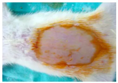
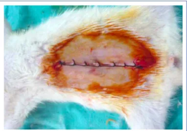
were performed by the same uninformed/blind surgeon, using six sutures. General anesthesia was administered with 50 mg/kg ketaminehydrochloride intraperitoneally and 5 mg/kg xylazinehydrochloride intramuscularly. Next, the anterior abdominal wall was shaved with an electric ra-zor and wiped with povidone iodine to obtain antiseptic conditions (Fig. 1). The peritoneal cavity was exposed with a 4 cm midline abdominal incision. The left colon was mo-bilized, and a full-layer cut was created in a 90° angle to the longitudinal axis of the colon. After the exploration of the abdominal cavity, animals with gastrointestinal anomalies and intra-abdominal tumors were excluded from the study. Next, end-to-end colocolic anastomosis was performed using a nonabsorbable mono filament 6/0 polypropylene inverting sutures (Fig. 2), and 0.1 cc of ABS was wiped on the anastomosis line. The abdominal wall was closed with 3/0 silk sutures (Fig. 3). At the end of the procedure, 15 ml of sterile saline was injected subcutaneously for
postoper-Figure 1. Shaved and cleansed abdominal wall.
ative hydration. Animals were fasted for postoperative 24 hours, after which they were started on standard diet and drinking water.
Evaluation of Adhesions
The uninformed/blind surgeon made the postmortem ex-amination that was conducted to grade adhesions on a scale from 0 to 3 according to the method introduced by van Deer Ham et al.[7] (Table 1).
Measurement of Colonic Anastomosis Bursting
Pressure
The uninformed/blind surgeon made the bursting pressure measurements, which were obtained within 3 minutes of sacrifice. First, the anastomosis integrity was determined by exposing the anastomosis line and removal of the adhe-sions in the surrounding tissues. Next, two cuts, one 2 cm proximal and the other 2 cm distal to the anastomosis site, were made to obtain a 4-cm-long colonic segment. Faucal content of the colon was removed with physiologic saline solution. The distal end of the excised segment was ligated using 4/0 silk sutures after an infusion set connected to a sphygmomanometer was inserted into the proximal end of the colon segment. The resected colon segment was then placed in a beaker filled with physiological saline solution
and inflated with air. The intraluminal pressure was in-creased in 10 mmHg increments and maintained for 10 sec-onds. The appearance of air bubbles was used to record the bursting pressure in mmHg. Notably, bursting occurred at the anastomosis line in all samples. The bursting pressure of rats with the anastomosis leakage was determined as 0 mmHg; these animals were excluded from the study. Next, the anastomosis segment was cut across the longitudinal axis and divided into two half rings; one half was placed in 10% formalin solution for histopathological evaluation, and the other half was stored at −80°C for the OH-proline measurement.
Histopathological Evaluation
The formalin-fixed colonic segments were sectioned, stained with hematoxylin and eosin, and analyzed with light microscopy at the 200× magnification by the same pathologist who was blinded to the treatment groups. The infiltration of inflammatory cells (leukocyte count), activity of fibroblast cells, neoangiogenesis, and collagen content were measured using the modified Ehrlich and Hunt scale by Philips et al.[8]
Tissue OH-Proline Assay
The tissue concentrations of OH-proline, which represent perianastomosis collagen levels, were measured using the spectrophotometric method by Bergman and Loxley. [9] The values were expressed as the microgram amount of OH-proline per milligram of tissue (μg/mg).
Statistical Analysis
Descriptive statistics including the frequency distribution and the mean and standard deviation were used. The Kru-skal–Wallis variance analysis was used to determine inter-group differences; paired inter-groups were compared using the Mann–Whitney U test for analyses yielding significant results. P-values less than 0.05 were considered statistical-ly significant. The Statistical Package for Social Sciences for Windows 11.0 was used for all statistical analyses.
Results
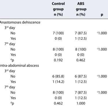
A statistically difference with regard to the anastomosis dehiscence and intra-abdominal abscess was not observed between the groups (Table 2).
Comparison of the tested variables between the ABS (Groups A–C) and control (Groups B–D) groups are shown in Table 3, 4. Acute inflammation, fibroblast activation, and neovascularization scores did not significantly differ be-tween the groups. However, collagenization scores were significantly higher on both Days 3 and 7 after surgery in the ABS group.
Table 1. Adhesion grading scale 0: No adhesions
1: Minimal adhesions, mainly between the omentum and the anastomosis site
2: Moderate adhesions, i.e., between the anastomosis site and the omentum or between the anastomosis site and a loop of the small bowel or the abdominal wall
3: Severe and extensive adhesions, i.e., between the anastomotic site and several loops of the small bowel and the abdominal wall, including the abscess formation Figure 3. Closed skin of the rat.
The mean values of the bursting pressure, adhesion score, and OH-proline level are presented in Tables 5, 6, and 7, re-spectively. The bursting pressure of one rat from the con-trol group on Day 3 was determined as 0 mmHg, and it was excluded from the study. In addition, briefly, the mean
bursting pressure on Day 3 was significantly higher than that on Day 7 in the ABS group (p=0.017) (Table 7). Overall, the bursting pressure was higher in animals treated with ABS than the control animals, although the difference was not statistically significant.
The OH-proline levels of both ABS groups were significant-ly higher than control groups. The mean OH-proline level on Day 7 was higher than that on Day 3 in the ABS-treated animals (p=0.038).
Discussion
Anastomotic leakage remains a challenge associated with morbidity and mortality in patients undergoing colorectal surgery. Leakage at the site of anastomosis was also re-ported to contribute to an increase in the length of hospi-tal stays and medical costs.[10–12] Treatment of anastomosis leakage is difficult and may require re-laparotomy, with fre-Table 2. Evaluation of anastomosis dehiscence, intra-abdominal
abscess, intestinal obstruction, adhesion score
Control ABS group group n (%) n (%) p Anastomoses dehiscence 3rd day No 7 (100) 7 (87.5) c1.000 Yes 0 (0) 1 (12.5) 7th day No 8 (100) 8 (100) c1.000 Yes 0 (0) 0 (0) dp 0.192 0.462 Intra-abdominal abscess 3rd day No 6 (85.8) 6 (87.5) c1.000 Yes 1 (14.2) 1 (12.5) 7th day No 8 (100) 7 (87.5) c1.000 Yes 0 (0) 1 (12.5) dp 0.462 1.000
aKruskal Wallis Test; bMann Whitney U Test; *p<0.05 ; cFisher-Freeman-Halton Exact Test; dFisher’s Exact Test.
Table 3. The comparison between ABS and control group via histopathological analysis in day3
Variable ABS Control p
(Day 3) (Day 3) (mean±SD) (mean±SD) Acute inflammation* 3.5±1.06 3.37±0.51 a0.256 Fibroblast activation* 2.5±0.75 2.25±0.46 a0.289 Collagen* 2.87±0.35 2.37±0.51 a0.046 Neovascularization* 2.75±0.46 2.62±0.51 a0.602 *Ehrlich and Hunt scale score. aKruskal-Wallis Test; SD: Standard Deviation.
Table 4. The comparison between ABS and control group via histopathological analysis in day 7
Variable ABS Control p
(Day 7) (Day 7) (mean±SD) (mean±SD) Acute inflammation* 3.5±0.53 3.75±0.46 a0.317 Fibroblast activation* 3.12±0.64 2.75±0.46 a0.199 Collagen* 3.37±1.06 2.75±0.46 a0.040 Neovascularization* 2.87±0.35 2.62±0.51 a0.264 *Ehrlich and Hunt scale score; aKruskal-Wallis Test; SD: Standard Deviation.
Table 5. The comparison between ABS and control group via bursting pressure, adhesion score, OH-pyroline level in day 3
Variable ABS Control p
(Day 3) (Day 3)
(mean±SD) (mean±SD)
Bursting pressure (mmHg) 91.25±39.07 61.25±9.9 b0.66
Adhesion score† 1.5±0.53 1.62±0.51 b1.00
OH-pyroline (µg/mg) 564.62±59.93 369.5±51.53 b0.001 †Adhesions were graded according to system developed by van Der Ham et al. bMann Whitney U Test; SD: Standard Deviation.
Table 6. The comparison between ABS and control group via bursting pressure, adhesion score, OH-pyroline level in day 7
Variable ABS Control p
(Day 7) (Day 7)
(mean±SD) (mean±SD)
Bursting pressure(mmHg) 193±28.75 165±53.45 b0.34
Adhesion score† 1.87±0.35 2.12±0.35 b0.31
OH-pyroline (µg/mg) 614.12±81.68 457.25±85.35 b0.007 † Adhesions were graded according to system developed by van Der Ham et al. bMann Whitney U Test; SD: Standard Deviation.
Table 7. The comparison between ABS day 3 and ABS day 7 group via bursting pressure, adhesion score, OH-pyroline level
Variable ABS ABS p
(Day 3) (Day 7)
(mean±SD) (mean±SD)
Bursting pressure (mmHg) 91.25±39.07 193±28.75 b0.017
Adhesion score† 1.5±0.53 1.87±0.35 b0.63
OH-pyroline (µg/mg) 564.62±59.93 614.12±81.68 b0.038 † Adhesions were graded according to system developed by van Der Ham et al. bMann Whitney U Test; SD: Standard Deviation.
quent necessity of stoma formation. Therefore, the preven-tion of anastomosis leakage remains the most critical issue. However, nonmodifiable risk factors such as an advanced age, comorbid conditions, and emergency surgery hinder these efforts,[3] highlighting the need for new treatment strategies to prevent anastomosis leakage. To this end, nu-merous experimental and clinical studies were conducted to achieve better wound healing after colon anastomosis.[3, 12–14] Numerous studies have demonstrated the hemostatic effect of ABS, which shortens the bleeding time and the amount of blood loss.[4–6, 17] The main mechanism underlying this ef-fect of ABS is an encapsulated protein network formation and increased erythrocyte aggregation.[15] Additionally, ABS increases vascular proliferation by reducing tissue necrosis. [16] Several studies have also shown the anti-inflammatory effects of ABS.[4–7, 16, 17] Interleukins are common to both the inflammatory/anti-inflammatory and the coagulation cas-cades that are targeted by ABS.[3] The most important factor in anastomotic healing is collagen, which forms the tensile strength of submucosal connective tissue.[18] Increased in-flammation may cause excessive collagenolysis, and the an-ti-inflammatory function of ABS might inhibit collagenolysis during tissue healing.[15] OH-proline levels can be used to as-sess collagen formation during wound healing. OH-proline is a component of collagen, which was shown to be positive-ly correlated with the collagen amount and strength of anas-tomosis.[19] In the current study, the OH-proline levels were significantly higher in the ABS-treated animals; furthermore, the OH-proline levels were higher on Day 7 compared to those on Day 3 among the animals treated with ABS. These results suggest that ABS might increase the strength of anas-tomosis by inducing collagen formation. ABS might also de-crease collagenolysis via an anti-inflammatory action, which requires further studies.
In clinical practice, the physical strength of anastomosis is not an ideal parameter to assess the healing of colonic anastomosis;[20] nonetheless, bursting pressure was used as an indirect method to assess the anastomotic integrity. We found that bursting pressure of the animals treated with ABS is higher than control groups, whereas there was no significant difference in the bursting pressures between the ABS-treated and the control groups.
Fibroblasts and neovascularization promote synthesis of collagen and mucopolysaccharides to increase anastomot-ic strength.[21] The first endothelial cells are seen 48 hours after the beginning of inflammatory process, and this in-flammatory process ends on the 6th or 7th day.[22] The fibri-nopurulent exudate fills anastomotic space in peritoneal sepsis. This exudate reduces the collagen synthesis and formation of new blood vessels and, as a result,
anastomo-sis heals by secondary intention.[23] In the presented study, fibroblast activity and neoangiogenesis were not statis-tically different between the control and ABS groups. We believe that the anti-inflammatory and antioxidant activity of ABS promotes the endothelial cell activity.
There was no difference in neovascularization and fibro-blast activity and indicators of fibrofibro-blast activity between the ABS and the control groups in the current study. How-ever, histopathological evaluation might not be ideal to assess overlooked ultrastructural changes, and an evalu-ation by electron microscopy may provide more informa-tion on fibrosis and neovascularizainforma-tion. In addiinforma-tion, the short duration of observation in the current study might not have allowed for ultrastructural changes to become evident. Future studies with a longer treatment time that utilize more advanced techniques such as electron mi-croscopy should allow for a better assessment of ultra-structural changes.
The current study findings did not reveal a clear clinical ap-plication for ABS in increasing the strength and safety of anastomosis; however, based on our finding of ABS-me-diated increase in collagenization. In accordance with the study by Cancan et al.[19] which investigated the same is-sue regarding ABS, we also revealed that ABS has a positive impact in the healing of colonic anastomosis performed under optimal surgical conditions, but further studies are required to elucidate the role of ABS in anastomotic heal-ing under septic conditions.
Conclusion
ABS increases the collagen formation and neovasculariza-tion, and it has a positive impact on the colon anastomosis healing in an experimental rat model of wound healing. Thus, ABS promotes colonic healing and provides a safer and stronger anastomosis than suturing alone.
Although further studies are needed to define the mech-anism of the ABS impact in the anastomotic healing of colon, our results are promising for clinical use of ABS to improve the safety of colonic anastomoses.
Disclosures
Ethics Committee Approval: This study was approved by the Biological Experiments with Living Animals Ethic Committe of Istanbul University, Istanbul, Turkey.
Peer-review: Externally peer-reviewed. Conflict of Interest: None declared.
Authorship Contributions: Concept – U.E.; Design – U.E., B.C.; Supervision – M.U.; Materials – U.E.; Data collection &/or process-ing – U.E.; Analysis and/or interpretation – U.E., M.F.F.; Writprocess-ing – U.E., M.F.F.; Critical review – B.C., M.U.
References
1. Bhandari A, Woodhouse M, Gupta S. Colorectal cancer is a leading cause of cancer incidence and mortality among adults younger than 50 years in the USA: a SEER-based analysis with comparison to other young-onset cancers. J Investig Med 2017;65:311–5. [CrossRef]
2. Neumann PA, Twardy V, Becker F, Geyer C, Schwegmann K, Mohr A, et al. Assessment of MMP-2/-9 expression by fluorescence en-doscopy for evaluation of anastomotic healing in a murine model of anastomotic leakage. PLoS One 2018;13:e0194249.
3. Akyuz C, Yasar NF, Uzun O, Peker KD, Sunamak O, Duman M, et al. Effects of melatonin on colonic anastomosis healing following chemotherapy in rats. Singapore Med J 2018;59:545–9. [CrossRef]
4. Haznedaroglu BZ, Beyazit Y, Walker SL, Haznedaroglu IC. Pleiotro-pic cellular, hemostatic, and biological actions of Ankaferd hemo-stat. Crit Rev Oncol Hematol 2012;83:21–34. [CrossRef]
5. Goker H, Haznedaroglu IC, Ercetin S, Kirazli S, Akman U, Ozturk Y, et al. Haemostatic actions of the folkloric medicinal plant extract Ankaferd Blood Stopper. J Int Med Res 2008;36:163–70. [CrossRef]
6. Işler SC, Demircan S, Cakarer S, Cebi Z, Keskin C, Soluk M, et al. Effects of folk medicinal plant extract Ankaferd Blood Stopper on early bone healing. J Appl Oral Sci 2010;18:409–14. [CrossRef]
7. van der Ham AC, Kort WJ, Weijma IM, van den Ingh HF, Jeekel H. Effect of antibiotics in fibrin sealant on healing colonic anastomo-ses in the rat. Br J Surg 1992;79:525–8. [CrossRef]
8. Phillips JD, Kim CS, Fonkalsrud EW, Zeng H, Dindar H. Effects of chronic corticosteroids and vitamin A on the healing of intestinal anastomoses. Am J Surg 1992;163:71–7. [CrossRef]
9. Bergman I, Loxley R. Two improved and simplified methods for the spectrophotometric determination of hydroxyproline. Ann Chem 1963;35:1961–5. [CrossRef]
10. Daglioglu YK, Duzgun O, Sarici IS, Ulutas KT. Comparison of plate-let rich plasma versus fibrin glue on colonic anastomoses in rats. Acta Cir Bras 2018;33:333–40. [CrossRef]
11. Ersoz N, Ozler M, Topal T, Uysal BZ, Poyrazoglu Y, Simsek K, et al. The effect of ozone treatment on experimental colon anastomo-sis in rats. Eur Surg 2016;48:122–8. [CrossRef]
12. Strunden MS, Heckel K, Goetz AE, Reuter DA. Perioperative fluid and volume management: physiological basis, tools and strate-gies. Ann Intensive Care 2011;1:2. [CrossRef]
13. Bai J, Huo X, Ma J, Lv Y, Yan X. Magnetic compression technique for colonic anastomosis in rats. J Surg Res 2018;231:24–9. [CrossRef]
14. Trotter J, Onos L, McNaught C, Peter M, Gatt M, Maude K, et al. The use of a novel adhesive tissue patch as an aid to anastomotic healing. Ann R Coll Surg Engl 2018;100:230–4. [CrossRef]
15. Aydın BK, Altan E, Acar MA, Erkoçak ÖF, Ugraş S. Effect of Ankaf-erd blood stopper® on tendon healing: an experimental study in a rat model of Achilles tendon injury. Eklem Hastalik Cerrahisi 2015;26:31–7. [CrossRef]
16. Akbal E, Köklü S, Karaca G, Astarci HM, Koçak E, Taş A, et al. Ben-eficial effects of Ankaferd Blood Stopper on caustic esophageal injuries: an experimental model. Dis Esophagus 2012;25:188–94. 17. Ozel-Demiralp D, Igci N, Ayhan B, Egin Y, Haznedaroglu IC, Akar
N. Prohemostatic and antithrombin activities of Ankaferd he-mostat are linked to fibrinogen gamma chain and prothrombin by functional proteomic analyses. Clin Appl Thromb Hemost 2012;18:604–10. [CrossRef]
18. Adam AB, Özdamar MY, Esen HH, Günel E. Local effects of epi-dermal growth factor on the wound healing in esophageal anas-tomosis: An experimental study. Int J Pediatr Otorhinolaryngol 2017;99:8–12. [CrossRef]
19. Cancan G, Teksoz S, Aytac E, Arikan AE, Erman H, Uzun H, et al. Effects of Ankaferd on anastomotic healing of colon. J Invest Surg 2014;27:1–6. [CrossRef]
20. Subhas G, Bhullar JS, Cook J, Shah A, Silberberg B, Andrus L, et al. Topical gentamicin does not provide any additional anas-tomotic strength when combined with fibrin glue. sAm J Surg 2011;201:339–43. [CrossRef]
21. Mariggiò MA, Cassano A, Vinella A, Vincenti A, Fumarulo R, Lo Muzio L, et al. Enhancement of fibroblast proliferation, collagen biosynthesis and production of growth factors as a result of com-bining sodium hyaluronate and aminoacids. Int J Immunopathol Pharmacol 2009;22:485–92. [CrossRef]
22. Oguido APMT, Hohmann MSN, Pinho-Ribeiro FA, Crespigio J, Do-miciano TP, Verri WA Jr, et al. Naringenin Eye Drops Inhibit Corneal Neovascularization by Anti-Inflammatory and Antioxidant Mech-anisms. Invest Ophthalmol Vis Sci 2017;58:5764–76. [CrossRef]
23. Saravanan M. A Comperative Study Between Single Versus Dou-ble Layered Intestinal Anastomosis. [Thesis]. The Tamil Nadu Dr. M.G.R. Medical University, India. 2015.