Full Terms & Conditions of access and use can be found at
https://www.tandfonline.com/action/journalInformation?journalCode=ibih20
Biotechnic & Histochemistry
ISSN: 1052-0295 (Print) 1473-7760 (Online) Journal homepage: https://www.tandfonline.com/loi/ibih20
Dexpanthenol reduces diabetic nephropathy and
renal oxidative stress in rats
B. Tutun, H. Elbe, N. Vardi, H. Parlakpinar, A. Polat, M. Gunaltili, M. M. Guclu
& E. N. Yasar
To cite this article: B. Tutun, H. Elbe, N. Vardi, H. Parlakpinar, A. Polat, M. Gunaltili, M. M. Guclu & E. N. Yasar (2019) Dexpanthenol reduces diabetic nephropathy and renal oxidative stress in rats, Biotechnic & Histochemistry, 94:2, 84-91, DOI: 10.1080/10520295.2018.1508746
To link to this article: https://doi.org/10.1080/10520295.2018.1508746
Published online: 15 Oct 2018.
Submit your article to this journal
Article views: 235
View related articles
View Crossmark data
Citing articles: 3 View citing articles
0. Taylor & Francis
~ Tllylorf.J,;i11caCr11u1,
ARTICLE
Dexpanthenol reduces diabetic nephropathy and renal oxidative stress in rats
B. Tutuna, H. Elbeb, N. Vardic, H. Parlakpinard, A. Polate, M. Gunaltilia, M. M. Guclua, and E. N. YasaraaMedical Faculty, Inonu University, Malatya, Turkey;bMedical Faculty, Department of Histology and Embryology, Mugla Sıtkı Kocman
University, Mugla, Turkey;cMedical Faculty, Department of Histology and Embryology, Inonu University, Malatya, Turkey;dMedical Faculty,
Departments of Pharmacology, Inonu University, Malatya, Turkey;eMedical Faculty, Physiology, Inonu University, Malatya, Turkey
ABSTRACT
Hyperglycemia increases reactive oxygen species (ROS) and the resulting oxidative stress con-tributes to the development of diabetic complications. Dexpanthenol (Dxp) is the biological active form of pantothenic acid. We investigated whether Dxp administration could decrease oxidative stress as a way to treat renal complications of diabetes mellitus (DM). Thirty-two male Wistar albino rats were divided into four groups: control, Dxp, DM and DM + Dxp. Experimental diabetes was induced by a single dose of streptozotocin (STZ). After administration of STZ, the DM + Dxp group was administered 500 mg/kg Dxp intraperitoneally every day for 6 weeks. At the end of the study, blood glucose levels were measured and rats were sacrificed. Kidneys were embedded in paraffin, sectioned and stained with hematoxylin and eosin, and periodic acid-Schiff. The mean malondialdehyde levels, glutathione peroxidase, superoxide dismutase and catalase activities, and total antioxidant and total oxidant status also were measured. The control group was normal in histological appearance. We observed congestion, inflammation, glomerulosclerosis, tubular des-quamation, loss of villi and hydropic degeneration in tubule cells in the DM group. Indicators of oxidative stress were elevated and antioxidant activity was reduced in the DM group compared to controls. In the DM + Dxp group, oxidative stress was decreased, antioxidant activity was increased and histopathological changes were reduced compared to the DM group. We found that Dxp exhibited ameliorative effects on STZ induced diabetic nephropathy by increasing antioxidant activity.
KEYWORDS
Dexpanthenol; diabetes; nephropathy; oxidative stress; rat
Diabetes mellitus (DM) is a metabolic disease that develops when insulin secretion by pancreatic β-cells is insufficient (Vardi et al. 2003). DM is prevalent worldwide (Patel et al. 2004; Liu et al. 2007) and its long term complications include renal failure, cardio-myopathy, vascular damage and visual degeneration owing to small and large blood vessel effects that are related directly to hyperglycemia (Militante et al.2000; Liu et al. 2007). Hyperglycemia is thought to promote oxidative stress by both enzymatic and non-enzymatic pathways, but its mechanism is unclear (Coskun et al.
2004; Wei et al.2009). Oxidative stress causes produc-tion of reactive oxygen species (ROS) that are toxic to cells, particularly to cell membranes, where these radi-cals interact with the lipid bilayer to produce lipid peroxides (Tatsuki et al. 1997). Oxidative stress plays an important role in the development and complica-tions of DM (Coskun et al. 2004). Metabolic abnorm-alities of diabetes cause overproduction of mitochondrial superoxide. Increased superoxide pro-duction causes tissue damage due to DM (Giacco and Brownlee 2010). Possible sources of oxidative stress in
DM may include auto-oxidation of glucose; shifts in redox balances; decreased tissue concentrations of low molecular weight antioxidants, such as GSH and vita-min E; and impaired activity of antioxidant defense enzymes such as superoxide dismutase (SOD) and cat-alase (CAT) (Matough et al. 2012).
Streptozotocin (STZ) is an antibiotic produced by Streptomyces achromogenes; it is the most commonly used agent to induce experimental diabetes (Yazdanparast et al. 2007; Rakieten et al. 2009). The mechanism by which STZ destroys β-cells of the pan-creas and induces hyperglycemia remains unclear (Coskun et al. 2004).
Dexpanthenol (provitamin B5; D-panthenol; (+)-2,4 -dihydroxy-N-(3-hydroxypropyl)-3,3 dimethyl-butyra-mide) (Dxp) is an alcoholic analog of pantothenic acid (PA) that is oxidized to PA in tissues (Altıntas et al. 2012). PA and its derivatives increase the level of GSH, coenzyme A (CoA), especially mitochondrial CoA, and adenosine-5ʹ- triphosphate (ATP) synthesis within the cell (Slyshenkov et al. 2001,2004). PA and its derivatives participate in cellular defense, in repair
CONTACTBugra Tutun [email protected] Department of Pediatrics, Adana City Hospital, Adana, Turkey
2019, VOL. 94, NO. 2, 84–91
https://doi.org/10.1080/10520295.2018.1508746
© 2018 The Biological Stain Commission
~
Taylor&
Francis~ Taylor&FrancisGroup
systems against OS and in the inflammatory response (Slyshenkov et al.1995).
We investigated the potential therapeutic effects of Dxp on STZ induced diabetic nephropathy by evaluat-ing both histopathological changes and biochemical changes by investigating malondialdehyde (MDA), superoxide dismutase (SOD), catalase (CAT), glu-tathione peroxidase (GSH-Px) levels, and total oxidant (TOS) and antioxidant status (TAS).
Material and methods
We used 32 140−220 g male Wistar albino rats purchased from the Experimental Animal Research Center of Inonu University. The animals were housed in individual cages for 10 days in a well ventilated room at 21 °C with a 12 h light:12 h dark cycle. Animals had access to standard rat chow and tap water ad libitum. The experiments were performed in accordance with the Guidelines for Animal Research from National Institute of Health and were approved by the Committee on Animal Research at Inonu University, Malatya, Turkey.
Experimental design
Rats were divided randomly into four groups of eight. Group 1 (control): rats were injected intraperitoneally (i.p.) with 0.9% saline. Group 2 (Dxp): rats were injected i.p. with 500 mg/kg/day Dxp (Bepanthene ampule, Bayer, Leverkusen, Germany Group 3 (DM): rats were injected i.p. with a single 50 mg/kg dose of STZ (Sigma, St. Louis, MO); STZ was freshly dissolved in 0.9% saline. Group 4 (DM + Dxp) rats were injected i.p. with a single 50 mg/kg dose of STZ after rats had been injected i.p. with 500 mg/kg/day Dxp for six weeks. Three days following STZ injection, blood glucose levels were measured using reagent strips (Accu-Check Active Glucose test strips; Roche, Mannheim, Germany) and a glucometer (Accu-Check Active; Roche) in samples obtained from the tail vein. Animals with hyperglycemia (plasma glucose level ≥ 270 mg/dl) were considered diabetic (Table 1). Treatments for control, Dxp and DM + Dxp groups were continued for six weeks.
Histology
At the end of the study, the rats were sacrificed by overdose of ketamine anesthesia. Blood glucose levels and body weights were measured. Kidneys were removed rapidly and divided into two portions. One portion was processed for histological evaluation, the other portion was prepared for biochemical evaluation. The portion for light microscopy was fixed in 10% formalin for 48 h. The specimens were washed, then dehydrated through a graded series of ethanols, cleared with xylene and embedded in paraffin. Blocks were sectioned at 5 μm using a rotary microtome. Sections were rehydrated in distilled water and stained with hematoxylin and eosin (H & E) and periodic acid-Schiff (PAS) (Bancroft and Gamble 2007), then exam-ined using a light microscope Ten different fields of the sections were examined for severity of kidney injury including infiltration of polymorphonuclear cells, con-gestion, desquamation, loss of microvilli and swelling of tubule cells. Kidney injury was graded semiquantita-tively as: 0, normal; 1, mild; 2, moderate; 3, severe for each criterion.
Glomerulosclerotic injury was graded semiquantita-tively using PAS stained sections as: 0, intact glomeruli (normal); 1, lesions affecting ≤ 25% of the glomerular area (mild sclerosis); 2, lesions affecting 25−50% of the glomerular area (moderate sclerosis); 3, lesions affect-ing ≥ 75% of the glomerular area (severe sclerosis) (Elbe et al.2015).
All sections were examined using a Leica DFC280 light microscope and a Leica Q Win and Image Analysis system (Leica Micros Imaging Solutions Ltd., Cambridge, UK).
Biochemistry
The kidney tissues were homogenized (PCV Kinematica Status Homogenizor) in 5 mM ice-cold phosphate-buffered saline, pH 7.4. The homogenate was sonified with an ultrasonifier (Branson sonifier 450, Branson Ultrasonics, Danbury, CT) using three cycles (20 sec sonications and 40 sec pause on ice). The homogenate was centrifuged at 30,000 x g for 30 min at 4 °C and cell-free supernatant was subjected to enzyme assay immediately.
The MDA content of tissue homogenates was deter-mined spectrophotometrically (Uchiyama and Mihara
1978) by measuring the presence of thiobarbituric acid (TBA) reactive substances. We added 3 ml 1% H3PO4
and 1 ml 0.6% aqueous TBA to 0.5 ml 10% homogenate of the tissue sample. The mixture was stirred and Table 1.Mean blood glucose levels of all groups.
Group 1 (control) Group 2 (Dxp) Group 3 (DM) Group 4 (DM + Dxp) 88.37 ± 2.22 86.75 ± 3.37 369.12 ± 36.45a 150.12 ± 18.09b
Data are mg/dl and are means ± SE of eight animals.a
p < 0.05 vs. groups 1
and 2,bp < 0.05 vs. group 3.
heated on a boiling water bath for 45 min. After cool-ing, 4 ml n-butanol was added, shaken and the butanol layer separated by centrifugation. We measured the aborbance of the butanol layer at 535 and 520 nm using a spectrophotometer (UV-1601; Shimadzu, Kyoto, Japan) and the difference in absorbances between the two measurements was considered the TBA value. MDA results were expressed as nmol/g protein in the homogenate.
We measured SOD activity using the method of Sun et al. (1988). The principle of the method is the inhibi-tion of nitroblue tetrazolium (NBT) reducinhibi-tion by the xanthine-xanthine oxidase system as a superoxide gen-erator. One unit of SOD was defined as the enzyme amount causing 50% inhibition in the NBT reduction rate. The SOD activity was calculated as U/g protein.
CAT activity was determined using the method of Aebi (1974). The assay is based on the determination of the rate constant, k (dimension: s-1) of H2O2 at
240 nm. The results were given as the constant rate (k)/g (U/g) of protein.
The GSH-Px activity was measured using the method of Paglia and Valentine (1967). An enzymatic reaction in a tube containing NADPH, GSH, sodium azide, and glutathione reductase was initiated by adding H2O2; the change in the absorbance at 340 nm was
measured using a spectrophotometer. The GSH-Px activity was calculated as U/mg protein.
Tissue TAS and TOS levels were assayed as described by Harma et al. (1978) using commercial assay kits (Rel Assay Diagnostics, Gaziantep, Turkey). The TAS assay relies on the ability of antioxidants in the sample to inhibit the formation of ABTSC from oxidation of ABTS (2.20-azino-di-(3-ethylbenz-thiazoline sulfonate)) by metmyoglobin, a peroxidase. An antioxidant at 1.65 mmol/l was used as a standard for the calculation of antioxidant levels in the samples. Values for TAS were expressed as mmol Trolox equivalent/l.
The TOS method is based on the oxidation of fer-rous ion to ferric ion in the presence of various oxida-tive species in acidic medium and the measurement of the ferric ion by xylenol orange. The test parameters were as follows: end point measurement, sample 10 ml, R1 200 ml, R2 50 ml, 10 min at 37 °C, read points 34, primary wavelength 560 nm and secondary wavelength 800 nm. The results were expressed as mmol H2O2
equivalent/l.
Statistical analysis
Statistical analysis was performed using the SPSS for Windows version 13.0 statistical program (SPSS Inc., Chicago, IL). All data are reported as means ± SE. Normality for continued variables in groups was deter-mined by the Shapiro Wilk test. If the variables did not show normal distribution (p < 0.05), Kruskal-Wallis and Mann-Whitney U tests were used for comparison of variables among the groups. Values for p≤ 0.05 were considered statistically significant.
Results
Blood glucose levels
The mean plasma glucose level of the control and Dxp groups was 88.37 ± 2.22 and 86.75 ± 3.37 mg/dl, respectively, before administration of STZ. After 72 h, plasma glucose levels were significantly increased in DM groups compared to the control and Dxp groups (p < 0.05 for all). The mean plasma glucose level of the DM group was 369.12 ± 36.45 mg/dl. At the end of the study, plasma glucose concentration was decreased sig-nificantly in the DM + Dxp group compared to the DM group (p < 0.05). The mean plasma glucose levels for all groups are shown in Table 1.
Body and kidney weight
The kidney weight of the DM group was significantly less than for the control and DM + Dxp groups (p < 0.05 for both). The mean body and kidney weights of all groups are shown in Table 2.
Histopathology
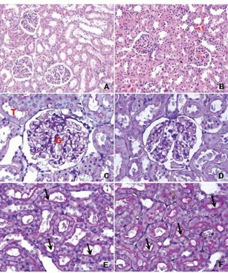
The histological appearance of the control group sec-tions was normal. The structure of glomeruli and prox-imal tubules was normal in PAS stained sections. The histological appearance of the Dxp group was similar to the control group (Figure 1); we found no statistically significant difference between the control and Dxp groups. In the DM group, however, infiltration, conges-tion, desquamation and swelling of tubule cells were observed in H & E stained sections (Figure 2a–d); the histopathological changes were significantly increased compared to the control group (p < 0.05). Table 2.Body and kidney weights.
Group 1 (control) Group 2 (Dxp) Group 3 (DM) Group 4 (DM + Dxp)
Body weight (g) 283.70 ± 33.50 275.70 ± 21.60 177.20 ± 33.70a 200.70 ± 44.10b
Kidney weight (g) 1.20 ± 0.11 1.12 ± 0.19 0.91 ± 0.18a 1.17 ± 0.09b
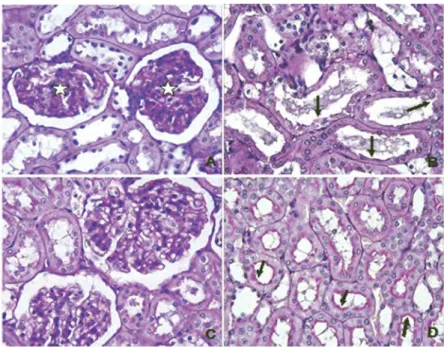
Histopathological changes were markedly reduced in the DM + Dxp group compared to the DM group (Figure 2e,f); we found no statistically significant differ-ence except for hydropic degeneration. Also, loss of villi and glomerulosclerotic changes were observed in PAS stained sections in the DM + Dxp group (Figure 3a,b). Glomerulosclerotic injury was increased in DM group compared to the control group (p < 0.0001) (Figure 3c,d). The glomerulosclerotic injury for the DM + Dxp group was decreased significantly compared to the DM group (p < 0.0001). The mean histopatholog-ical damage score and glomerulosclerotic index for all groups are shown inTables 3and4.
Biochemistry
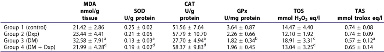
The MDA level was significantly higher for the DM group (32.58 ± 7.91 nmol/g tissue) than for the control group (21.42 ± 2.86 nmol/g tissue) (p < 0.05) and a statistically significant decrease in SOD and CAT levels
was detected in DM group (SOD: 0.13 ± 0.03 U/g protein, CAT: 27.70 ± 4.94 U/g protein) compared to the control group (SOD: 0.25 ± 0.02 U/g protein, CAT: 51.56 ± 7.64 U/g protein) (p < 0.05). The GSH-Px level in the DM group (1.82 ± 0.34 U/mg protein) was significantly decreased compared to the control group (3.64 ± 0.87 U/mg protein) (p < 0.05). The kidney MDA level was significantly lower for the DM + Dxp group (21.99 ± 4.28 nmol/g tissue) than for the DM group (p < 0.05). SOD and CAT levels were signifi-cantly higher for the DM + Dxp group (SOD: 0.19 ± 0.02 U/g protein, CAT: 58.37 ± 9.83 U/g protein) than for the DM group (SOD: 0.13 ± 0.03 U/g protein, CAT: 27.70 ± 4.94 U/g protein) (p < 0.05). The differ-ence in GSH-Px level between the DM + Dxp and DM groups was not statistically significant. We found a significant difference in TOS level between the DM + Dxp and DM groups (p < 0.05); the DM group exhibited a higher level of TOS. The biochemical results for all groups are shown in Table 5.
Figure 1.a) Control group: normal histological appearence of kidney tissue. H & E. x 20. b) Dxp group: normal histological appearence. H & E. x 20. c) Control group: intact glomerulus (G) and tubules (T). PAS. x 40. d) Dxp group: glomeruli were normal. PAS. x 40. e) Control group: villi of proximal tubules generally were normal (arrows). PAS. x 40. f) Dxp group: proximal tubules were normal (arrows). PAS. x 40. All magnifications refer to objective lens only.
Discussion
Hyperglycemia causes oxidation of glucose, glycation of proteins and activation of the polyol pathway. These changes accelerate generation of ROS and increase oxida-tive modification of lipids, DNA and proteins. Oxidaoxida-tive stress appears to play an important role in the develop-ment of complications of diabetes such as nephropathy and other long term problems (Osawa and Kato2005).
The characteristics of diabetic nephropathy include glomerular hyperfiltration, glomerular and renal hyper-trophy, increased urinary albumin excretion, increased basement membrane thickness and mesangial expan-sion (Lakshmanan et al. 2011; Zhou et al. 2012). We found that diabetic rats exhibited signs of diabetic nephropathy including inflammatory cell infiltration, congestion, tubule desquamation, swelling of tubular cells and an increased glomerulosclerotic index.
Dxp is oxidized to PA within tissues. PA and its deriva-tives increase the level of GSH; coenzyme A (CoA), espe-cially mitochondrial CoA; and adenosine-5ʹ- triphosphate
(ATP) synthesis within the cell (Slyshenkov et al. 2001,
2004). PA and its derivatives participate in cellular defense, in repair systems against OS and in the inflammatory response (Slyshenkov et al.1995). PA is required for nor-mal epithelial functions owing to its role in metabolic pathways (Bayrak et al.2012). The protective effect of PA and its derivatives against cell damage caused by ROS has been reported elsewhere (Ermis et al.2013). We found that after daily 500 mg/kg Dxp injections i.p. for 6 weeks, all features of diabetic renal injury were decreased. Altintas et al. (2012) reported that Dxp treatment decreased ischemic renal injury and apoptosis. Ermis et al. (2013) reported that inflammation and collagen deposition induced by a bleomycin administration was decreased by Dxp. Zakaria et al. (2011) reported that Dxp produced amelioration of cerebral ischemia reperfusion induced brain injury (Bayrak et al. 2012). Gulle et al. (2014) reported that Dxp exhibited beneficial properties including normalization of cytokine levels and pancreaticβ-cell and hepatocyte protection in diabetic rats. Demirci et al. (2014) Figure 2.a) DM group: congestion (arrows) was observed. H & E. x 20. b) DM group: inflammation area was evident (star). H & E. x 20. c) DM group: tubuler desquamation (arrows) and inflammation (star) were observed. H & E. x 20. d) DM group: cellular swelling in tubules (arrows) were observed. H & E. x 40. e) DM + Dxp group: tubule desquamation and cellular swelling were reduced compared to the DM group. H & E. x 20. f) DM + Dxp group: congestion and hydropic degeneration were reduced. H & E. x 40. All magnifications refer to objective lens only.
reported that cardiovascular tissue damage was reduced by Dxp administration in DM. Gulle et al. (2014) and Demirci et al. (2014) reported that Dxp may be a preventive agent against DM. We have found no report of histological study of effects of Dxp in diabetic nephropathy
Increased oxidative stress may play a role in the patho-genesis and progression of diabetic nephropathy (Maheshwari et al. 2016; Das and Ghosh 2017; Karapinar OS et al.2017). Both increased production of oxidants and decreased action of antioxidants participate in increasing oxidative stress in diabetic nephropathy. Elevated extra- and intracellular glucose concentrations cause oxidative stress (Matough et al. 2012). MDA, a product of lipid oxidation, is a commonly used index of oxidative stress (Draper and Hadley1990).
Etensel et al. (2007) investigated the effect of 250 or 500 mg/kg doses of Dxp on tissue damage and lipid oxidation in a rat testis torsion model and found a significant decrease in serum MDA levels in the 500 mg/kg Dxp group. We observed a significant increase in the amount of MDA in the kidneys of diabetic rats. Our finding is consistent with earlier studies (Obrosova et al.2003; Gui et al.2012).
The activity of the antioxidant enzymes, SOD, CAT and GSH-Px, decreased significantly in the kidney of diabetic rats. Our observations are consistent with earlier reports (Makni et al.2010; Gui et al.2012). We found that Dxp exhibited an antioxidant effect and decreased TOS significantly. GSH-Px is a component of the protective mechanism of cells against lipid oxidation and oxidative stress, both of which occur in diabetic nephropathy (Aouacheri et al.2015; Bhakkiyalakshmi et al.2016).
The mechanism of the antioxidant effect of Dxp is poorly understood. Dxp increased GSH levels within the tissue, which is crucial for protection against oxida-tive stress and play a role in inflammatory response (Karapinar et al. 2017). Our findings suggest that Dxp Figure 3.a) DM group: glomerulosclerosis was observed (stars). PAS. x 40. b) DM group: loss of villi in proximal tubules (arrows). PAS. x 40. c) DM + Dxp group: glomerulosclerosis was reduced compared to the DM group. PAS. x 40. d) DM + Dxp group: reduced loss of villi compared to the DM group. (arrows). PAS. x 40. All magnifications refer to objective lens only.
Table 3.Histopathological damage scores.
Infiltration Congestion Desquamation Loss of microvilli Hydropic degeneration
Group 1 (control) 0.50 ± 0.18 0.62 ± 0.18 1.00 ± 0.00e 0.50 ± 0.18g 0.62 ± 0.18h,k
Group 2 (Dxp) 1.00 ± 0.26 0.87 ± 0.22 1.12 ± 0.22f 0.62 ± 0.18 0.87 ± 0.22
Group 3 (DM) 2.00 ± 0.26a 2.00 ± 0.37c 2.37 ± 0.18 2.00 ± 0.26 2.12 ± 0.12m
Group 4 (DM + Dxp) 1.62 ± 0.26b 1.37 ± 0.18d 2.00 ± 0.00 1.75 ± 0.16 1.12 ± 0.12
Data are means ± SE of eight animals.ap = 0.002 vs. group 1,bp = 0.006 vs. group 1,cp = 0.012 vs. group 1,dp = 0.018 vs. group 1,ep < 0.0001 vs. groups 3
and 4,fp = 0.003 vs. groups 3 and 4,gp = 0.002 vs. groups 3 and 4,hp < 0.0001 vs. group 3,kp = 0.044 vs. group 4,mp = 0.001 vs. group 4.
Table 4.Glomerulosclerotic injury scores.
Group 1 (control) Group 2 (Dxp) Group 3 (DM) Group 4 (DM + Dxp) Sclerotic injury 0.39 ± 0.46 0.82 ± 0.06 1.90 ± 0.07a 1.32 ± 0.78b
Data are means ± SE of eight animals.ap < 0.0001 vs. groups 1 and 4,bp
< 0.0001 vs. group 1.
acts as a free radical scavenger. Dxp also reduced infil-tration of polymorphonuclear cells; therefore, it plays a role in the inflammatory response.
We found that STZ induced DM caused a decrease in GSH-Px levels and Dxp treatment increased GSH-Px levels in diabetic animals. We found increased TOS levels and decreased TAS levels in the DM group. TOS levels in the DM + Dxp group were reduced compared to the DM group; however, no significant increase in TAC levels was detected. Our findings are consistent with earlier reports (Altintas et al.2012; Ermis et al.2013; Turgut et al.2013; Cagın et al.2016) that suggest that DM increases oxidative stress, whereas Dxp decreases it.
Dxp is a safe and readily available agent that appears to ameliorate diabetic renal injury based on both bio-chemistry and histopathology. The beneficial changes in biochemical characteristics that we observed were consistent with changes in the histopathological appearance of the tissue. Dxp may be useful as an adjuvant for treatment of diabetic nephropathy.
Disclosure statement
No potential conflict of interest was reported by the authors.
Funding
Our project was supported by Turkish Scientific and Technological Research Council (TUBITAK) under Domestic University Students Research Projects Support Programme, 2209-A/2012.
References
Aebi H.1974. Catalase. In: Bergmeyer HU, editor. Methods of enzymatic analysis. New York: Academic Press; p. 673–677. Altıntas R, Parlakpinar H, Beytur A, Vardi N, Polat A, Sagir M, Odabas GP.2012. Protective effect of dexpanthenol on ischemia-reperfusion-induced renal injury in rats. Kid Blood Press Res. 36:220–230.
Aouacheri O, Saka S, Krim M, Messaadia A, Maidi I.2015. The investigation of the oxidative stress-related parameters in type 2 diabetes mellitus. Can J Diabetes. 39:44–49. Bancroft JD, Gamble M.2007. Bancroft’s theory and practice
of histological techniques. 6th ed. New York: Elsevier. Chapters 9 and 11: p. 121–161
Bayrak O, Seckiner I, Solakhan M, Karakok M, Erturhan SM, Yagci F.2012. Effects of intravesical dexpanthenol use on lipid peroxidation and bladder histology in a chemical cystitis animal model. Urology. 79:1023–1026.
Bhakkiyalakshmi E, Sireesh D, Sakthivadivel M, Sivasubramanian S, Gunasekaran P, Ramkumar KM.
2016. Anti-hyperlipidemic and anti-peroxidative role of pterostilbene via Nrf2 signaling in experimental diabetes. Eur J Pharmacol. 777:9–16.
Cagın YF, Parlakpinar H, Vardi N, Polat A, Atayan Y, Erdogan MA, Tanbek K.2016. Effects of dexpanthenol on acetic acid induced colitis in rats. Exp Ther Med. 12:2958–2964. Coskun O, Ocakcı A, Bayraktaroglu T, Kanter M. 2004.
Exercise training prevents and protects streptozotocin-induced oxidative stress andβ-cell damage in rat pancreas. Tohoku J Exp Med. 203:145–154.
Das K, Ghosh M.2017. Structured DAG oil ameliorates renal injury in streptozotocin-induced diabetic rats through inhibition of NF-κB and activation of Nrf2 pathway. Food Chem Toxicol. 100:225–238.
Demirci B, Demir O, Dost T, Birincioglu M.2014. Protective effect of vitamin B5 (dexpanthenol) on cardiovascular damage induced by streptozocin in rats. Bratisl Lek Listy. 115:190–196.
Draper H, Hadley M.1990. Malondialdehyde determination as index of lipid peroxidation. Methods Enzymol. 186:421–431. Elbe H, Vardi N, Esrefoglu M, Ates B, Yologlu S, Taskapan C.
2015. Amelioration of streptozotocin-induced diabetic nephropathy by melatonin, quercetin, and resveratrol in rats. Hum Exp Toxicol. 34:100–113.
Ermis H, Parlakpinar H, Gulbas G, Vardi N, Polat A, Cetin A, Kilic T, Aytemur ZA.2013. Protective effect of dexpanthe-nol on bleomycin induced pulmonary fibrosis in rats. Naunyn Schmiedebergs Arch Pharmacol. 386:1103– 1110.
Etensel B, Özkisacik S, Özkara E.2007. Dexpanthenol attenu-ates lipid peroxidation and testicular damage at experi-mental ischemia and reperfusion injury. Pediatr Surg Int. 23:177–181.
Giacco F, Brownlee M. 2010. Oxidative stress and diabetic complications. Circ Res. 107:1058–1070.
Gui D, Guo Y, Wang F, Liu W, Chen J, Chen Y, Huang J, Wang N.2012. Astragaloside IV, a novel antioxidant, pre-vents glucose-induced podocyte apoptosis in vitro and in vivo. PLoS One. 7:e39824.
Gulle K, Ceri NG, Akpolat M, Arasli M, Demirci B.2014. The effects of dexpanthenol in streptozotocin-induced diabetic rats: histological, histochemical and immunological evi-dences. Histol Histopathol. 29:1305–1313.
Harma M, Harma M, Erel O.1978. Increased oxidative stress in patients with hydatidiform mole. Swiss Med Wkly. 133:563–566.
Table 5.Mean oxidant and antioxidant parameters.
MDA nmol/g tissue SOD U/g protein CAT U/g protein GPx U/mg protein TOS mmol H2O2eq/l TAS mmol trolox eq/l
Group 1 (control) 21.42 ± 2.86 0.25 ± 0.02 51.56 ± 7.64 3.64 ± 0.87 14.47 ± 4.40 0.74 ± 0.08
Group 2 (Dxp) 23.44 ± 4.41 0.21 ± 0.05 57.79 ± 10.70 2.26 ± 0.66 12.10 ± 1.92 0.74 ± 0.09
Group 3 (DM) 32.58 ± 7.91a 0.13 ± 0.03a 27.70 ± 4.94a 1.82 ± 0.34b 18.91 ± 3.31c 0.57 ± 0.12a
Group 4 (DM + Dxp) 21.99 ± 4.28d 0.19 ± 0.02d 58.37 ± 9.83d 1.96 ± 0.45 13.04 ± 3.25d 0.65 ± 0.14
Karapinar OS, Pinar N, Ozgur T, Ozcan O, Bayraktar HS, Kurt RK, Nural O.2017. The protective role of dexpanthe-nol on the endometrial implants in an experimentally induced rat endometriosis model. Reprod. Sci. 24:285-290. Lakshmanan AP, Watanabe K, Thandavarayan RA, Sari FR, Harima M, Giridharan VV, Soetikno V, Kodama M, Aizawa Y. 2011. Telmisartan attenuates oxidative stress and renal fibrosis in streptozotocin induced diabetic mice with the alteration of angiotensin-(1,7) mas receptor expression associated with its PPAR-c agonist action. Free Rad Res. 45:575–584.
Liu B, Liu W-S, Han B-Q, Sun -Y-Y. 2007. Antidiabetic effects of chitooligosaccharides on pancreatic islet cells in streptozotocin-induced diabetic rats. World J Gastroenterol. 13:725–731.
Maheshwari R, Balaraman R, Sen AK, Shukla D, Seth A.
2016. Effect of concomitant administration of coenzyme Q10 with sitagliptin on experimentally induced diabetic nephropathy in rats. Ren Fail. 13:1–10.
Makni M, Sefi M, Fetoui H, Garoui EM, Gargouri NK, Boudawara T, Zeghal N. 2010. Flax and pumpkin seeds mixture ameliorates diabetic nephropathy in rats. Food Chem Toxicol. 48:2407–2412.
Matough FA, Budin SB, Hamid ZA, Alwahaibi N, Mohamed J. 2012. The role of oxidative stress and anti-oxidants in diabetic complications. Sultan Qaboos Univ Med J. 12:5–18.
Militante JD, Lombardini JB, Schaffer SW.2000. The role of taurine in the pathogenesis of the cardiomyopathy of insulin-dependent diabetes mellitus. Cardiovasc Res. 46:393–402. Obrosova IG, Fathallah L, Liu E, Nourooz-Zadeh J. 2003.
Early oxidative stress in the diabetic kidney: effect of DL-alpha-lipoic acid. Free Rad Biol Med. 34:186–195. Osawa T, Kato Y.2005. Protective role of antioxidative food
factors in oxidative stress caused by hyperglycemia. Ann NY Acad Sci. 1043:440–451.
Paglia DE, Valentine WN.1967. Studies on the quantitative and qualitative characterization of erythrocyte glutathione peroxidase. J Lab Clin Med. 70:158–170.
Patel R, Yago MD, Manas M, Martinez EV, Shervington A, Singh J. 2004. Mechanism of exocrine pancreatic insuffi-ciency in streptozotocin-induced diabetes mellitus in rat: effect of cholecystokinin-octapeptide. Molec Cell Biochem. 261:83–89.
Rakieten N, Rakieten ML, Nadkarni MV.2009. Studies on the diabetogenic action of streptozotocin. Cancer Chemother Rep. 29:91–98.
Slyshenkov VS, Rakowska M, Moiseenok AG, Wojtczak L.
1995. Pantothenic acid and its derivatives protect Ehrlich ascites tumor cells against lipid peroxidation. Free Rad Biol Med. 19:767–772.
Slyshenkov VS, Piwocka K, Sikora E, Wojtczak L. 2001. Pantothenic acid protects jurkat cells against ultraviolet light-induced apoptosis. Free Rad Biol Med. 30:1303–1310. Slyshenkov VS, Dymkowska D, Wojtczak L.2004. Pantothenic acid and pantothenol increase biosynthesis of glutathione by boosting cell energetics. FEBS Lett. 569:169–172. Sun Y, Oberley L, Li Y. 1988. A simple method for clinical
assay of superoxide dismutase. Clin Chem. 34:497–500. Tatsuki R, Satoh K, Yamamoto A, Hoshi K, Ichihara K.1997.
Lipid peroxidation in the pancreas and other organs in streptozotocin diabetic rats. Jpn J Pharmacol. 75:267–273. Turgut O, Ay AA, Turgut H, Ay A, Kafkas S, Dost T.2013. Effects of melatonin and dexpanthenol on antioxidant parameters when combined with estrogen treatment in ovariectomized rats. Age. 35:2229–2235.
Uchiyama M, Mihara M.1978. Determination of malonalde-hyde precursor in tissues by tiobarbituric acid test. Anal Biochem. 34:271–278.
Vardi N, Ucar M, Iraz M, Ozturk F. 2003. Morphological changes of rat endocrine pancreas in experimental dia-betes. Turk J Med Sci. 23:27–32.
Wei W, Liu Q, Tan Y, Liu L, Li X, Cai L. 2009. Oxidative stress, diabetes, and diabetic complications. Hemoglobin. 33:370–377.
Yazdanparast R, Ardestani A, Jamshidi S.2007. Experimental diabetes treated with Achillea santolina: effect on pancreatic oxidative parameters. J Ethnopharmacol. 112:13–18.
Zakaria MM, Hajipour B, Khodadadi A, Afshari F. 2011. Ameliorating effects of dexpanthenol in cerebral ischaemia reperfusion induced injury in rat brain. J Pak Med Assoc. 61:889–892.
Zhou Q, Liu K, Wu H, Chen L, Pouranan V, Yuan M, Xiao Z, Peng W, Xiang A, Tang R, et al. 2012. Spironolactone rescues Dot1a-Af9-mediated repression of endothelin-1 andımproves kidney ınjury in streptozotocin-ınduced dia-betic rats. PloS One. 7:e47360.