ISSN 1684–5315 © 2011 Academic Journals
Full Length Research Paper
Screening of the antioxidant, antimicrobial and DNA
damage protection potentials of the aqueous
extract of Asplenium ceterach DC
.Seyda Berk
1, Bektas Tepe
1*, Serdal Arslan
1, Cengiz Sarikurkcu
2 1Department of Molecular Biology and Genetics, Faculty of Science, Cumhuriyet University, TR-58140, Sivas, Turkey.
2
Department of Chemistry, Faculty of Science and Literature, Mugla University, TR-48000, Mugla, Turkey.
Accepted 17 June, 2011
In this study, in vitro antioxidant, antimicrobial and DNA damage protection potentials of the aqueous extract of Asplenium ceterach was firstly evaluated in addition to its total phenolic and flavonoid contents. Antioxidant activity was determined by five complementary test systems named βββ-β
carotene/linoleic acid, 1,1-diphenyl-2-picrylhydrazyl (DPPH) free radical scavenging, reducing power, chelating effect and phosphomolibdenum methods. Except for chelating effect assay, the extract exhibited remarkable activity potential. Antimicrobial activity was determined by agar-well diffusion and minimum inhibitory concentration tests. In this case, Shigella dysenteriae and Staphylococcus aureus were found to be the most sensitive microorganisms. According to the electrophoretic pattern of pBR322 plasmid DNA after treatment with UV and H2O2, supercoiled DNA was successfully protected in
the presence of 20 mg/ml or above concentrations of aqueous extract.
Key words: Antioxidant activity, antimicrobial activity, DNA damage protection, Asplenium ceterach.
INTRODUCTION
Antioxidants are compounds which, when present at low concentrations (as compared to those of oxidisable substrates), significantly delay, or even inhibit oxidation of the said substrates (Wood et al., 2006). In biological systems, antioxidants are assumed to protect cells against oxidative stress which might otherwise lead to cell damage (Buhler and Miranda, 2004; Fennema, 1996; Rivero-Perez et al., 2005; Valls-Belleset al., 2002). Coronary heart diseases, ulcers, cancers and neurode-generative diseases (e.g. Parkinson’s and Alzheimer’s), are but a few examples of human diseases and health conditions that can effectively be prevented (or, at least, delayed) via regular and balanced inclusion of antioxidants in one’s diet (Bamforth, 2002).
In the last decade, it was recognized that the hydroxyl radicals derived from superoxide radicals and hydrogen
*Corresponding author. E-mail: [email protected]. Tel: + 90 346 219 10 10. Ext: 2907. Fax: + 90 346 219 11 86.
peroxide is the most potent reactive oxygen radical which causes DNA damage by converting guanine into 8-hydroxyguanine (Gutteridge, 1984). In order to find new compounds to control oxidative DNA damage, which has been particularly implicated in carcinogenesis (Feig et al., 1994), an investigation was made on the effects of extracts obtained from some medicinal and/or aromatic plants on DNA cleavage.
Antioxidants also provide protection against the hazardous effects of ultraviolet radiation. Ultraviolet radiation causes damage to the skin, which may result in both precancerous and cancerous skin lesions and acceleration of skin ageing. Topical administration of enzymatic and non-enzymatic antioxidants is an effective strategy for protecting the skin against UV-mediated oxidative damage.
Secondary metabolites produced by plants constitute a major source of bioactive substances. The scientific interest in these metabolites has increased today with the search of new therapeutic agents from plant source, due to the increasing development of the resistance pattern of
microorganisms to most currently used antimicrobial drugs. According to World Health Report of infectious diseases 2000, overcoming antibiotic resistance is one of the major issues of the WHO for the present millennium. Hence, the last decade witnessed an increase in the investigation of plants as a source of human disease management (Prashanth et al., 2001). Furthermore, it is estimated that two-thirds of the world population rely on traditional remedies due to the limited availability and high prices of most pharmaceutical products (Tagboto and Townson, 2001).
Asplenium ceterach (syn. Ceterach officinarum) is a fern species commonly known as rusty back. It is characterized by a short rhizome which gives rise to several green fronds that have a pinnated lamina with trichomes on the abaxial (lower) surface, but not the adaxial (upper) one. These trichomes (hairs) are orange-brown in color, hence the name "rusty back". The petiole is shorter than the corpus of the leaf. This species is found in Western and Central Europe, including the Mediterranean region. It is associated with fissures in carbonate rocks and also grows on the mortar of stone and brick walls. This fern species has been used medicinally as a diuretic (GRIN, 2010).
We reported herein, the investigation of the aqueous extracts of antioxidant, antimicrobial and DNA damage protection potentials of A. ceterach from Turkey. As far as our literature survey could ascertain, no report is available in the literature for the biological activities of this plant species evaluated here.
MATERIALS AND METHODS
Plant material and preparation of extracts
A. ceterach was collected from Sogutlugol plateau, Duzici, Osmaniye (C6), Turkey (923 m.) in 14-04-2009. Plant sample was deposited at the herbarium of Biology Department, Cumhuriyet University (CUFH Voucher No: ED 15628). Five grams of powdered aerial parts of the plant material was suspended and extracted with 100 ml of distilled water with shaking at 25°C for 24 h. The extract was centrifuged at 58,300 g for 30 min and the supernatant was freeze dried under vacuum to obtain a powder of the water extracts. Extract yield was calculated as 6.42% (w/w).
Total antioxidant activity by ββββ-carotene–linoleic acid method In this assay, antioxidant capacity is determined by measuring the inhibition of the volatile organic compounds and the conjugated diene hydroperoxides arising from linoleic acid oxidation (Dapkevicius et al., 1994). A stock solution of β-carotene–linoleic acid mixture was prepared as follows: 0.5 mg β-carotene was dissolved in 1 ml of chloroform (HPLC grade). 25 µl linoleic acid and 200 mg Tween 40 was added. Chloroform was completely evaporated using a vacuum evaporator. Then, 100 ml of oxygenated distilled water was added with vigorous shaking; 2.5 ml of this reaction mixture was dispersed to test tubes and 0.5 ml of the extracts (2 mg/ml) in water were added and the emulsion system was incubated for up to 2 h at 50°C. The same procedure was repeated with the positive control BHT, BHA and a blank. After
this incubation period, absorbance of the mixtures was measured at 490 nm. Measurement of absorbance was continued until the color of β-carotene disappeared. The bleaching rate (R) of β-carotene was calculated according to Equation 1:
R = ln (a/b)/t (1) Where, ln = natural log, a = absorbance at time 0, b = absorbance at time t (120 min) (Cheung et al., 2003). The antioxidant activity (AA) was calculated in terms of percent inhibition relative to the control using Equation (2):
AA = [(Rcontrol – RSample) / Rcontrol] x 100 (2) Antioxidative activities of the extracts were compared with those of BHT and BHA at 2 mg/mland blank consisting of only 0.5 ml water.
Scavenging effect on 1,1-diphenyl-2-picrylhydrazyl (DPPH)
The hydrogen atoms or electrons donation ability of the corresponding extracts and some pure compounds were measured from the bleaching of purple colored methanol solution of DPPH. The effect of water extracts on DPPH radical was estimated according to Hatano et al. (1988). 1 ml of various concentrations (0.2 to 1.0 mg/ml) of the extracts in water was added to 1 ml of DPPH radical solution in methanol (final concentration of DPPH was 0.2 mM). The mixture was shaken vigorously and allowed to stand for 30 min; the absorbance of the resulting solution was measured at 517 nm with a spectrophotometer (Shimadzu UV-1601, Kyoto, Japan). Inhibition of free radical DPPH in percent (I%) was calculated in the following way:
I% = 100 x (AControl – ASample) /AControl
Where, AControl is the absorbance of the control reaction (containing all reagents except the test compound), and ASample is the absorbance of the test compound. BHT and BHA were used as control.
Reducing power
The reducing power was determined according to the method of Oyaizu (1986). Each extract (0.2 to 1.0 mg/ml) in water (2.5 ml) was mixed with 2.5 ml of 200 mM sodium phosphate buffer (pH 6.6) and 2.5 ml of 1% potassium ferricynide and the mixture was incubated at 50°C for 20 min. Then, 2.5 ml of 10% trichloroacetic acid were added, and the mixture was centrifuged at 200 g (MSE Mistral 2000, London, UK) for 10 min. The upper layer (2.5 ml) was mixed with 2.5 ml of deionized water and 0.5 ml of 0.1% ferric chloride. Finally, the absorbance was measured at 700 nm against a blank. BHT and BHA were used as control.
Chelating effects on ferrous ions
The chelating effect was determined according to the method of Dinis et al. (1994). Briefly, 1 ml (2 mg/ml) of the extracts in water was added to 1 ml of water and a solution of 2 mM FeCl2 (0.05 ml).
The reaction was initiated by the addition of 5 mM ferrozine (0.2 ml). Then, the mixture was shaken vigorously and left at room temperature for 10 min. Absorbance of the solution was measured spectrophotometricaly at 562 nm. The inhibition percentage of ferrozine-Fe2+ complex formation was calculated by using the formula given below:
Where, AControl is the absorbance of the control (the controlcontained FeCl2 and ferrozine complex formation molecules) and ASample is the absorbance of the test compound. EDTA was used as a control.
Phosphomolibdenum method
Antioxidant activity of the aqueous extract of A. ceterach by this way was determined according to the method of Prieto et al. (1999). An aliquot of 0.1 ml of the sample solution containing aqueous extract of A. ceterach was combined in an Eppendorf tube with 1.0 ml of reagent solution (0.6 M sulfuric acid, 28 mM sodium phosphate, and 4 mM ammonium molybdate). The tubes were capped and incubated in a thermal block at 95°C for 90 min, after the samples had cooled to room temperature, the absorbance of the aqueous solution was measured at 695 nm against a blank. A typical blank solution contained 1.0 ml of the reagent solution and the appropriate volume of the same solvent used for the sample, and it was incubated under the same conditions as the rest of the samples. For samples of unknown composition, water soluble anti-oxidant capacities were expressed as equivalents of ascorbic acid.
Assay for total phenolics
Total phenolic constituent of the water extracts were determined by employing the methods given in the literature (Chandler and Dodds, 1983; Slinkard and Singleton, 1977) involving Folin-Ciocalteu reagent and gallic acid as standard. 1 ml of extract solution containing 2000 µg extract was added to a volumetric flask. 45 ml distilled water and 1 ml Folin-Ciocalteu reagent was added and flask was shaken vigorously. After 3 min, 3 ml of Na2CO3 (2%)
solution was added and the mixture was allowed to stand for 2 h by intermittent shaking. Absorbance was measured at 760 nm. The concentrations of phenolic compounds were calculated according to the following equation that was obtained from the standard gallic acid graph:
Absorbance = 0.0258 gallic acid (µg) - 0.005 (R2: 0.9967)
Assay for total flavonoids
Total flavonoid content was determined using the Dowd method as adapted by Arvouet-Grand et al. (1994). Briefly, 1 ml of 2% aluminium trichloride (AlCl3) in methanol was mixed with the same
volume of the water extracts (2000 µg). Absorption readings at 415 nm were taken after 10 min against a blank sample consisting of a 1 ml extract solution with 1 ml methanol without AlCl3. The
concentrations of flavonoid compounds were calculated according to the following equation that was obtained from the standard quercetin graph:
Absorbance = 0.0322 quercetin (µg) – 0.005 (R2: 0.997)
Antimicrobial activity Microbial strains
Aqueous extract of the plant species was tested against a panel of microorganisms including Salmonella typhi (NCTC 9394), Pseudomonas aeruginosa (ATCC 27853), Shigella boydii (NCTC 9359), Shigella dysenteriae (NCTC 9762), Bacillus subtilis (ATCC 6633), Klebsiella pneumoniae (NCTC 5046), Staphylococcus aureus (ATCC 25923), Escherichia coli (ATCC 35218), Proteus vulgaris (RSHM 96022), Corynebacterium diphteriae (RSHM 633) and Canfida albicans (ATCC 10231). Bacterial strains were cultured
overnight at 37°C in Mueller Hinton agar (MHA). C. albicans was cultured overnight at 30°C in Sabouraud dextrose agar.
Agar well diffusion method
In agar-well diffusion method, aqueous extract was weighed and dissolved in phosphate buffer saline (PBS; pH 7.0 to 7.2) and dimethylsulphoxide (DMSO) (Merck, Germany) (10 mg/ml), respectively, followed by sterilisation using a 0.45 µm membrane filter (NCCLS, 1999). Each microorganism was suspended in sterile saline and diluted at ca. 106 colony forming unit (cfu) per ml. They
were “flood-inoculated” onto the surface of Mueller Hinton agar (MHA). The wells (six mm in diameter) were cut from the agar and 0.06 ml of the extract solution was put into them. After incubation for 24 h at 37°C, all plates were examined for any zone of growth inhibition, and the diameter of these zones were measured in millimetres. All the tests were performed in triplicate. Gentamycin and nystatin were used as positive control agents in parallel to the experiments.
Minimum inhibitory concentration (MIC)
A broth microdilution broth susceptibility assay was used as recommended by NCCLS, for the determination of the MIC (NCCLS, 1999). All tests were performed in Mueller Hinton broth (MHB; BBL) supplemented with Tween 80 (Merck, Germany) detergent (final concentration of 0.5%, v/v) with the exception of the yeasts (Sabouraud dextrose broth-SDB + Tween 80). Bacterial strains were cultured overnight at 37°C in MHA and the yeasts were cultured overnight at 30°C in SDB. Test strains were suspended in MHB to give a final density of 5 x 10⁵ cfu ml-1 and these were confirmed by viable counts. Geometric dilutions ranging from 0.036 to 72.00 mg ml1- of the extract were prepared in a 96-well microtiter plate, including one growth control (MHB + Tween 80) and one sterility control (MHB + Tween 80 + test oil). Plates were incubated under normal atmospheric conditions at 37°C for 24 h for bacteria and at 30°C for 48 h for the yeasts. The bacterial growth is indicated by the presence of a white “pellet” on the well bottom.
DNA damage protection potential
DNA damage protection activities of the extracts were evaluated on pBR322 plasmid DNA (vivantis). Plasmid DNA was oxidized with H2O2 + UV treatment in the presence of extracts and checked on
1% agarose gels according to Russo et al. (2000) after some modifications. In brief, the experiments were performed in a volume of 10 µl in a microfuge tube containing 3 µl pBR322 plasmid DNA (172 ng/µl), 1 µl of 30% H2O2, and 5 µl of extract in the
concentrations of 5, 10, 20, 40 and 50 mg/ml, respectively. The reactions were initiated by UV irradiation and continued for 5 min on the surface of a UV transilluminator (DNR-IS) with an intensity of 8000 µW/cm2 at 302 nm at room temperature. After irradiation, the reaction mixture (10 µl) along with gel loading dye (6×) was loaded on a 1% agarose gel for electrophoresis. Untreated pBR322 plasmid DNA was used as a control in each run of gel electrophoresis along with partially treated plasmid, that is, only UV or only H2O2 treatment. Gels were stained with EtBr and
photo-graphed with the Gel documentation system (DNR-IS, MiniBIS Pro).
RESULTS AND DISCUSSION Antioxidant activity
Table 1. Antioxidant activity, total phenolic and flavanoid content of the aqueous extract of A. ceterach1.
Sample Test system
β ββ
β-carotene/linoleic acid assay (Inhibition, %)
A. ceterach 94.62 ± 0.13 BHA 86.48 ± 1.93 BHT 92.14 ± 0.15 DPPH assay (%) 0.2 mg/ml 0.4 mg/ml 0.8 mg/ml A. ceterach 57.86 ± 1.29 85.52 ± 0.32 84.73 ± 0.23 BHA 92.83 ± 0.84 - - BHT 81.41 ± 0.00 - -
Reducing power (absorbance at 700 nm) 0.2 mg/ml 0.4 mg/ml 1.0 mg/ml
A. ceterach 0.328 ± 0.014 0.743 ± 0.035 1.793 ± 0.012 BHA 2.303 ± 0.064 - - BHT 1.258 ± 0.121 - - Chelating effect (%) A. ceterach 9.52 ± 2.61 EDTA 99.74 ± 0.15
Phosphomolibdenum method (µµµg ascorbic acid/mg extract) µ
A. ceterach 167.55 ± 5.64
Total phenolic content (µµµg GAEs/mg extract)µ 2 Total flavonoid content (µµµg QEs/mg extract)µ 3
95.14 ± 2.46 29.69 ± 0.11
1 Values expressed are means ± S.D. of three parallel measurements; 2GAEs, gallic acid equivalents; 3QEs, quercetin
equivalents.
easily oxidized by the oxygen in the air. This auto-oxidation leads to the occurrence of chain reactions with the formation of coupled double bonds, and at a later stage also to obtain secondary products, such as alde-hydes, ketones and alcohols. Using the β-carotene/ linoleic acid method, aqueous extract of A. ceterach showed remarkable antioxidant activity (Table 1). As can be seen from the table, antioxidant activity was measured as 94.62% which is almost equal to the synthetic antioxidant BHA and BHT.
The radical scavenging of the extracts was tested using a methanolic solution of the ‘‘stable’’ free radical, DPPH. Unlike laboratory-generated free radicals such as the hydroxyl radical and superoxide anion, DPPH has the advantage of being unaffected by certain side reactions, such as metal ion chelation and enzyme inhibition (Amarowicz et al., 2004). A freshly prepared DPPH solution exhibits a deep purple color with absorption maximum at 517 nm. This purple color generally fades/ disappears when an antioxidant is present in the medium. Thus, antioxidant molecules can quench DPPH free radicals (by providing hydrogen atoms or by electron donation, conceivably via a free-radical attack on the DPPH molecule) and convert them to a colorless/
bleached product (2,2-diphenyl-1-hydrazine, or a substituted analogous hydrazine), resulting in a decrease in absorbance at 517 nm. Hence, the more rapidly the absorbance decreases, the more potent the antioxidant activity of the extract. Free radical scavenging is one of the known mechanisms by which antioxidants inhibit lipid oxidation. This test is a commonly employed assay in antioxidant studies of specific compounds or extracts across a short time scale. The radical scavenging activity values of water extracts from the species evaluated here are presented in Table 1. From the analysis of Table 1, we can conclude that the scavenging effects of aqueous extract on DPPH radicals increased dose dependently. Free radical scavenging capacity of the extract was almost equal for the concentrations of 0.4 and 0.8 mg/ml (approximately 85%). However, the scavenging effects of BHA and BHT (0.2 mg/ml) were 92.83 ± 0.84 and 81.41 ± 0.00, respectively.
In this study, assay of reducing activity was based on the reduction of Fe3+/ferricyanide complex to the ferrous form in the presence of reductants (antioxidants) in the tested samples. The Fe2+ was then monitored by measuring the formation of Perl’s Prussian blue at 700 nm (Oyaizu, 1986).
Table 1 shows the reducing power of the aqueous extract as a function of their concentration. The reducing power of the extract increased with concentration. Reducing power of A. ceterach was found as 1.793 ± 0.012 nm at 1.0 mg/ml concentration. Data obtained from the synthetic antioxidants BHA and BHT were also recorded as 2.303 ± 0.064 and 1.258 ± 0.121 nm, respectively at 0.2 mg/ml concentration.
Metal ions can initiate lipid peroxidation and start a chain reaction that leads to the deterioration of food (Gordon, 1990). The catalysis of metal ions also correlates with incidents of cancer and arthritis (Halliwell et al., 1995). Ferrous ions, the most effective pro-oxi-dants, are commonly found in food systems (Yamaguchi et al., 1998). In this study, the chelating ability of the water extracts toward ferrous ions was also investigated. Table 1 also shows the chelating effects of the aqueous extract of A. ceterach. In this study, EDTA was also used as standard on the ferrous ions. The extract showed weak chelating capacity (9.52% ± 2.61). Chelating effect of EDTA was also found as 99.74% ± 0.15.
Ferrous ions could stimulate lipid peroxidation by Fenton reaction, and also accelerate peroxidation by decomposing lipid hydroperoxides into peroxyl and alkoxyl radicals that can themselves abstract hydrogen and perpetuate the chain reaction of lipid peroxidation (Halliwell, 1991). Chelating agents may serve as secon-dary antioxidants because they reduce the redox potential thereby stabilizing the oxidized form of the metal ions (Gordon, 1990).
The formation of a green colored complex of phosphate and Mo (V) was presented by Fiske and Subbarrow (1925) as the basis of a spectrophotometric method to determine inorganic phosphate. This method was later revised and modified by Chen et al. (1956). The requirement of a reducing agent to produce Mo (V) from the Mo (VI) supplied with the reagent mixture was suggested by Prieto et al. (1999) in the modification of this method for the determination of any reducing species. As can be seen from the Table 1, ascorbic acid content of the aqueous extract of A. ceterach was determined as 167.55 ± 5.64 µg/mg.
Phenolic compounds such as flavonoids, phenolic acids and tannins are considered to be major contributors to the antioxidant capacity of plants. These antioxidants also possess diverse biological activities, such as anti-inflammatory, anti-atherosclerotic and anti-carcinogenic. These activities may be related to their antioxidant activity (Chung et al., 1998). Thus, the total phenolic content of the samples was also evaluated, using the Folin–Ciocalteu method. As can be seen from Table 1, amounts of phenolic and flavonoid compounds were determined as 95.14 ± 2.46 and 29.69 ± 0.11 µg/mg, respectively.
When the results obtained from the total phenolic assay was compared with those found in other studies in the literature, polyphenolic compounds seems to have
important role in stabilizing lipid oxidation and to be associated with antioxidant activity (Gulcin et al., 2003; Yen et al., 1993). The phenolic compounds may contri-bute directly to antioxidative action (Duh et al., 1999). It is suggested that polyphenolic compounds have inhibitory effects on mutagenesis and carcinogenesis in humans, when up to 1.0 g is ingested daily from a diet rich in fruits and vegetables (Tanaka et al., 1998).
Antimicrobial activity
Antimicrobial activity of the aqueous extract of A.
ceterach was determined by two complementary test
systems named agar-well diffusion and minimum inhibitory concentration. Results obtained from these systems are presented in Table 2. As can be seen from the table, no activity was observed against P. aeruginosa,
K. pneumoniae, E. coli, P. vulgaris, C. diphteriae and C.
albicans. On the other hand, among the tested
microorganisms, the most sensitive one was S. aureus of which the MIC value was 9.00 µg/ml. This was closely followed by S. dysenteriae (18.00 µg/ml). Antimicrobial activities of gentamycin and nystatin were found as positive controls in parallel experiments.
It is not possible to make an exact decision on the selectivity of the extract, especially towards some micro-organisms. In general, Gram positive microorganisms are more sensitive against the antimicrobial agents than those of Gram negatives. On the other hand, when compared with the standard therapy methods (for example antibiotic treatments), activity of the plant species studied could be assumed to be moderate.
DNA damage protection potential
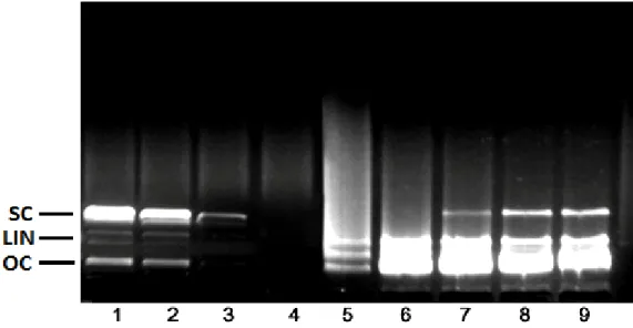
Figure 1 shows the electrophoretic pattern of DNA after UV-photolysis of H2O2 in the absence and presence of the aqueous extract of A. ceterach. DNA derived from pBR322 plasmid showed two bands on agarose gel electrophoresis (Figure 1, column 1); the faster moving band corresponded to the native form of supercoiled circular DNA (scDNA) and the slower moving band was the open circular form (ocDNA). The UV irradiation of DNA in the presence of H2O2 (Figure 1; column 3) resulted in the cleavage of ocDNA to a faint linear DNA and smears on the agarose gel, indicating that the OH· generated from UV-photolysis of H2O2 produced DNA strand scission.
As can be seen from the Figure 1, columns 5 to 9 shows the DNA damage protection potential of the aqueous extract of A. ceterach. In the presence of 5.0 mg/ml extract (Figure 1; column 5), a smear was observed on the agarose gel. In Figure 1 (column 6), two major bands named ocDNA and linear DNA were pro-tected by the presence of 10 mg/ml extract concentration.
Table 2. Antimicrobial activity of the aqueous extract of A. ceterach1.
Microorganism A. ceterach Antibiotics
4
A.W.D.2 M.I.C.3 Gentamycin Nystatin
S. typhi - 36.00 10.00 ± 0.45 n.t.5 P. aeruginosa - - 20.00 ± 1.06 n.t. S. boydii 13.00 ± 0.25 36.00 12.6 ± 0.20 n.t. S. dysenteriae 19.00 ± 1.40 18.00 13.5 ± 0. 00 n.t. B. subtilis 10.00 ± 0.40 72.00 29.00 ± 1.15 n.t. K. pneumoniae - - 20.00 ± 0.70 n.t. S. aureus 24.00 ± 0.74 9.00 23.00 ± 0.76 n.t. E. coli - - 16.00 ± 0.96 n.t. P. vulgaris - - 22.00 ± 1.40 n.t. C. diphteriae - - 23.00 ± 0.10 n.t. C. albicans - - n.t. 25.00 ± 0.90 1
Values expressed are means ± S.D. of three parallel measurements; 2A.W.D., agar well diffusion diameter of inhibition zone including well diameter of 6 mm; 3M.I.C., minimum ınhibitory concentration (as µg/ml);
4
concentrations of the antibiotics: 30 µg/well; 5 n.t., not tested.
Figure 1. Electrophoretic pattern of pBR322 plasmid DNA after treatment with UV and H2O2 in the presence
of A. ceterach aqueous extract. Column 1, plasmid DNA (3 µl) + dH2O (6 µl); Column 2, plasmid DNA (3 µl) +
dH2O (6 µl) + UV; Column 3, plasmid DNA (3 µl) + dH2O (6 µl) + H2O2 (1 µl); Column 4, plasmid DNA (3 µl) +
dH2O (6 µl) + UV + H2O2 (1 µl); Column 5, plasmid DNA (3 µl) + aqueous extract (5 mg/ml) (5 µl) + + UV +
H2O2 (1 µl); Column 6, plasmid DNA (3 µl) + aqueous extract (10 mg/ml) (5 µl) + + UV + H2O2 (1 µl); Column
7, plasmid DNA (3 µl) + aqueous extract (20 mg/ml) (5 µl) + + UV + H2O2 (1 µl); Column 8, plasmid DNA (3
µl) + aqueous extract (40 mg/ml) (5 µl) + + UV + H2O2 (1 µl); Column 9, plasmid DNA (3 µl) + aqueous
extract (50 mg/ml) (5 µl) + + UV + H2O2 (1 µl).
On the other hand, the addition of 20, 40 and 50 mg/ml A. ceterach extract to the reaction mixture conferred significant protection to the damage of native supercoiled DNA (Figure 1, columns 7, 8 and 9).
Conclusions
As can be seen from the results presented, aqueous
extract of A. ceterach showed remarkable antioxidant and DNA damage protection potential. Antioxidant and antimicrobial properties of plant species are among the major research topics for many scientists. By these studies, thousands of plant species have been identified for their antioxidant and antimicrobial potentials. How-ever, there are still many plant species that are still unidentified from this point of view. Moreover, DNA damage potentials of the plants are not evaluated
sufficiently and many plant species published for their biological activities have remained unexplored in terms of their DNA damage protection potentials. By exploring this activity, it is possible to suggest new and alternative phytochemicals to the food and cosmetic industries. To identify the phytochemicals responsible of the activities reported here, further chromatographic analyses need to be carried out.
ACKNOWLEDGEMENTS
The results presented here basically originated from Mrs. Seyda Berk’s M.Sc. thesis, which is not financially supported by any institution. Authors wish to thank Dr. Erol Donmez for the identification of the plant material collected.
REFERENCES
Amarowicz R, Pegg RB, Rahimi-Moghaddam P, Barl B, Weil JA (2004). Free-radical scavenging capacity and antioxidant activity of selected plant species from the Canadian prairies. Food Chem. 84: 551-562. Arvouet-Grand A, Vennat B, Pourrat A, Legret P (1994).
Standardisation d’un extrait de propolis et identification des principaux constituants. J. Pharm. Belg. 49: 462-468.
Bamforth CW (2002). Nutritional aspects of beer-a review. Nutr. Res. 22: 227-237.
Buhler DR, Miranda C (2004). Antioxidant activities of flavonoids. OR, USA: Department of Environmental and Molecular Toxicology, Oregon State University. <http://lpi.oregonstate.edu/f-w00/flavonoid.html/> Accessed 9.12.2004.
Chandler SF, Dodds JH (1983). The effect of phosphate, nitrogen and sucrose on the production of phenolics and solasidine in callus cultures of Solanum lacinitum. Plant Cell Rep. 2: 105.
Chen PS, Toribara TY, Warner H (1956). Microdetermination of phosphorus. Anal. Chem. 28: 1756-1763.
Cheung LM, Cheung PCK, Ooi VEC (2003). Antioxidant activity and total phenolics of edible mushroom extracts. Food Chem. 81: 249-255.
Chung KT, Wong TY, Huang YW, Lin Y (1998). Tannins and human health: a review. Crit. Rev. Food Sci. Nutr. 38: 421-464.
Dapkevicius A, Venskutonis R, Van Beek TA, Linssen PH (1998). Antioxidant activity of extracts obtained by different isolation procedures from some aromatic herbs grown in Lithuania. J. Sci. Food Agric. 77: 140-146.
Dinis TCP, Madeira VMC, Almeida LM (1994). Action of phenolic derivates (acetoaminophen, salycilate, and 5-aminosalycilate) as inhibitors of membrane lipid peroxidation and as peroxyl radical scavengers. Arch. Biochem. Biophys. 315: 161-169.
Duh PD, Tu YY, Yen GC (1999). Antioxidant activity of water extract of harn jyur (Chrysanthemum morifolium Ramat). Lebensm. Wiss. Technol. 32: 269-277.
Feig DI, Reid TM, Loeb LA (1994). Reactive oxygen species in tumorigenesis. Cancer Res. 54: S1890-S18904.
Fennema OR (1996). Food Chemistry (3rd ed.). New York: Marcel Dekker. pp. 780-782.
Fiske CH, Subbarow IP (1925). The colorimetric determination of phosphorus. J. Biol. Chem. 66: 375-379.
Germplasm Resources Information Network (GRIN) (2010). Taxon: Asplenium ceterach L. United States Department of Agriculture, Agric. Res. Serv. Beltsville Area. http://www.ars-grin.gov/cgi-bin/npgs/html/taxon .p. 411750.
Gordon MH (1990). The mechanism of antioxidant action in vitro. In: Hudson, B.J.F. (Ed.), Antioxidants. Elsevier Appl. Sci., London New York. pp. 1-18.
Gulcin Y, Buyukokuroglu ME, Oktay M, Kufrevioglu OY (2003). Antioxidant and analgesic activities of turpentine of Pinus nigra Arn. subsp. pallsiana (Lamb.) Holmboe. J. Ethnopharm. 86: 51-58. Gutteridge JMC (1984). Lipid peroxidation initiated by
superoxide-dependent hydroxyl radicals using complexed iron and hydrogen peroxide. FEBS. Lett. 172: 245-249.
Halliwell B (1991). The biological toxicity of free radicals and other reactive oxygen species. In O.I. Aruoma, & B. Halliwell (Eds.), Free radicals and food additives London: Taylor & Francis Ltd. pp. 37-57. Halliwell B, Murcia HA, Chirco S, Aruoma OI (1995). Free radicals and
antioxidants in food an in vivo: what they do and how they work. CRC Crit. Rev. Food Sci. Nutr. 35: 7-20.
Hatano T, Kagawa H, Yasuhara T, Okuda T (1988). Two new flavonoids and other constituents in licorice root: their relative astringency and radical scavenging effects. Chem. Pharm. Bull. 36: 1090-1097. NCCLS (National Committee for Clinical Laboratory Standards) (1999).
Performance standards for antimicrobial susceptibility testing. 9th International Supplement Wayne Pa. M100-S9.
Oyaizu M (1986). Studies on products of browning reactions: antioxidative activities of browning reaction prepared from glucosamine. Jpn. J. Nutr. 44: 307-315.
Prashanth D, Asha MK, Amit A (2001). Antibacterial activity of Punica granatum. Fitoter, 72: 171-173.
Prieto P, Pineda M, Aguilar M (1999). Spectrophotometric quantitation of antioxidant capacity through the formation of a phosphomolibdenum complex: Specific application to the determination of vitamin E. Anal. Biochem. 269: 337-341.
Rivero-Perez MD, Perez-Magarino S, Gonzalez-Sanjose ML, Valls V, Cordoner P, Muniz P (2005). Inhibition of induced DNA oxidative damage by beers: Correlation with the content of polyphenols and melanoidins. J. Agric. Food Chem. 53: 3637-3642.
Russo A, Acquaviva R, Campisi A, Sorrenti V, Di Giacomo C, Virgata G, Barcellona ML, Vanella A (2000). Bioflavonoids as antiradicals, antioxidants and DNA cleavage protectors. Cell Biol. Toxicol. 16: 91-98.
Slinkard K, Singleton VL (1977). Total phenol analyses: Automation and comparison with manual methods. Am. J. Enol. Vitic. 28: 49-55. Tagboto S, Townson S (2001). Antiparasitic properties of medicinal
plants and other natural occurring products. Adv. Parasitol. 50: 199-295.
Tanaka M, Kuei CW, Nagashima Y, Taguchi T (1998). Application of antioxidative maillrad reaction products from histidine and glucose to sardin products. Nippon Suisan Gakk. 54: 1409-1414.
Valls-Belles V, Muniz P, Gonzalez P, Gonzalez-Sanjose ML, Beltran S (2002). Mechanism of protection by epicatechin against tert-butylhydroperoxide induced oxidative cell injury in isolated rat hepatocytes and calf thymus DNA. Process. Biochem. 37: 659-664. Wood LG, Gibson PG, Garg ML (2006). A review of the methodology for
assessing in vivo antioxidant capacity. J. Sci. Food Agric. 86: 2057-2066.
Yamaguchi T, Takamura H, Matoba T, Terao J (1998). HPLC method for evalution of the free radical-scavenging activity of foods by using 1,1-dicrylhydrazyl. Biosci. Biotechnol. Biochem. 62: 1201-1204. Yen GC, Duh D, Tsai CL (1993). Relationship between antioxidant