559
Abstract
Background: Bisphosphonate (BP)-related osteonecrosis of the jaw (BRONJ) disease is rare, but there are serious side-effects of BP therapy in patients. In some patients, surgery is needed and could not be cured. A standard test is not available showing the risk of jaw osteonecrosis in routine use. The measurement of serum C-terminal telopeptide (CTX) levels has been used in diseases of BRONJ resorption and antiresorptive therapy.
Aim: This paper is aimed at investigating the relationship between traumatic procedures and presence of BP-related osteonecrosis.
Materials and Methods: Thirty male Wistar albino rats with weighing 200 ± 20 g were used for the experimental procedures. Rats were randomly divided into three groups each containing 10 rats as follows: Group 1 (traumatic extraction group), Group 2 (atraumatic extraction group), and Group 3 (control group). All groups, zoledronic acid (ZA) (0.3 mg/kg/week)[1]
was diluted with physiological saline and given subcutaneously for 2 months. After the 2 months, Group 1 was subjected to traumatic extraction of right first lower molars, and Group 2 was subjected to atraumatic extractions of the right first lower molars. Group 3 was subjected to no extractions as a control group. Animals were euthanized 32 days after tooth extractions, and the ZA administration protocol was maintained until the animals’ death. After sacrifice, blood samples were collected for C-terminal cross-linking telopeptide of type I collagen (CTX-1) levels, clinical and radiological findings were recorded. Results: The bone resorption marker CTX-1 showed a significant difference among the groups. CTX-1 was measured significantly higher in blood samples of Group 2 (4.15 ± 0.34; P = 0.001) than Group 1 (3.77 ± 0.34; P = 0.0001). No, statistically significant changes were found between Groups 1 and 2 as for clinical and radiological assessment. Conclusion: This study provides preliminary observations for the development of an animal model of BRONJ. Although clinical and radiological findings were not relevant, serum CTX values are reliable biochemical markers for predicting BRONJ and also atraumatic surgical procedures are important to prevent BRONJ.
Key words: Bisphosphonates, bone, osteonecrosis, prevention, serum C-terminal telopeptide level
Date of Acceptance: 23-Apr-2014
Address for correspondence: Dr. Kamil Serkan Ağaçayak,
Department of Maxillofacial Surgery, Faculty of Dentistry, Dicle University, 21280 Diyarbakır, Turkey.
E-mail: [email protected]
Introduction
Bisphosphonate (BP) is chemical analog of inorganic pyrophosphate. The BPs are widely used in the treatment of osteoporosis. BPs such as zoledronic acid (ZA) are used to treat bone disease in both multiple myeloma and breast cancer patients with bone metastasis.[2] BP‑related
osteonecrosis of the jaw (BRONJ) is a well‑known adverse reaction of BPs including ZA.[3] BRONJ develops after oral
trauma and is manifested by poor wound healing and soft tissue breakdown followed by necrosis of intra‑oral bone.[4]
Oxidative stress induces DNA damage and ZA treatment
Experimental investigation of relationship between
trauma and bisphosphonate‑related osteonecrosis
KS Ağaçayak, H Yuksel1, S Atılgan, M Koparal, MC Uçan, M Özgöz2, F Yaman, Y Atalay3, İ Acıkan
Department of Oral and Maxillofacial Surgery, Faculty of Dentistry, Dicle University, Diyarbakır, 1Department of
Biochemical, Faculty of Medicine, Dicle University, Diyarbakır, 2Department of Periodontology, Faculty of Dentistry,
Akdeniz University, Antalya, 3Department of Oral and Maxillofacial Surgery, Faculty of Dentistry, Kocatepe University,
Afyonkarahisar, Turkey
Access this article online Quick Response Code:
Website: www.njcponline.com DOI: 10.4103/1119-3077.141417 PMID: *******
560
induces a DNA damage response in oral keratinocytes that activates the ubiquitin‑mediated proteolysis of cell cycle regulators, resulting in cell cycle arrest and repressive effects on cell viability, proliferation, and epithelial turnover.[5]
The ubiquitin system is at the core of protein turnover as the main pathway for the degradation of proteins related to oxidative stress.[6] The BPs may induce the degenerative
effects through decrease of antioxidant, antioxidant trace element and increase of lipid peroxidation in oral epithelium. The most widely used serologic biochemical marker for stratifying the risk of BRONJ among BP users is the C‑terminal cross‑linking telopeptide of type I collagen (CTX‑1).[7] In situations of increased bone turnover,
type I collagen is degraded by osteoclasts, which releases C‑terminal telopeptide (CTX) molecules. Collagen I is one of the most abundant constituents of bone, reaching up to 90‑98% of the organic matrix.[8] It has been demonstrated
that declines in serum levels of CTX can be quantified within weeks of initiation of BP therapy.[9]
This paper is aimed at investigating the relationship between traumatic procedures and presence of BP‑related osteonecrosis.
Materials and Methods
Study design
Thirty male Wistar albino rats weighing 200 ± 20 g were used for the experimental procedures. Animals were housed in individual cages with bedding. Standard rat food and tap water were available ad libitum for the duration of the experiments unless otherwise noted. The animal room was maintained at a constant temperature of 22.0 ± 0.6°C by the air conditioner. A 12/12‑h light/dark cycle was maintained; with lights on at 0600 h. Experimental protocol of the study was approved by the Ethical Committee of University Medical Faculty of our tertiary center (2013‑31). Animals were maintained and used in accordance with the animal welfare act and guide for the care and use of laboratory animals. Rats were randomly divided into three groups as follows:
• Group 1 (traumatic extraction group) (n = 10) • Group 2 (atraumatic extraction group) (n = 10)and • Group 3 (no extraction, control group) (n = 10). An experimental model of BRONJ was induced in all 30 rats by weekly subcutaneous (SC) doses of ZA (Zomebon, Koçak Farma, İstanbul, Turkey) (0.3 mg/kg/week)[1] for 60 days.
The drug was dissolved in sterile physiological saline (0.9% NaCl) and diluted to the given concentration.
Surgical procedure
After 60 days of the pharmacologic therapy, Group 1 was subjected to traumatic extractions, and Group 2 was subjected to atraumatic extractions of the right first lower
molars (M1) by the same professional. Group 3 were subjected to no extractions. Teeth extraction was performed under general anesthesia by a combination of ketamine chloridrate (Ketalar; Eczacibasi, Turkey 0.08 mL/100 g body weight) and xylazine 2% (Rompun; Bayer, Germany; 0.04 mL/100 g body weight).
For dental extraction the rats of Group 1 (traumatic group) were placed in a dorsal position and fixed in a special device. The surrounding gingivae were carefully detached from the lower first molars with a dental explorer. Then, with a Hollenback Carver, the tooth was luxated and separated into two segments (mesial and distal) that were removed with a forceps adapted around the cervical line of the segments. In addition, the interdental septum was removed in this group. The gingivae were not sutured after tooth extraction. In Group 2 (atraumatic group), initially the rats were placed in a dorsal position and fixed in a special device. The surrounding gingivae were carefully detached from the lower first molars with a dental explorer. Then, with a Hollenback Carver, the tooth was luxated and separated into two segments (mesial and distal) that were removed with a forceps adapted around the cervical line of the segments. Irregular bone edges were shaved. Gingivae were sutured after tooth extraction. The rats of Group 3 (control group) were not submitted to tooth extraction.
After the surgical procedure, all animals received an intramuscular dose of ceftriaxon, 25 mg/kg (Rocephin®,
Roche, Turkey) and carprofen, 4 mg/kg (Rimadyl®,
Pfizer, Turkey). The ZA administration protocol was maintained until the animals’ death and animals were euthanized by cardiac puncture under anesthesia 32 days after tooth extractions.
Study data collected
Blood samples were collected on the time of sacrifice by cardiac puncture and centrifuged for plasma separation. For biochemical examination serum were frozen at −20°C until the measurement. Serum CTX‑1 level was analyzed according to manufacturer’s instructions by using CTX‑1 ELISA kit (Hangzhou Eastbiopharm Co., Ltd., China). Serum concentrations of CTX‑1 were expressed in a unit of ng/mL. Groups 1 and 2 rats were observed intraorally and findings were recorded after sacrifice procedure. Observed clinical findings were graded as follows:
• Grade 0 = complete mucosal healing
• Grade 1 = unhealed mucosa and bone trabeculae • Grade 2 = bone sequestrum and pus.
At the end of all procedures, cephalometric X‑ray of Group 1 and Group 2 rats was assessed for osteomyelitis. Radiological findings were graded as follows:
• Grade 0 = radiolucent view smaller than tooth extraction socket
561
• Grade 1 = radiolucent view same as tooth extraction socket
• Grade 2 = radiolucent view larger than tooth extraction socket.
All observations and scorings were performed by the same researcher.
Statistical analysis
The statistical calculations were carried out with the SPSS 18 software (SPSS 18 for Windows, SPSS Inc., Chicago, IL, USA). Clinical observations and radiological evaluation were compared among groups using Chi‑square test. Mann‑Whitney U‑tests for paired comparison were used to estimate the differences CTX values in among of the groups. Statistical significance level was accepted at
P < 0.05.
Results
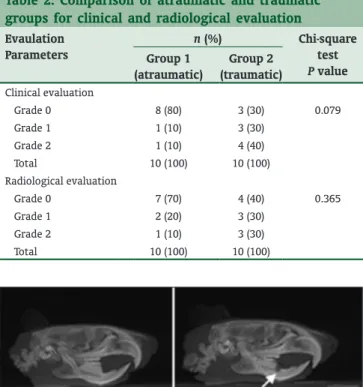
None of the rats died from all groups during the course of the experiment. All rats of all groups were not occurred unwanted condition until the end of the experiment, and none of the rats were excluded from the study. In general, animals tolerated the procedure well and demonstrated good hemostasis and rapid recovery from anesthesia. Gross evidence of the extraction site healing was seen in the majority of Group 2 animals. In contrast, in Group 1 open wounds and exposed bone were noted in rats.
Bone turnover marker
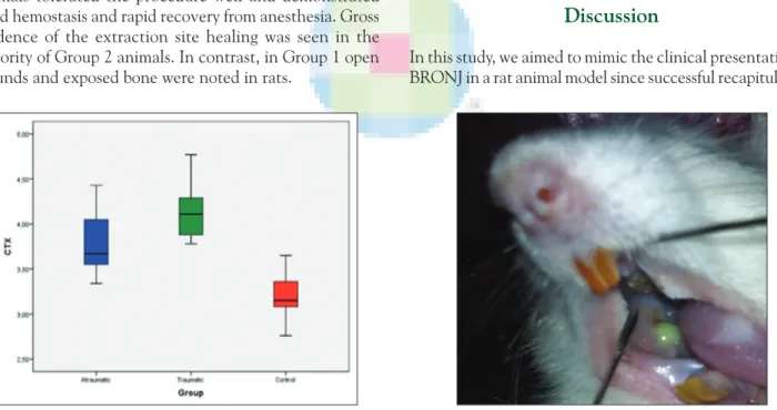
The bone resorption marker CTX‑1 showed a significant difference among groups (P = 0.0001). In comparison to the control group (Group 3) (3.19 ± 0.27), CTX‑1 increased in Group 1 (3.77 ± 0.34; P = 0.0001) and Group 2 (4.15 ± 0.34; P = 0.001) [Table 1 and Figure 1]. However, CTX‑1 was significantly higher in Group 2 than Group 1 [Table 1 and Figure 1].
Clinical and radiological assessment
No, statistically significant changes were found for clinical and radiologic findings between Group 1 and Group 2 (P ˃ 0.05) [Table 2]. As for the clinical evaluation, complete mucosal healing was observed in 80% of the rats in atraumatic extraction group while observed in 30% of the rats in traumatic extraction group. Conversely, bone sequestrum and pus were observed in 10% of the rats in atraumatic group, whereas observed in 40% of the rats in traumatic group [Figure 2]. As for the radiological evaluation, radiolucent view smaller than tooth extraction socket was observed in 70% of the rats in atraumatic extraction group, while observed in 40% of the rats in traumatic extraction group. Conversely, radiolucent view larger than tooth extraction socket was observed in 10% of the rats in atraumatic group, while observed in 30% of the rats in traumatic group [Figure 3].
Discussion
In this study, we aimed to mimic the clinical presentation of BRONJ in a rat animal model since successful recapitulation
Table 1: Comparison of CTX‑1 values with groups Evaulation parameter Group 1 (atraumatic) Group 2 (traumatic) Group 3 (control) Chi‑square test P value CTX 3.77±0.34 4.15±0.34 3.19±0.27 0.0001 P (Group 1 vs. Group 2)=0.021 P (Group 2 vs. Group 3)=0.001 P (Group 1 vs. Group 3)=0.0001 CTX=C-terminal telopeptide
Figure 1: Comparison of C-terminal cross-linking telopeptide of
562
of a BRONJ‑like indication in a rodent model will be useful for studying pathogenesis, as well as preventative and treatment strategies for BRONJ. Compatible with the literature, we have determined that in traumatic group of rats receiving ZA, CTX‑1 levels were higher than that of atraumatic rats and control group cases. However, although the clinical and radiological data were better on atraumatic group, the difference between traumatic and atraumatic cases was not statistically significant.
As can be seen with the increasing number of reports on BRONJ from the scientific community, many efforts have been made to obtain experimental models of these lesions.[10,11] Several hypotheses concerning the pathogenesis
of BRONJ have been proposed, but none has been accepted completely. However, analysis of data retrieved from clinical studies suggests that some cofactors may play a relevant role in the development of these lesions, even in the presence of BRONJ with no obvious comorbidity factors.[12] The
models of osteonecrosis of the jaw (ONJ)‑like lesions have been published in the literature, being developed by the association of BP therapy, tooth extraction and other cofactors, such as concomitant steroid use, vitamin D deficiency or increased socket damage.[11,13,14]
Although BRONJ can occur spontaneously, most of the reports have been associated with surgical procedures.[15,16]
In this study, we opted to use tooth extraction as a trigger, based on the fact that analysis of data retrieved from clinical
and in vivo studies suggested a strong relationship between tooth extraction and BRONJ.[17,18] The clinical diagnosis
of BRONJ is defined as an area of exposed bone in the maxillofacial region which did not heal within 8 weeks in a patient who is or was exposed to a BP and did not have radiation therapy to the craniofacial region.[19] Although
the alveolar healing phases are similar in both humans and rats, they occur more rapidly in rodents than in humans, lasting about one‑third of the time required for human healing.[20,21] Therefore, the 4 weeks bone exposure in
rats observed in this study could be considered a clinical equivalent concept of ONJ induced by BP in rats. To support this assumption, studies have shown transient impairment of BPs in alveolar socket healing, and these effects have not been extended beyond 14 days.[22] When
the rats were investigated in this regard it has been determined that rats with Grades 1 and 2 clinical picture can be diagnosed with BRONJ and the ratios of rats with BRONJ in traumatic and atraumatic groups respectively were 70% and 20%. Although there was not a statistically significant difference in regards to the clinical and radiological findings of these two groups, when the groups were evaluated for the presence of BRONJ clinically, the difference was statistically significant (P = 0.0001). Similarly, Marx had reported that in 110 (72.3%) of 152 BRONJ cases history tooth extraction was present with periodontal inflammation.[23]
Zoledronic acid has a strong affinity to bone mineral and exerts antiresorptive effects by targeting osteoclasts.[24]
Since osteoclasts play an important role in hematopoiesis, chronically suppressed osteoclasts would likely alter cellular responses during wound healing.[25] This could
be in connection with the development of the BRONJ. In this study, high dose of ZA was administered for an extended duration; both the dose and duration of the drug are considered as known risk factors for BRONJ. However, with this high doses and long duration of ZA treatment, in atraumatic rats BRONJ was investigated in 20% of cases demonstrating that ZA treatment is not the only factor determining BRONJ development. In literature, the risk of BRONJ development for patients on intravenous BPs is estimated at between 1% and 11%, which increases after longer treatment time and in patients on ZA.[26,27] The
higher ratios of BRONJ cases in atraumatic group in our study may be associated with longer treatment durations and/or ZA.
Bisphosphonates have been administered by non‑intravenous (IV) route in many other rodent studies.[28]
While the drug is usually administered IV in humans, we administered the drug SC in rats. In our experiments, we use SC injections, which could mean that less BP is entering the systemic circulation. With higher doses or intravenous administration, this could hypothetically constitute a negative influence on bone formation. Further, a prolonged
Table 2: Comparison of atraumatic and traumatic groups for clinical and radiological evaluation Evaulation Parameters n (%) Chi‑square test P value Group 1 (atraumatic) Group 2 (traumatic) Clinical evaluation Grade 0 8 (80) 3 (30) 0.079 Grade 1 1 (10) 3 (30) Grade 2 1 (10) 4 (40) Total 10 (100) 10 (100) Radiological evaluation Grade 0 7 (70) 4 (40) 0.365 Grade 1 2 (20) 3 (30) Grade 2 1 (10) 3 (30) Total 10 (100) 10 (100)
Figure 3: Cephalometric X-ray showing no lesion in the control
group (a), and necrosis on the teeth extraction area (b) b
563
effect of the drug with slow‑release effects could be the effect of a SC deposition compared to an intravenous administration.
Serum CTX values have been used as biochemical markers of bone formation and resorption. The first clinical application of CTX measurement for predicting BRONJ was reported by Marx et al.[29] Conversely, Kunchur et al.
concluded that CTX is not predictive of the development of BRONJ.[9] Biochemical markers of bone turnover provide
insight into the dynamic changes of the skeleton and are primarily used as research tools to study the pathogenesis and treatment of bone diseases.[30] Research using bone
biomarkers has suggested their clinical use to monitor the effect of antiresorptive therapy, predict bone loss and fracture in osteoporosis, predict complications of metastatic bone disease, and to identify the progression of joint damage in rheumatoid arthritis and the extent of bone involvement in metastatic cancer and multiple myeloma.[31,32] Bone
biomarkers have been reported to be especially relevant in patients who have a history of oral BP use. Because of differences in bony metabolism and aging rates between rats and humans, even a short exposure to ZA was sufficient to elicit changes in serum CTX level compared to untreated controls. Thus, in an attempt to increase the model’s reliability, we reduced the number of variables and demonstrated the development of experimental BRONJ‑like lesions using the combination of BPs therapy and different tooth extraction methods (traumatic or atraumatic) in this study.
The main limitation of this study was that we did not examine the necrosis of mandible histopathologically in the diagnosis of BRONJ. However, BRONJ was diagnosed clinically according to its previous definitions. On the other hand, although the clinical and radiological findings of the atraumatic group were better, the difference between traumatic and atraumatic groups was not statistically significant, which in turn may be the result of the low number of rats included in the study. Larger studies may give more precise data about this topic.
Conclusion
This study provides preliminary observations for the development of an animal model of BRONJ. Although clinical and radiological findings were not relevant, CTX‑1 values and the prevalence of clinically diagnosed BRONJ cases significantly differed in extraction groups with or without trauma. The results also suggest that traumatic extractions with periodontal changes may predispose patients to higher CTX levels and also BRONJ. We concluded that tooth extraction with trauma effects on levels of serum CTX, which may be a marker for development jaw osteonecrosis in patients undergoing BP treatment.
References
1. Yamashita J, Koi K, Yang DY, McCauley LK. Effect of zoledronate on oral wound healing in rats. Clin Cancer Res 2011;17:1405‑14.
2. Coscia M, Quaglino E, Iezzi M, Curcio C, Pantaleoni F, Riganti C, et al. Zoledronic acid repolarizes tumour‑associated macrophages and inhibits mammary carcinogenesis by targeting the mevalonate pathway. J Cell Mol Med 2010;14:2803‑15. 3. Kuroshima S, Go VA, Yamashita J. Increased numbers of nonattached osteoclasts after long‑term zoledronic acid therapy in mice. Endocrinology 2012;153:17‑28. 4. Ravosa MJ, Ning J, Liu Y, Stack MS. Bisphosphonate effects on the behaviour of oral epithelial cells and oral fibroblasts. Arch Oral Biol 2011;56:491‑8. 5. Ohnuki H, Izumi K, Terada M, Saito T, Kato H, Suzuki A, et al. Zoledronic acid
induces S‑phase arrest via a DNA damage response in normal human oral keratinocytes. Arch Oral Biol 2012;57:906‑17.
6. Demasi M, Laurindo FR. Physiological and pathological role of the ubiquitin‑proteasome system in the vascular smooth muscle cell. Cardiovasc Res 2012;95:183‑93.
7. O’Connell JE, Ikeagwani O, Kearns GJ. A role for C‑terminal cross‑linking telopeptide level to predict the development of bisphosphonate‑related osteonecrosis of the jaws following oral surgery? Ir J Med Sci 2012;181:237‑42. 8. Lazarovici TS, Mesilaty‑Gross S, Vered I, Pariente C, Kanety H, Givol N, et al. Serologic bone markers for predicting development of osteonecrosis of the jaw in patients receiving bisphosphonates. J Oral Maxillofac Surg 2010;68:2241‑7. 9. Kunchur R, Need A, Hughes T, Goss A. Clinical investigation of C‑terminal cross‑linking telopeptide test in prevention and management of bisphosphonate‑associated osteonecrosis of the jaws. J Oral Maxillofac Surg 2009;67:1167‑73.
10. Aghaloo TL, Kang B, Sung EC, Shoff M, Ronconi M, Gotcher JE, et al. Periodontal disease and bisphosphonates induce osteonecrosis of the jaws in the rat. J Bone Miner Res 2011;26:1871‑82.
11. Hokugo A, Christensen R, Chung EM, Sung EC, Felsenfeld AL, Sayre JW, et al. Increased prevalence of bisphosphonate‑related osteonecrosis of the jaw with vitamin D deficiency in rats. J Bone Miner Res 2010;25:1337‑49.
12. Marx RE, Sawatari Y, Fortin M, Broumand V. Bisphosphonate‑induced exposed bone (osteonecrosis/osteopetrosis) of the jaws: Risk factors, recognition, prevention, and treatment. J Oral Maxillofac Surg 2005;63:1567‑75. 13. López‑Jornet P, Camacho‑Alonso F, Molina‑Miñano F, Gómez‑García F,
Vicente‑Ortega V. An experimental study of bisphosphonate‑induced jaws osteonecrosis in Sprague‑Dawley rats. J Oral Pathol Med 2010;39:697‑702. 14. Biasotto M, Chiandussi S, Zacchigna S, Moimas S, Dore F, Pozzato G,
et al. A novel animal model to study non‑spontaneous bisphosphonates
osteonecrosis of jaw. J Oral Pathol Med 2010;39:390‑6.
15. Conte‑Neto N, Bastos AS, Spolidorio LC, Marcantonio RA, Marcantonio E Jr. Oral bisphosphonate‑related osteonecrosis of the jaws in rheumatoid arthritis patients: A critical discussion and two case reports. Head Face Med 2011;7:7. 16. Ruggiero SL, Dodson TB, Assael LA, Landesberg R, Marx RE, Mehrotra B, et al. American Association of Oral and Maxillofacial Surgeons position paper on bisphosphonate‑related osteonecrosis of the jaw‑2009 update. Aust Endod J 2009;35:119‑30.
17. Thumbigere‑Math V, Sabino MC, Gopalakrishnan R, Huckabay S, Dudek AZ, Basu S, et al. Bisphosphonate‑related osteonecrosis of the jaw: Clinical features, risk factors, management, and treatment outcomes of 26 patients. J Oral Maxillofac Surg 2009;67:1904‑13.
18. Treister N, Sheehy N, Bae EH, Friedland B, Lerman M, Woo S. Dental panoramic radiographic evaluation in bisphosphonate‑associated osteonecrosis of the jaws. Oral Dis 2009;15:88‑92.
19. Advisory Task Force on Bisphosphonate‑Related Ostenonecrosis of the Jaws, American Association of Oral and Maxillofacial Surgeons. American Association of Oral and Maxillofacial Surgeons position paper on bisphosphonate‑related osteonecrosis of the jaws. J Oral Maxillofac Surg 2007;65:369‑76.
20. Bodner L, Kaffe I, Littner MM, Cohen J. Extraction site healing in rats. A radiologic densitometric study. Oral Surg Oral Med Oral Pathol 1993;75:367‑72. 21. Okamoto T, de Russo MC. Wound healing following tooth extraction.
Histochemical study in rats. Rev Fac Odontol Aracatuba 1973;2:153‑69. 22. Aguirre JI, Altman MK, Vanegas SM, Franz SE, Bassit AC, Wronski TJ. Effects
of alendronate on bone healing after tooth extraction in rats. Oral Dis 2010;16:674‑85.
23. Marx R. Oral and Intravenous Bisphosphonate‑Induced Osteonecrosis of the Jaws. Chicago: Quintessence Publishing Co.; 2007.
564
24. Green JR. Chemical and biological prerequisites for novel bisphosphonate molecules: Results of comparative preclinical studies. Semin Oncol 2001;28:4‑10. 25. Kollet O, Dar A, Shivtiel S, Kalinkovich A, Lapid K, Sztainberg Y, et al. Osteoclasts degrade endosteal components and promote mobilization of hematopoietic progenitor cells. Nat Med 2006;12:657‑64.
26. Durie BG, Katz M, Crowley J. Osteonecrosis of the jaw and bisphosphonates. N Engl J Med 2005;353:99‑102.
27. Hoff AO, Toth BB, Altundag K, Johnson MM, Warneke CL, Hu M, et al. Frequency and risk factors associated with osteonecrosis of the jaw in cancer patients treated with intravenous bisphosphonates. J Bone Miner Res 2008;23:826‑36.
28. Hardt AB. Bisphosphonate effects on alveolar bone during rat molar drifting. J Dent Res 1988;67:1430‑3.
29. Marx RE, Cillo JE Jr, Ulloa JJ. Oral bisphosphonate‑induced osteonecrosis: Risk factors, prediction of risk using serum CTX testing, prevention, and treatment. J Oral Maxillofac Surg 2007;65:2397‑410.
30. Looker AC, Bauer DC, Chesnut CH 3rd, Gundberg CM, Hochberg MC, Klee G,
et al. Clinical use of biochemical markers of bone remodeling: Current status
and future directions. Osteoporos Int 2000;11:467‑80.
31. Robins SP. Collagen turnover in bone diseases. Curr Opin Clin Nutr Metab Care 2003;6:65‑71.
32. Brown JE, Cook RJ, Major P, Lipton A, Saad F, Smith M, et al. Bone turnover markers as predictors of skeletal complications in prostate cancer, lung cancer, and other solid tumors. J Natl Cancer Inst 2005;97:59‑6.
How to cite this article: Agaçayak KS, Yuksel H, Atilgan S, Koparal M, Uçan MC, Özgöz M, et al. Experimental investigation of relationship between trauma and bisphosphonate-related osteonecrosis. Niger J Clin Pract 2014;17:559-64.