PERIPHERAL NERVE REGENERATION BY SYNTHETIC PEPTIDE NANOFIBERS
A THESIS SUBMITTED TO
THE GRADUATE SCHOOL OF ENGINEERING AND SCIENCE OF BILKENT UNIVERSITY
IN PARTIAL FULFILLMENT OF THE REQUIREMENTS FOR THE DEGREE OF
MASTER OF SCIENCE IN
MATERIALS SCIENCE AND NANOTECHNOLOGY
By Mevhibe Geçer
ii
PERIPHERAL NERVE REGENERATION BY SYNTHETIC
PEPTIDE NANOFIBERS
By Mevhibe Geçer August, 2016
We certify that we have read this thesis and that in our opinion it is fully adequate, in scope and in quality, as a thesis for the degree of Master of Science.
______________________________ Mustafa Özgür Güler (Advisor)
______________________________ Ayşe Begüm Tekinay (Co-Advisor)
______________________________
Çağlar Elbüken
______________________________ Yusuf Şükrü Çağlar
Approved for the Graduate School of Engineering and Science:
________________________________________ Levent Onural
iii
ABSTRACT
PERIPHERAL NERVE REGENERATION BY SYNTHETIC
PEPTIDE NANOFIBERS
Mevhibe Geçer
M.S. in Materials Science and Nanotechnology Advisor: Mustafa Özgür Güler
Co-Advisor: Ayşe Begüm Tekinay August, 2016
The peripheral nervous system (PNS) has a complex structure that consists of high numbers of nerve cells and communication networks between the central nervous system and the body parts. Unlike the central nervous system, the PNS exhibits a considerable capacity for regeneration; however, peripheral nerve injuries can nevertheless cause lifelong disability. Various methods are currently available for the treatment of nerve injuries, but autologous nerve grafting is considered as ‘the gold standard’. Donor site morbidity, neuroma formation and failure of functional recovery are some limitations of this technique, especially when used for the repair of long nerve gaps. Polymeric nerve conduits are clinically available alternatives to nerve grafting, and function by guiding the axonal growth and isolating the regenerating axon from the inhibitory environment present in the post-injury neuroma. In this thesis, we used peptide amphiphile molecules (PAs) that can self-assemble into the nanofibers and mimic both the structure and function of healthy ECM of nerve cells for sciatic nerve regeneration. Two bioactive PAs, LN-PA (derived from laminin) and GAG-PA
iv
(derived from glycosaminoglycan), were tested for their ability to induce neural regeneration in a rat sciatic nerve model. Hollow nerve conduits were filled with peptide nanofiber gels, and electrophysiology and histology results were compared with autologous graft treated groups. Our results show that bioactive peptide nanofibers are able to boost regeneration and functional motor and sensory recovery. Electromyography results demonstrated that better signal transmission was observed in peptide nanofiber treated groups compared with empty conduits and autograft treated groups. Histological assessments also confirmed that bioactive peptide nanofiber treated groups exhibited better axonal regeneration. These results suggest that these biologically active PA nanofiber gels may be used as a biomaterial for peripheral nerve regeneration in clinical practice.
Keywords: Extracellular matrix, peptide nanofibers, peripheral nerve regeneration,
v
ÖZET
SENTETİK PEPTİT NANOFİBERLERLE PERİFERİK SİNİR
REJENERASYONU
Mevhibe Geçer
Malzeme Bilimi ve Nanoteknoloji, Yüksek Lisans Tez danışmanı: Mustafa Özgür Güler
Eş Danışman: Ayşe Begüm Tekinay Ağustos, 2016
Periferik sinir sistemi çok sayıda sinir hücresi ve merkezi sinir sistemi ile vücudun diğer organları arasındaki iletişim ağını kapsayan oldukça kompleks bir yapıya sahiptir. Periferik sinir hasarları ömür boyu sürebilecek sakatlıklara neden olmasına rağmen, merkezi sinir sisteminin aksine, periferik sinir sisteminde rejenerasyon mümkündür. Günümüzde çeşitli tedavi yöntemleri uygulanmaktadır fakat otogreft yöntemi altın standart olarak bilinmektedir. Donör bölgede görülen morbidite, nöroma oluşumu ve fonksiyonel iyileşmedeki başarısızlıklar, uzun sinir boşluklarını tedavi etmek için otogreft yöntemi kullanıldığında meydana gelebilecek sorunlardan birkaçıdır. Alternatif olarak klinik uygulamalarda polimerik sinir tüpleri aksonların uzaması için bir yönlendirme ve inhibe edici ortamdan izole etme amacıyla kullanılmaktadır. Bu tezde, siyatik sinir rejenerasyonu için, kendiliğinden bir araya gelerek nanofiberler oluşturabilen ve hem fonksiyonel hem de yapısal olarak sağlıklı sinir hücrelerinin hücreler arası matrikslerini taklit edebilen peptit amfifil molekülleri kullanılmıştır. Bu peptit amfifillerden, LN-PA molekülünün biyoaktif sekansı laminin
vi
proteininden ve GAG-PA molekülünün biyoaktif sekansı glikozaminoglikandan türetilmiştir ve rat siyatik sinir hasarı modelinde rejenerasyona olan etkisini belirleyebilmek için kullanılmıştır. İçleri boş olan sinir tüpleri bu peptit nanfiber jeller ile doldurulmuştur, elde edilen elektrofizyoloji ve histoloji sonuçları otogreft uygulanan gruplar ile karşılaştırılmıştır. Sonuçlarımıza göre kullanılan biyoaktif peptit nanofiberlerin sinir rejenerasyonunu, motor ve duyusal fonksiyonların geri kazanımını desteklediği gösterilmiştir. Elektromiyografi sonuçlarına göre sinyal iletimi peptit nanofiber uygulanan gruplarda, boş tüp ve otogreft uygulaması yapılan gruplara göre daha iyidir. Ayrıca histoloji sonuçları ile aksonal rejenerasyonun biyoaktif peptit gruplarında daha iyi olduğu desteklenmiştir. Biyolojik olarak aktif peptit amfifil nanofiber jeller, klinik uygulamalarda periferik sinir rejenerasyonu için bir biyomalzeme olarak kullanılabilir.
Anahtar kelimeler: Hücreler arası ortam, peptit nanofiberler, periferik sinir
vii
ACKNOWLEDGEMENTS
Firstly, I would like to thank my advisors Prof. Mustafa Özgür Güler and Prof. Ayşe Begüm Tekinay for giving this unique opportunity to win valuable experiences. I also would like to thank all NBT and BML members for their precious friendships, contributions and providing such nice environment.
I would like to thank Prof. Dr. Ümit Hıdır Ulaş, Assoc. Prof. Dr. Fatih Zor and Assist. Prof. Dr. Hakan Akgün for their supports and collaborations for in vivo experiments. I would like to thank Dr. Büşra Mammadov for her guidance and helpful discussions. Especially my dear friend Nuray Gündüz, I will never forget your companionship during my three years and I hope that it will take to the bitter end. I also would like to thank Mustafa Güler, Zeynep Erdoğan and Zeynep Ergül Ülger for their technical supports and assistances. I would like to thank Melike Sever for her collaboration during my master studies and always being helpful to me.
Especially I am thankful to Alper Devrim Özkan, Meryem Hatip, Aygül Zengin, Yasin Tümtaş, Seher Üstün Yaylacı, Ayşe Özdemir, Aref Khaliy, Hepi Hari Susapto, Elif Arslan, Berna Şentürk, Gülcihan Gülseren, Gülistan Tansık, Şehmus Tohumeken, Fatih Yergöz, Aslı Çelebioğlu, Yelda Ertaş and Zeynep Aytaç.
I would like to thank the Scientific and Technological Research Council of Turkey, TÜBİTAK BİDEB 2210-C fellowship.
The most intimate thank goes to my dear husband Muhammed Abdullah Geçer. I will never end my master thesis without him and junior Muhammed. I also would like to
viii
thank all Yakut and Geçer family members. They always support and make me feel special.
ix
CONTENTS
ABSTRACT ... iii ÖZET ... v ACKNOWLEDGEMENT ... vii CONTENTS ... ix LIST OF FIGURES ... xiLIST OF TABLES ... xvi
CHAPTER 1 INTRODUCTION ... 1
1.1. PERIPHERAL NERVE INJURY AND REPAIR ... 2
1.1.1. The Gold Standard of Peripheral Nerve Injury: Autograft... 7
1.1.2. Regeneration within Nerve Conduits ... 8
1.2. ECM MOLECULES AND THEIR FUNCTIONS IN PERIPHERAL NERVE REGENERATION ... 13
1.2.1. Neurotrophic Factors ... 16
1.3. INNOVATIVE DESIGN OF ECM MIMICING SCAFFOLDS ... 17
1.4. SELF-ASSEMBLED PEPTIDE NANOFIBERS AS SCAFFOLDS IN PERIPHERAL NERVE REGENERATION ... 21
CHAPTER 2 SCIATIC NERVE REGENERATION INDUCED BY GLYCOSAMINOGLYCAN AND LAMININ MIMETIC PEPTIDE NANOFIBER GELS ... 26
2.1. INTRODUCTION ... 27
2.2. MATERIALS AND METHODS ... 30
2.2.1. Materials ... 30
2.2.2. Synthesis and Purification of Peptide Amphiphile Molecules ... 31
2.2.3. Physical, Mechanical and Chemical Characterization of Self-assembled Nanofiber Network ... 33
2.2.3.1 Scanning Electron Microscopy (SEM) ... 33
x 2.2.4. Surgical Procedure ... 34 2.2.5. Electrophysiological Investigation ... 35 2.2.6. Histological Analysis ... 36 2.2.7. Ultrastructural Investigation ... 37 2.2.8. Statistical Analysis ... 38
2.3. RESULTS AND DISCUSSION ... 39
2.3.1. Synthesis and Purufication of Peptide Amphiphile Molecules ... 39
2.3.2. Characterization of Peptide Amphiphile Nanofibers ... 46
2.3.3. Electrophysiological Assessment ... 50
2.3.4. Histological Assessment of Sciatic Nerve Tissues ... 53
2.3.5.Ultrastructural Assessment of Nerve Regeneration... 62
CHAPTER 3 CONCLUSION AND FUTURE PERSPECTIVES ... 80
xi
LIST OF FIGURES
Figure 1.1. Schematic view of the degeneration and the regeneration steps related with
peripheral nerve injury. A: First few days after injury, the initial signs of axonal degeneration are present. B: Wallerian degeneration continues to progress, penetrating macrophages are recruited to remove cell debris. C: A large number of axonal sprouts pass through the injury site and reach the distal site of the axon with the assistance of Schwann cells in the bands of Bügner. D: Successful elongation and regeneration allows the restoration of signal transmission. E: Inappropriate regeneration and elongation of sprouts from the proximal stump to the distal stump of the injured axon results in neuroma formtion. Adapted from Ref. [2] with permission. ... 3 Figure 1.2. An ideally designed nerve conduit should havea well-defined set of
features to permit the outgrowth and regeneration of peripheral nerve cells. Copyright © 2006 Foundation for Cellular and Molecular Medicine/Blackwell Publishing Ltd. Reproduced from Ref. [44] with permisson. ... 9 Figure 1.3. An illustration of the extracellular matrix, consisting of collagen fiber
bridges that extend over cells through integrins located on the plasma membrane (A). Scanning electron micrograph of engineered self-assembled nanofiber scaffold (B) mimicing the native ECM. Reproduced with permission from Pearson Education, Inc., publishing as Benjamin Cummings. ... 20 Figure 1.4. Chemical structure of IKVAV-PA molecule, showing four different
regions (A). Graphical illustration of peptide amphiphile molecule and its self-assembly into a complex nanostructure (B). SEM image of peptide nanofiber scaffold demonstrtes the similarity of the network structure of the naturel ECM (C). TEM
xii
image of IKVAV nanofibers (D). Copyright © 2001 American Association for the Advancement of Science. Reproduced with permisson from Ref. [109]. ... 23 Figure 2.1. Images of electrophysiological assessment shows supramaximal stimulus
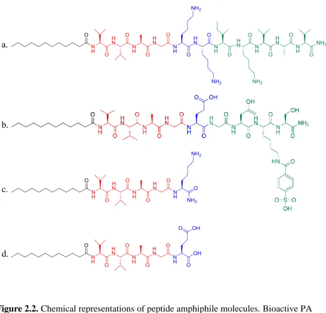
(a) and the location of needle electrodes on solues muscles (a-b). ... 35 Figure 2.2. Chemical representations of peptide amphiphile molecules. Bioactive PA
molecules; LN-PA (a) and GAG-PA (b). Positively charged PA molecule K-PA (c) and negatively charged molecule E-PA (d). Lauric acid tails are shown as black, beta sheet building blocks are red, charged units are blue and bioactive building blocks are shown as green.. ... 41 Figure 2.3. Liquid chromatography (A) and mass spectrometry (B) analysis of
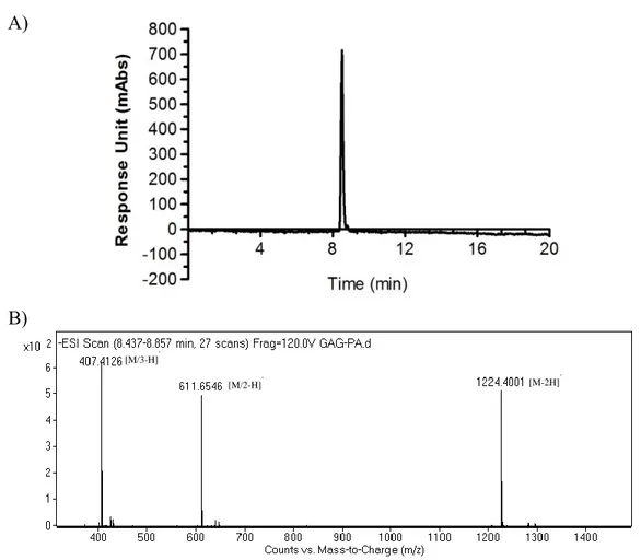
LN-PA . ... 42 Figure 2.4. Liquid chromatography (A) and mass spectrometry (B) analysis of
GAG-PA . ... 43 Figure 2.5. Liquid chromatography (A) and mass spectrometry (B) analysis of E-PA.
... 44 Figure 2.6. Liquid chromatography (A) and mass spectrometry (B) analysis of K-PA.
... 45 Figure 2.7. Scanning electron microscopy images of nanofibrous scaffold PA gels
surfaces. LN-PA/GAG-PA (A, B), K-PA/E-PA (C, D). Scale bars are 1 µm (A, C) and 5 µm (B, D) ... 47 Figure 2.8. Storage and loss modulus measurements of peptide amphiphile gels by
oscillatory rheology. Rheology results showed that the mixture of peptide amphiphiles are soft materials suitable for neural tissue applications ... 49
xiii
Figure 2.9. Functional evaluation of transected nerve by the measurement of
amplitude. ... 51 Figure 2.10. The results of evoked potential at 12 weeks after surgery. ... 52
Figure 2.11. Hematoxylin & Eosin (H&E) staining of sciatic nerve tissue specimens:
(p) proximal, (d) distal. Scale bars are 50 µm. ... 57 Figure 2.12. Immunohistochemical staining of sciatic nerve tissue specimens with anti
S-100 antibody: (p) proximal, (d) distal. Scale bars are 50 µm. ... 60 Figure 2.13. Immunohistochemical staining of sciatic nerve tissue specimens with anti
β-III-Tubulin antibody: (p) proximal, (d) distal. Scale bars are 50 µm. ... 61 Figure 2.14. Toluidine Blue-O stained semi-thin transverse sections of resin
embedded tissues. Schwann cells are indicated with white arrows. LN-PA/GAG-PA (A-C), Healthy (D) groups. Scale bars are 100 µm.. ... 64
Figure 2.15. Toluidine Blue-O stained semi-thin transverse sections of resin
embedded tissues. K-PA/E-PA (A-C), Healthy (D) groups. Scale bars are 100 µm. 65 Figure 2.16. Toluidine Blue-O stained semi-thin transverse sections of resin
embedded tissues. Undefined morphological structures are indicated with black arrows. Sucrose (A-C), Healthy (D) groups. Scale bars are 100 µm. ... 66 Figure 2.17. Toluidine Blue-O stained semi-thin transverse sections of resin
embedded tissues. Several stages of degeneration are indicated with white arrows. Autograft (A-C), Healthy (D) groups. Scale bars are 100 µm. ... 67 Figure 2.18. Quantification of the number myelinated axons/mm2 from semi-thin sectioned sciatic nerve tissues stained with toluidine blue-O.Statistical analysis was performed with GraphPad Prism 5.01 applying One-way ANOVA... 68
xiv
Figure 2.19. Transmission electron micrograph (TEM) of LN-PA/GAG-PA filled
conduit treated group. Scale bars are 200 nm (A) and 500 nm (B-D). ... 70 Figure 2.20. Transmission electron micrograph (TEM) of K-PA/E-PA filled conduit
treated group (A, B) and scanning transmission electron micrograph (STEM) of K-PA/E-PA filled conduit treated group (C, D). Scale bars are 200 nm (A), 500 nm (B-D). ... 71
Figure 2.21. Transmission electron micrograph (TEM) of sucrose filled conduit
treated group (A, B) and scanning transmission electron micrograph (STEM) of sucrose filled conduit treated group (C, D). Scale bars are 200 nm (A), 500 nm (B, C) and 2 µm (D). ... 72 Figure 2.22. Transmission electron micrograph (TEM) of autograft treated group (A,
B) and scanning transmission electron micrograph (STEM) of autograft treated group
(C, D). Scale bars are 200 nm (A, B), 500 nm (D) and 2 µm (C). ... 73
Figure 2.23. Transmission electron micrograph (TEM) of healthy group (A-C) and
scanning transmission electron micrograph (STEM) of healthy group (D). Scale bars are 500 nm (A-C) and 2 µm (D). ... 74 Figure 2.24. Quantification axonal diameter of myelinated fibers on ultrathin
transverse sections. Statistical analysis was performed with GraphPad Prism 5.01 applying One-way ANOVA (p<0.05 (*), p<0.01 (**), p<0.001 (***)) ... 76 Figure 2.25. Myelination thickness quantification of ultrathin transverse sections.
Statistical analysis was performed with GraphPad Prism 5.01 applying One-way ANOVA (p<0.001 (***)). ... 77 Figure 2.26. Representative diagram indicating the critical nerve tissue fiber
xv
Figure 2.27. G ratio quantification of ultrathin transverse sections. Statistical analysis
was performed with GraphPad Prism 5.01 applying One-way ANOVA (p<0.05 (*),
xvi
LIST OF TABLES
Table 1.1 FDA-approved nerve guidance conduits with the name of company,
degradation time, material and max gap length. Reproduced with permission from Ref. [65]. ... 12 Table 2.1. Sequence, molecular weight and net charge at pH 7 of peptide amphiphile
1
CHAPTER 1
INTRODUCTION
2
1.1. PERIPHERAL NERVE INJURY AND REPAIR
The peripheral nervous system (PNS) contains the primary, sensory, motor, and autonomic neurons that are outside of the central nervous system (CNS), Schwann cells and ganglionic satellite cells. The brain is the principal organ that determines how an organism reacts to its external environment; however, this critical function relies strongly on the signals relayed by the peripheral nervous system. Peripheral neurons are composed of a cell body and an axon that can reach lengths in excess of one meter. Axons consist of short segments wrapped by an insulating sheath, called the myelin sheath, which is produced by Schwann cells and plays an important role during the nerve regeneration process. Axons are located in fascicles and surrounded by a perineurial sheath, while bundles of perineurial sheaths are in turn protected by an epineurial layer to form a complete peripheral nerve. Sensory neurons, which are responsible for carrying signals to the CNS, and motor neurons, which carry the messages from the CNS to internal organs, are the principal types of peripheral neurons.
Signal transmission throughout motor and sensory neurons enables the connection between the central nervous system and peripheral target organs. Peripheral nerve injuries (PNIs) disrupt this connection and are typically caused by physical trauma, such as vehicle accidents, falls and fractures. PNIs occur in every 1/40 trauma patients [1] and generally cause lifelong loss in nerve functions, potentially impairing the function of the target organ innervated by the injured neurons.
3
Figure 1.1. Schematic view of the degeneration and the regeneration steps associated
with peripheral nerve injury. A: First few days after injury, the initial signs of axonal degeneration are present. B: Wallerian degeneration continues to progress, penetrating macrophages are recruited to remove cell debris. C: A large number of axonal sprouts pass through the injury site and reach the distal site of the axon with the assistance of Schwann cells in the bands of Büngner. D: Successful elongation and regeneration allows the restoration of signal transmission. E: Inappropriate regeneration and elongation of sprouts from the proximal stump to the distal stump of the injured axon results in neuroma formation. Adapted from Ref. [2] with permission.
4
Furthermore, PNI interferes with the quality-of-life and socio-economic circumstances of patients [3, 4]. According to a survey published in 2006, more than 100,000 patients undergo surgery in the USA and Europe each year for the treatment of nerve injuries [5].
Peripheral nerve injuries generally result in either good regeneration or painful neuroma formations that may result in severe muscle fiber atrophy. According to Sunderland [6], first and second degree peripheral nerve injuries (neurapraxia and axonotmesis) can be treated. The functional restoration is complete and morphological and physical damage is fully recoverable. Contrary to these milder conditions, the treatment of more severe injuries (i.e. those in which the endoneurial layer of the peripheral nerve is damaged, the nerve environment is ischemic, the axons undergo swelling and the myelin sheath cannot reverse its original morphology and function) are outside the scope of current medical treatments. In these injuries, reinnervation does not occur and impulse conduction is permanently interrupted.
Total functional recovery following nerve injury involves a complex series of degenerative and regenerative events (Figure 1.1). Before regeneration, the site of injury first experiences the local fragmentation of myelin sheaths, which directly stimulates the regeneration process (Figure 1.1A). In the first 6 hours following injury, nerve cell nuclei migrate to the edge of cells while Nissl granules and endoplasmic reticuli disassemble and disperse. This process is also known as chromatolysis and is necessary for the isolation of the neurons from the inhibitory environment prior to the recovery phase. However, cell survival cannot be ensured in severe nerve injuries; for instance, the frequency of apoptosis-related cell death in dorsal root ganglion (DRG)
5
neurons is between 20 to 50% under such cases [7]. In general, the steps of neuronal cell death are not understood in depth, but it is accepted that the microenvironment of the injury area is crucial for cell survival. For instance, many studies have showed that the isolated central neurons have the ability to regenerate when cultured in a PNS milieu, while isolated peripheral nerves do not regenerate in a CNS microenvironment [8, 9]. This regenerative difference between the PNS and the CNS microenvironments has been studied in detail and found to depend on the inductive roles of Schwann cells, ECM molecules and neurotrophic factors in the peripheral nerve milieu. In addition, growth factors such as fibroblast growth factor (FGF), nerve growth factor (NGF), brain-derived neurotrophic factor (BDNF) and other molecules enhance cell survival in the peripheral nervous system [10-12].
Morphologically, visible structural changes occur in the neuronal cell body while chromatolytic changes occur at the proximal injury site, where neuronal nuclei protrude from the cell bodies and the native nucleoproteins reassemble into Nissl granules (Figure 1.1B). During the chromatolytic process, numerous cellular activities are changed to induce RNA synthesis and reduce neurotransmitter production. Thus, neurons at the injury site are ready to produce the vast amount of proteins and lipids that are required for axonal outgrowth during the regeneration process. Fully functional recovery may take weeks to months and is reported to involve the reorganization and reconstitution of physical, morphological and functional connections at the injury site [13]. In peripheral nerve injuries [14], Schwann cells have a crucial role regarding the removal of axonal and myelin debris, the presence of which prevents axonal outgrowth in the distal nerve stump and interferes with the
6
secretion of ECM molecules and trophic factors [14], including laminins [15], the inflammatory cytokine IL-6 [16, 17], NGF [18-22] and FGF-2. Especially in the distal nerve region, Schwann cells and macrophages take part in removing tissue and cell debris and this process triggers cellular and functional changes related with Wallerian degeneration [23] (Figure 1.1A and 1B). Schwann cells proliferate and organize as columns, called bands of Büngner (Figure 1.1C). These bands are used as guides for the sprouting of axons across the basal laminae of Schwann cells, and serve as an early marker of neural regeneration [24, 25]. At the regenerating distal nerve site, interactions between mature Schwann cells and axons facilitate the remyelination process and create small internodes (Figure 1.1C). However, connective tissue scarring and impaired regeneration of seriously injured axons may cause abnormalities in the elongation process (Figure 1.1E). These types of PNI lead the formation of a neuroma in the proximal segment. Such abnormal formations have been accepted as an underlying reason of spontaneous neuropathic pain syndrome [26]. This ineffective regeneration causes an incomplete reinnervation, altering the alignment of Schwann cells in the distal nerve stump and inducing the production and deposition of endoneurial collagens. This denervation process negatively affects the function of the target end organ, and must generally be corrected through surgical intervention. There are various methods for the microsurgery repair of degraded PNI environments, including autograft/allograft transplantation, direct repair and hollow nerve conduits (NGC). Small nerve gaps can regenerate by themselves; however, transection gaps of over 5 mm in length are best repaired surgically by connecting the ends of nerve using sutures. This method is known as direct nerve repair [27-30].
7
1.1.1. The Gold Standard of Peripheral Nerve Injury:
Autograft
The autograft technique has served as a ‘gold standard’ for the treatment of peripheral nerve injuries for the last 50 years [28, 30]. Simple nerve grafting methods were initially described in the late 19th century, but autografting was only perfected decades later by Millesi, who optimized the technique and demonstrated its effectiveness over epineural suturing. Nerve grafts are preferably derived from less essential nerves, such as sural or cutaneous nerves, of the patient's own body [31]. Nevertheless, the operation has a rate of success under 50% [32, 33] and is associated with numerous limitations, such as donor site morbidity, formation of neuromas, possible loss of sensation and function, insufficient graft length, the necessity of secondary surgeries, mismatch of donor nerve owing to morphological differences, high cost to healthcare providers and the extension of the recovery time [33-37].
Autograft technique can be used for PNI treatment when the critical length of nerve gap is shorter than 5 cm, and beyond this length, allograft is an inevitable treatment [38]. However, this technique necessitates the use of immunosuppressives up to 2 years after surgery, and patients are prone to the threats coming from microenvironment which eventually results in tumor formation [39]. Consequently, these unfavorable consequences of autograft and allograft techniques are reasons to focus of efforts on alternative engineering strategies or devices for PNI repair. The use of nerve guidance conduits (NGCs) is the primary artificial tissue engineering technique.
8
1.1.2. Regeneration within Nerve Conduits
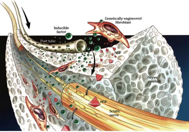
Biochemically engineered nerve conduits have been found to be effective alternatives to autografting for the repair of damaged nerves. Hollow nerve conduits were first used for bridging a 30 mm nerve gap in a dog, using a bone conduit [40]. These hollow tubes present numerous advantages for PNI treatment, including isolation from the scar tissue, decreased formation of neuromas and scars and the aggregation of huge amounts of trophic factors, which allows these materials to provide an axon-guiding channel that connects the distal and proximal injury sites [7]. However, it should be noted that these NGCs are incapable of facilitating nerve repair over gap distances of 40 mm [41]. Nevertheless, the use of NGCs creates a well-defined microenvironment to modulate the neuronal repair process, allowing functional recovery of PNIs in clinical settings [42, 43].
An ideal nerve conduit should exhibit certain parameters to optimally facilitate the regeneration and reassociation of both elongated nerve fibers and components of the peripheral nerve system (Figure 1.2) [44]. These nerve conduits should permit the transition of neurotrophic factors released by Schwann cells and macrophages [45]. In addition, biocompatibility is a critical feature for any material intended for biological applications, and potential nerve conduits should therefore exert minimal toxicity and immunogenicity in living systems. Biodegradability is another desired feature, as the gradual removal of the scaffold material enhances the completion of the recovery process. Furthermore, a sufficient level of porosity should be maintained in the conduit environment to ensure the adequate distribution and diffusion of both endogenous and exogenous factors [46].
9
Figure 1.2 An ideally designed nerve conduit should have a well-defined set of
features to permit for the outgrowth and regeneration of peripheral nerve cells. Copyright © 2006 Foundation for Cellular and Molecular Medicine/Blackwell Publishing Ltd. Reproduced from Ref. [44] with permission.
The materials of nerve conduits are generally classified as synthetic [47] or natural [48]. Polyphosphoesters, aliphatic polyesters, hydrogel-based materials, polyurethanes and piezoelectric polymers are synthetic materials, while polysaccharides such as chitosan, collagen and decellularized scaffolds are animal derived natural materials. Polylactic acid (PLA), polyglycolic acid (PGA) [49, 50], polycaprolatone (PCL) [51-53], polylactide-caprolactone (PLCL), poly(lactic-coglycolic acid) (PLGA) [37, 54, 55], poly(3-hydroxybutyrate) (PHB), and various combinations of the PGA and PLCL
10
are some examples for aliphatic polyesters, which constitute one of the sub-classes of polymers. These polymers can be manufactured as fibers by electrospinning technique [56]. Collagen [57], laminin [58], chitosan [59] and other naturally derived polymers can also be used for the fabrication of nerve conduits.
It is well-established that peripheral nerve regeneration can be enhanced by using NGCs modified with biological and chemical moieties, soluble factors and natural or synthetic materials. As one example, a polysulfone NGC filled with laminin and NGF-loaded microtubules in a hydrogel matrix was found to enhance the regeneration of transected rat sciatic nerves with nerve gaps of 20 mm in length [60]. Biofunctionalization of hollow NGCs with growth factors or filling them with cells or stem cells are other strategies for bridging nerve gaps between injured nerve fibers [61]. These strategies can enhance axonal regrowth into the conduit and can also be used for the treatment of spinal cord injury [62].
The use of stem cells is another potential method for the enhancement of PNI repair. Stem cells are frequently delivered following their embedding in natural or synthetic platforms such as hydrogels or collagens that can be filled into NGCs. Chitosan NGCs filled with neural progenitor cells have been demonstrated to promote regeneration after spinal cord injury, and this conduit could also be functionalized with laminin to enhance neural adhesion and cell survival [63].
The US Food and Drug Administration (FDA) is a federal agency that is responsible for protecting public health in accordance with legal regulations. Many nerve guidance conduits and wraps have been approved by the FDA for use in the treatment of peripheral nerve injuries. FDA evaluates clinical products and devices with respect to
11
their safety, reliability and efficacy, and regards the use of nerve conduits as an effective means of enhancing peripheral nerve regeneration.
Neurolac® is one of the FDA-approved NGCs. Manufactured by Polyganics company, it is up to 3 cm in length and 1.5-10 mm in diameter (Table 1.1). This NGC is a synthetic, transparent and biodegradable scaffold that is produced from poly(DL-lactide-ε-caprolactone); it is hydrolysed within the body within 2 to 3 years. The use of Neurolac® is recommended for up to 2 cm gaps in PNIs. The advantages of
12
Table 1.1 FDA-approved nerve guidance conduits with the name of company, degradation time, material and max gap length.
Reproduced with permission from Ref. [65].
Product Name Material Degradation Time Company Max Gap Length
Neurolac Poly(DL-lactic-co-1-
caprolactone) (PLCL) 2-3 years
Polyganics Inc., The
Netherlands 3 cm
Neurogen Collagen type I 4 years Integra Neurosciences,
Plainsboru, NJ, USA 3 cm
Neuromend Collagen type I 4-8 months Collagen Matrix, Inc.,
Franklin Lakes, NJ, USA 2.5 cm
NeuraWrap Collagen type I 4 years Integra Neurosciences,
Plainsboru, NJ, USA 4 cm Neuromatrix/Neuroflex Collagen type I 4-8 months Collagen Matrix, Inc.,
Franklin Lakes, NJ, USA 2.5 cm Neurotube Woven polyglycolic acid
(PGA) 6-12 months
Synovis Micro Companies Alliance, Birmingham, AL, USA 3 cm Salubridge/Hydrosheath or Salutunnel Salubria—polyvinyl alcohol
(PVA) hydrogel Non-biodegradable
Salumedica LLC, Atlanta,
GA, USA 6.35 cm
Surgisis Nerve Cuff/ Axoguard
Porcine small intestinal
submucosa (SIS) matrix Not reported
Cook Biotech Products, West
Lafayette, IN, USA
4 cm AxonScaff/ Cellscaff or
StemScaff
Polyhydroxybuturate
13
1.2. ECM MOLECULES AND THEIR FUNCTIONS
IN PERIPHERAL NERVE REGENERATION
Extracellular matrix (ECM) is a collection of a unique set of collagens, non-collagenous glycoproteins and proteoglycans that are produced by cells. The precise composition of the ECM is under strict regulatory control, and the expression levels of its constituent molecules are restricted to particular time periods both during development and in adulthood. The primary role of ECM molecules in the nervous system is the regulation of cell migration, survival, differentiation and axonal elongation in addition to structural support [66].
After PNI, the distal stump of the injury site begins to experience Wallerian degeneration, which involves the removal of cell debris and recruitment of macrophages to the site of injury, while the proximal nerve site initiates the regeneration process. However, the signals that trigger the regeneration of the proximal site have not been fully investigated [67]. PNI triggers both the endogenous and exogenous growth capacity of damaged neurons, which may induce the regeneration of the PNS [68]. Extracellular matrix proteins, neurotrophic factors and cell adhesion molecules all promote the successful axonal regeneration by activating the endogenous growth capability of the neural cell body.
This growth capacity is well-studied in a group of primary sensory neurons, the dorsal root ganglion cells (DRG). Myelin-associated glycoprotein (MAG) is able to inhibit DRG neurite outgrowth in vitro, but lesions in peripheral axons enhance the endogenous outgrowth capacity to cancel the inhibitory effects of MAG [69, 70].
14
Dibutyryl-cAMP, an analog of intracellular cyclic adenosinemonophosphate (cAMP), can also enhance axon growth capacity of the DRG after injury [69-71]. The activation of protein kinase A (PKA) by cAMP can cause the axonal elongation of sensory neurons cultured on MAG [69, 70]. In addition, PKA and cAMP can positively regulate the expression level of Arginase I, which is an enzyme related with polyamine synthesis after PNI. Therefore, the upregulation of Arginase I can abolish the inhibitory effect of MAG or myelin [72]. cAMP response element binding protein (CREB) expression is regulated transcriptionally by cAMP, which can reduce the inhibitory effect of MAG or myelin and result in the upregulation of neurite outgrowth and axonal regeneration in vivo [73]. c-Jun is another transcriptionally downregulated gene in PNI, and the knockout of c-Jun promotes neural regeneration after injury [74]. Laminin, an ECM glycoprotein, has a crucial role in axonal regeneration and is required for proper myelination by Schwann cells [75]. Laminin 8 (α4β1γ1) and laminin 2 (α2β1γ1) are two of the 15 accepted isoforms that are expressed largely in the endoneurial layer of PNS [15]. Synthesis of laminin is upregulated by Schwann cells at the injury site after PNI and this upregulation promotes regeneration [21, 76]. Consequently, laminin has an indispensable effect for neural regeneration. In vitro inhibition of neurite growth was shown by an anti-α2 laminin chain blocker after sciatic nerve injury [77]. The α chain of laminin, which contains the RGD and IKVAV sequences, has also been shown to promote neurite outgrowth in the PC-12 cell line [78] while antibodies against the IKVAV site disable the function of laminin by binding to its signaling sequences. Laminin interacts with cells through both integrins and α-dystroglycans [79, 80] and its interaction with integrins is principally mediated by the two integrins α1β1 and α3β1, in PC-12 cells [81]. ]. Laminin has an important
15
effect on Schwann cell behavior, including myelin production and ensheathment. Abrupt changes in laminin levels may therefore result in improper Schwann cell differentiation and hypomyelination [82].
Proteoglycans are strongly glycosylated proteins that constitute the majority of ECM
components and receptors, and are found commonly in the basal lamina. These
proteins have an important role for neuronal and axonal guidance. They are
categorized into two major groups: heparan sulfate proteoglycans (HSPGs) and
chondroitin sulfate proteoglycans (CSPGs). Cell surface proteoglycans bind to the
ECM molecules and they are implicated as receptors for growth factors [83]. For
example, FGF binds to both the core protein and glycosaminoglycan (GAG) moieties
of heparan sulfate (HS) proteoglycans [84]. As a result of this interaction, proteolysis
of FGF is blocked by proteoglycans and the ECM functions as a reservoir for FGF
[85]. In addition to this interaction between FGF and HS, many other growth factors
have been suggested to serve as mitogens for Schwann cells, triggering cellular
proliferation by binding to HS [86]. HS chains present on different types of
proteoglycans can interact with various growth factors, such as bFGF, aFGF, G-CSF,
INF-1, and Ile-3 [87].
Schwann cells promote the regeneration in the PNS by three ways: increasing the synthesis of cell adhesion molecules (CAMs) such as N-cadherin, L2/HNK-1 and NCAM; regulating the basement proteins such as laminin, fibronectin, HSPGs and tenascin; and releasing neurotrophic factors such as NGF, FGF-2, IGF, CNTF, GDNF, and BDNF. As a result, laminin plays a significant role in both the formation of neurite outgrowth and the proliferation and survival of Schwann cells.
16
1.2.1. Neurotrophic Factors
Neurotrophic factors and neurite growth inducing factors are fundamental for neural cell survival, axonal outgrowth and the regulation of Schwann cell behavior after PNI. Injury induced neuronal death will decrease the potency for axonal growth. The axonal outgrowth-promoting effect of neurotrophic factors and neurite outgrowth promoting factors, including NGF, neurotrophin 3 (NT-3), and brain-derived neurotrophic factor (BDNF), were shown by various in vivo studies and resulted in a comprehensive understanding of the signaling pathways involved [88]. The inductive effect of NGF on axonal growth was shown by both in vivo and in vitro studies [89]. Briefly, the NGF signaling pathway upregulates the activation of phosphatidylinositide 3-kinases (PI3K), while PI3K inhibits glycogen synthase kinase 3 (GSK-3β) to promote axonal regeneration by regulating cytoskeleton proteins [88].
NGF exhibits an inducer effect on neurite outgrowth of DRGs by modulating the activity of laminin, but neither laminin nor NGF cannot induce sufficient axonal growth in PNS by themselves [90, 91]. Tyrosine kinase receptor B (trkB) and trkC are used for inducing the biological activities of BDNF, NT-3, and NT-4/5 [92, 93]. FGF-2 is also upregulated after PNI and the regulation of Schwann cell proliferation and differentiation is stimulated by FGF-2 [94]. The binding of neurotrophin factors to their receptors triggers the phosphorylation of tyrosine residues, resulting in cell proliferation and differentiation, while FGF-2 inhibits the expression of myelin gene and myelin zero protein [95]. Therefore, after sciatic nerve injury, FGF-2 boosts the number of axons while reducing the myelin sheath thickness. Ciliary neurotrophic factor (CNTF) is a survival factor enhancing synthesis of neurotransmitters and
17
promoting neural outgrowth after injury in the peripheral nervous system [96]. Although it has not so far been demonstrated to be effective in promoting nerve repair [97], it is plausible that this factor shows regenerative and functional impact in tandem with other neurotrophic factors [98].
TGF-β helps the Schwann cells to stay in their proliferative state by inhibiting myelin production through development. Furthermore, after nerve injury, the release of TGF-β is increased in the distal nerve stump by macrophages and Schwann cells, and the induced release of TGF-β modulates Schwann cell behavior during axonal outgrowth [99, 100]. The binding of neurotrophic factors to their receptors triggers the phosphorylation of tyrosine residues, resulting in the proliferation and differentiation of Schwann cells and neurons.
1.3. INNOVATIVE NANODESIGN OF ECM MIMICING
SCAFFOLDS
Tissue engineering and regenerative medicine provide new strategies for the fabrication of biological scaffolds. However, the effective repair of damaged tissues and organs necessitates the development of top-down and the bottom-up processing methods that seamlessly integrate design principles associated with nanotechnology, material science and tissue engineering [101]. The applicability of new strategies often depends on two important factors, which are: (i) the biocompatibility of newly designed scaffolds, which should above all not be harmful to the body, (ii) their suitability for the differentiation of cells, and especially stem cells and primary cell
18
lines. Overall, when developing new strategies for the regeneration of damaged tissues or organs, the ideal material is a scaffold that can mimic the ECM of the host cells or tissue in their native microenvironment (Figure 1.3). The ECM can modulate and regulate the functions of cells by releasing and producing biologically active molecules. This allows the facile modulation of cells and their behavior, as biological, mechanical, developmental and pathological responses can be altered greatly through external cues that occur in the cellular milieu [102].
Another critical characteristic of nanomaterials used for ECM mimicry is porosity. Cells in their native microenvironment are found in a highly ordered and ultracomplex structure at the nanoscale, including different sizes of pores and fibers. Consequently, to guide regeneration and provide proper cell distribution in 3D-scaffold, materials should be engineered in terms of these design criteria.
ECM hydrogels have porous and hydrated structures, making them promising alternatives for mimicking the native microenvironment of cells. Self-assembled peptide nanofiber gels are one example of hydrogels and can be used for nerve regeneration due to their ease of modification and capacity to maintain cells or any soluble factors in a liquid environment. Biologically active molecules can be used for the surface modifications of hydrogels to enhance their biocompatibility and establish a variety of interactions between the material-cell/tissue interface, creating a matrix similar to the ECM of nervous tissue. Long nerve gaps have been treated by using hydrogels containing collagen I and laminin to promote axonal regeneration in peripheral nerve injuries [103].
19
Studies about tissue engineering are mainly focused on the synthesis of polymers and proteins as scaffolds [104]. Furthermore, these biological scaffolds can be designed innovatively to promote and regulate cell differentiation, proliferation and adhesion without adding any animal-derived factors to the material. This new approach is also used for nerve tissue engineering. This design technique depends on the optimization of the topographical, mechanical, biological and chemical properties of the scaffold to enhance the proliferation and elongation of axons.
Mimicking the ECM by using biomaterials is an effective means to control and direct neural growth and adhesion. This modified microenvironment can be promoted by co-culture technique with cells such as endothelial cells, Schwann cells or oligodendrocytes, and studies have demonstrated major enhancements in neural adhesion, axonal outgrowth and myelination of axons under such conditions [105, 106].
Complex tissues include different types of cells that require a precise set of cell-cell and cell-matrix interactions to continue their function, which can be provided by nanotechnology in a 3D scaffold. The use of nanotechnology allows the implantation of nanoscale cues in scaffolds, which can greatly alter the behavior of cells and enhance a given tissue's capacity for regeneration. However, nanofiber scaffolds are largely under-researched materials for tissue engineering applications, and further research is required to realize their full potential. For ECM applications, the most effective nanofiber scaffolds are typically fabricated using three methods: self-assembly, electrospinning and phase separation.
20
Figure 1.3. An illustration of the extracellular matrix, consisting of collagen fiber bridges that extend over cells through integrins located
on the plasma membrane (A). Scanning electron micrograph of self-assembled nanofiber scaffold (B) mimicking the native ECM. Reproduced with permission from Pearson Education, Inc., publishing as Benjamin Cummings.
A
B
21
1.4. SELF-ASSEMBLED PEPTIDE NANOFIBERS AS
SCAFFOLDS
FOR
PERIPHERAL
NERVE
REGENERATION
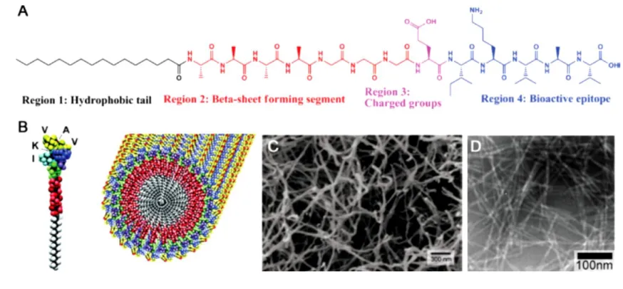
Self-assembly is an inspirational characteristic of nature and allows the design of new materials as bioactive scaffolds through a bottom-up approach. Many complex tissues and scaffolds can be produced by mimicking self-assembled peptide nanofibers and their hierarchical order, which allow these materials to form highly complex scaffold systems. In addition, peptide amphiphile building blocks can be synthesized chemically using commonly available chemical methods. A broad variety of PA types can self-assemble into micelles, high-aspect ratio nanofibers and various other morphologies under specific conditions. The final structure of the PA assembly can be controlled by modulating the kinetics of the assembly mechanism through changes in pH, temperature and solvent exposure. Peptides are short sequences and functional building blocks that can be used to design biodegradable and biocompatible nanostructures for regenerative medicine applications. The sequence of peptide amphiphile molecules can be determined according to the requirements of the cell type and tissue of interest. Mimicking the biological, chemical and mechanical properties of the natural ECM is the best route for developing novel materials for various applications.
Peptide amphiphiles generally have 4 different regions: a hydrophobic region that contains specific alkyl chains attached to the peptide sequence, a β-sheet forming region that contains 4-8 hydrophobic amino acids (such as valine and alanine) directly
22
adjacent to the alkyl chain and is important for determining the mechanical properties of the nanofiber gel [107], a charged amino acid region for good solubility and ease of purification, and lastly a bioactive epitope region that contains a peptide sequence intended to emulate some function of the natural ECM of the tissue of interest [108] (Figure 1.4).
The assembly of peptide amphiphiles depends on three important driving forces: the electrostatic interactions between oppositely charged groups, hydrogen bonding among β-sheet forming peptide sequences and the hydrophobic interactions of the alkyl chains. In an aqueous environment, individual PA molecules form PA nanofibers where the hydrophobic core contains alkyl chains trapped in nanofibers and the hydrophilic periphery presents the bioactive epitopes to the external environment without altering the cylindrical geometry of PA nanofibers.
23
Figure 1.4. Chemical structure of an IKVAV-PA molecule, showing four different regions (A). Graphical illustration of peptide
amphiphile molecule and its self-assembly into a complex nanostructure (B). SEM image of a peptide nanofiber scaffold demonstrates the similarity of the network structure to the natural ECM (C). TEM image of IKVAV nanofibers (D). Copyright © 2001 American Association for the Advancement of Science. Reproduced with permission from Ref. [109].
24
The structure of peptide amphiphile nanofibers is similar to the fibrous organization of the natural ECM, allowing the former to serve as a suitable replacement for the latter. In addition, PA networks are able to stimulate cell signaling pathways while physically supporting the cells they contain, effectively performing the roles of the ECM. As such, self-assembled PA nanofibers provide both functional and morphological mimicry of the fibrous extracellular matrix. These nanofiber scaffolds can also promote neuronal migration, proliferation and adhesion. There is a number of peptide sequences in the literature for the treatment of peripheral nerve injuries, including RGD (Arg-Gly-Asp), IKVAV (Ile-Lys-Val-Ala-Val), YIGSR (Tyr-Ile-Gly-Ser-Arg, derived from the laminin β chain) and RNIAEIIKD (Arg–Asn–Ile–Ala–Glu– Ile–Ile–Lys–Asp–Ile, derived from the laminin γ chain and primary cell binding domains) [110, 111]. In addition to these peptide sequences, the N-cadherin mimetic sequence HAV (His-Ala-Val) is an important signaling motif in both neurons and glia [110]. Integrins are major components of the cell membrane and play vital roles in the attachment of cells to the ECM, and PA scaffolds are able to emulate these interactions. RGD is one of the most common sequences for biological adhesion, and has been derived from fibronectin [112]. The laminin-derived IKVAV sequence is also important for cell attachment, migration and growth of neural cells [113, 114]. A gradient of IKVAV-containing peptides was also shown to modulate the development of growth cones in DRG neurons [115]. The direction of axonal elongation was slow when faced with the gradient, and the growth cone reversed directions below a certain concentration threshold.
Self-assembled PA nanofiber gels also function as platforms for the encapsulation, controlled release and delivery of small hydrophobic molecules. A number of
25
therapeutic drugs and molecules can be incorporated into PA gels to provide biofunctionality, enhance bioactivity and reduce the immune response. Dexamethasone, an anti-inflammatory drug, was loaded into PA gels for controlled release and shown to reduce the immune reaction [116]. Furthermore, PA gels can be used as a gene delivery platform, e.g. for antisense oligonucleotides as a novel approach for gene therapy [117]. Hedgehog signaling pathway is also important in nerve regeneration following injury, and sonic hedgehog homolog (SHH) protein plays a critical role in this pathway. SHH-incorporated PA gels were shown to decrease apoptosis and promote nerve regeneration after cavernous nerve injury [118].
26
CHAPTER 2
SCIATIC NERVE REGENERATION INDUCED
BY GLYCOSAMINOGLYCAN AND LAMININ
MIMETIC PEPTIDE NANOFIBER GELS
This chapter of thesis is partially submitted in the following article; “Mammadov, B., Sever, M., Gecer, M., Zor, F., Ozturk, S., Akgun, H., Ulas, U.H., Guler, M.O., and Tekinay, A.B., Sciatic Nerve Regeneration Induced by Glycosaminoglycan and Laminin Mimetic Nanofiber Gels. Acta Biomaterialia, 2016.”
27
2.1. INTRODUCTION
Peripheral nerve lesions often lead to loss of motor and sensory function of affected area in patient’ body need to be reconstructed by surgery. In Europe, over 300,000 people have accidents resulting in PNI and the number of these cases increase every year [119]. These injuries effect the patients’ lives negatively socio-economically. Many patients require medical operations for treatment and usually a second operation is inevitable. More than 100,000 patients have an operation because of PNI in US and Europe according to the survey in 2006 [5]. Defects occurred in motor and sensory functions result in the disability of affected limb and neuropathies causing long-term disability. In contrast to long nerve gap, nerve injury with a short nerve gap can be treated by end to end coaptation, but numerous obstacles hamper functional recovery and axonal regeneration. Target organ atrophy and errors in regeneration cause the denervation of the targets and devastating alteration in proliferation, regeneration and production of myelin sheaths of Schwann cells.
Different types of microsurgery techniques are used for the treatment of PNI. These are direct repair (end to end suturing), grafting techniques, (autograft/allograft transplantation) and hollow nerve guidance conduits (NGCs) [27-30]. Deformed nerve gaps can be regenerated by themselves or by using end to end coaptation technique when nerve gap length is less than 1 cm, but long nerve gaps which are 1-5 cm in length can be treated by autograft or allograft (implantation of cadaveric nerve) transplantation methods. Autograft technique is accepted as ‘the gold standard’; however, donor site morbidity, neuroma formation, target organ atrophy and
28
neuropathic pain can occur after applying this technique [120]. Although every other person can recuperate by autograft transplantation operation [106], this nerve grafting technique has been the most preferable for PNI treatment. This successful outcome is owing to the presence of Schwann cells and basal lamina of endoneurial layers. These two factors play an important role for supplying neurotrophic factors and adhesion moieties located on endoneurial layer to promote axonal regeneration. The use of allograft transplantation technique has an advantage to provide an internal scaffold in a 3D manner for Schwann cells and axons; however, the cadaveric transplant can cause immunological response by transferring pathogens to host. As an alternative to these techniques, hollow nerve guidance conduits are more recently researched and used as a template for cell culture and the three dimensional microenvironment for cell adhesion, growth and orientation. NGCs are used for promoting successful regeneration in both the CNS and PNS. These conduits can function as a guidance channel for axon sprouting from the regenerating nerve site, proximal nerve stump, a barrier for infiltration of fibrous tissue, and a permissive environment for neurotrophic factors secreted by Schwann cells at injury site [45]. In spite of functional recovery in PNI, hollow NGCs cannot reach to regeneration level of autologous nerve grafting method [121]. This poor functional outcome resulting from the use of hollow tubes can be overcome by designing biomaterials to modify and functionalize hollow NGCs. Functionalization of hollow NGCs has emerged as an alternative to the autologous nerve grafting technique. This strategy should have properties that allow regeneration of injuries and reconstruction of damaged tissues, and also better mimic of PNS. Especially, internal surface of conduits can be modified by multiple molecules, including neuroinductive moieties, physical guidance cues and neurotrophic factors.
29
Currently, self-assembled peptide amphiphiles are able to accomplish these design concerns and also perfect candidates for biofunctionalization of nerve conduits. Peptide amphiphiles (PAs) can spontaneously self-assemble into nanofibers due to their molecular multifunctionality including hydrophobic and hydrophilic characteristics with the additional property of presenting biologically active moieties on the external surface of nanofibers [109].
In this thesis, I investigated the use of peptide nanofiber gels filled in polymeric NGCs for peripheral nerve regeneration by using histological analyses and transmission electron microscopy. Peptide amphiphiles were designed to form nanofibers as scaffolds presenting bioactive epitopes associated with nerve regeneration including amino acid sequences derived from laminin and mimicking heparan sulfate. LN-PA molecules, which carry IKVAV sequence, has been shown to promote neurite outgrowth and play an important role in improvement of peripheral nerve regeneration [75]. Laminin is mainly secreted by Schwann cells, required for proper myelination and its disturbance causes inappropriate Schwann cell differentiation and hypomyelination [82]. Highly glycosylated HSPGs are naturally found in basement membrane of neural cells. Both the core protein and glycosaminoglycans (GAG) moieties of HSPGs are known as receptors for growth factors [83, 84]. Especially, bFGF binds to GAG moieties on HS, and bFGF is protected from proteolysis owing to this binding [85]. As well as bFGF, Schwann cells release various types of growth factors that enhance axonal regeneration and control the proteolysis mechanism of neurotrophic factors [45]. Dual effect of LN-PA and GAG-PA nanofiber gels on improvement of neurite outgrowth has been shown in vitro [122]. Neurolac® TW
30
hollow polymeric nerve conduits were used and functionalized with peptide nanofiber gels and fully transected sciatic nerve model was investigated as in vivo model.
2.2. MATERIALS AND METHODS
2.2.1. Materials
9-fluorenylmethoxycarbonyl (Fmoc) and tert-butoxycarbonyl (Boc) protected amino acids [4-[a-(20,40-dimethoxyphenyl]enoxy]acetamidonorleucyl-MBHA resin (Rink amide MBHA resin), Fmoc-Glu(OtBu)-Wang resin and 2-(1H-Benzotriazol-1-yl)-1,1,3,3-tetramethyluronium hexafluorophosphate (HBTU) were purchased from NovaBiochem and ABCR. Dichloromethane (DCM), acetic anhydride, diisopropyl-ethylamine (DIEA), piperidine, dimethylformamide (DMF), trifluoroacetic acid (TFA), triisopropylsilane (TIS) were purchased from Sigma-Aldrich. Toluidine Blue and osminium tetroxide (OsO4) were also purchased from Sigma-Aldrich. The other
chemicals were purchased from Fischer, Merck, Alfa Aesar or Sigma-Aldrich. Araldite 502 kit was purchased from Electron Microscopy Sciences. All diamond knives were purchased from Diatome. Neurolac® TW conduits were purchased from Polyganics. β-III-tubulin and S-100 antibodies were purchased from Millipore.
31
2.2.2. Synthesis and Purification of Peptide Amphiphile
Molecules
Four different PA molecules were synthesized by solid phase peptide synthesis protocol. LN-PA (Lauryl-VVAGKKIKVAV-Am), GAG-PA (Lauryl-VVAGEGD (ρ-sulfobenzoate)-Am and K-PA (Lauryl-VVAGK-Am) were synthesized on Rink amide MBHA resin, only E-PA (Lauryl-VVAGE) was synthesized on Fmoc-Glu(OtBu) Wang resin. During the synthesis; Fmoc protected amino acids, Rink Amide resin for solid support and HBTU for activation of protected groups of amino acids were used. The removal of Fmoc protecting group was performed by 20% piperidine/dimethylformamide (DMF) solution for 20 min. Coupling reaction of 2 mole equivalents of Fmoc protected amino acids was performed by 1.95 mole equivalents of HBTU, and 3 mole equivalents of N, N-diisopropylethylamine (DIEA) for 1 mole equivalent of functional sites on the solid resin, in DMF solution for 4 h. At the end of each coupling reaction, Kaiser test was used for testing coupling reaction. To prevent coupling from remaining free amino groups, 10% acetic anhydride solution in DMF was used for 30 min. After each step, reaction solution was washed with 3 times DMF, 3 times DCM and 3 times DMF again. Except from other three peptide amphiphile molecules, in order to synthesize GAG-PA, sulfobenzoic acid coupling to side chain of lysine residue was performed. To achive this reaction, Mtt removal was performed by shaking reaction mixture for 5 min with TFA: TIS: H2O: DCM in the
ratio of 5:2.5:2.5:90. Cleavage of the peptide molecules from the resins was accomplished with a mixture of TFA: TIS: H2O in the ratio of 95:2.5:2.5 for 2 h. The
32
ice-cold diethyl ether was added to precipitate peptide solution and incubated overnight at -20 °C. Centrifugation was done to completely precipitate peptide molecules, then diethyl ether was poured out of falcons and remained ether was evaporated. Peptide molecules was dissolved in the ddH2O and frozen -80 °C,
lyophilized for 2-3 days and saved at -20 °C.
Synthesized peptide molecules were characterized by liquid chromatography-mass spectrometry (LC-MS) to determine the purity of the molecules. Agilent LC-MS equipped with Agilent 6530 Q-TOF with an ESI source and Zorbax SB-C8 4.6 mm x 100 mm column was used for positively charged peptide amphiphile molecules. On the other hand, an Agilent Zorbax Extend-C18 2.1 mm x 50 mm column was used for negatively charges peptide amphiphile molecules. LC-MS method was optimized as gradient of water (0.1% formic acid or 0.1% NH4OH) and acetonitrile (0.1% formic
acid or 0.1% NH4OH) that was determined as a mobile phase.
A preparative reverse phase HPLC system (Agilent 1200 series) equipped with a Zorbax SB-C8 (21.2 mm x 150 mm) column was used to purify positively charged PA molecules for acidic conditions while Zorbax Extend-C18 21.2 mm x 150 mm column was used for negatively charged PA molecules for basic conditions. The same optimized gradient of water 0.1% TFA or 0.1% NH4OH) and acetonitrile (0.1% TFA
33
2.2.3. Physical, Mechanical and Chemical Characterization
of Self-assembled Nanofiber Network
2.2.3.1. Scanning Electron Microscopy (SEM)
Peptide amphiphile nanofiber networks were visualized by using a scanning electron microscope. Gel solutions were prepared as a 1 wt % concentration and they were oppositely mixed in a 120 µL final volume. Gels were incubated at 37 ºC for 30 min to enhance gelation process. After obtaining PA gels on the silicon wafers, serial dehydration steps were performed. Samples were incubated by increasing ethyl alcohol concentration as 20%, 40%, 60%, 80%, 90%, 100% for 3 min and lastly, in 100% ethyl alcohol were used for overnight incubation at 4 ºC. Gels were dried by using Tousimis Autosamdri®-815 critical point dryer for 2 h to conserve the network structure. After drying process, samples were coated 6 nm Au/Pd and images were taken using a FEI Quanta 200 FEG scanning electron microscope equipped with ETD (Everhart Thornley Detector) detector under high vacuum.
2.2.3.2. Oscillatory Rheology
Investigation of viscoelastic properties of peptide amphiphile gels was performed by using oscillatory rheology measurements with Anton Paar Physica RM301 Rheometer with a 25 mm parallel plate configuration. PA gels were prepared as 125 µL/PA molecules at concentration of 4 mM LN-PA, 4 mM GAG-PA, 4 mM K- PA and 2 mM E-PA dissolved in 0.25 M isotonic sucrose solution. Two oppositely charged PA molecules were mixed and carefully transfer to the center of the plate and incubated
34
for 15 min. After the system reached a plateau, measurement position was set to 0.5 mm.
2.2.4. Surgical Procedure
Twenty eight ten weeks old male Sprauge-Dawley rats were used for this study. Animals were divided into four experimental groups with seven rats. Full transaction injury was performed and 10 mm nerve tissue was excised. 2.5 mm nerve parts from both proximal and distal parts were inserted into 15 mm nerve conduit, and thus, 10 mm gap was left after suturing. In order to obtain biofunctional LN-PA+GAG-PA gels, 100 µL from 4 mM LN-PA and 4 mM GAG-PA were injecting into inert conduits while 100 µL from 4 mM K-PA and 2 mM E-PA were injected into inert conduit and used as an epitope-free control. All PA molecules dissolved into 0.25 M isotonic sucrose solution. For non-treated controls, nerve conduits were filled with sucrose and for autograft controls, sciatic nerves were excised then reversed and sutured back to nerve stumps. All surgical operations were performed on the right hind limbs and left hind limbs were used as healthy control. All procedures were approved by Animal Ethics Committee of Gülhane Military School.
35
Figure 2.1. Images of electrophysiological assessment shows supramaximal stimulus
(a) and the location of needle electrodes on soleus muscles (a-b).
2.2.5. Electrophysiological Investigation
The assessment of electrophysiological recovery was investigated by using electromyography (EMG) device. 12 weeks after the surgery, rats were anesthetized by a subcutaneous injection of xylazine (20 mg/kg) and ketamycin (80 mg/kg) then both sciatic nerves were explored. Needle electrodes were placed to soleus muscles (Figure 2.1., b.) and supramaximal stimulus (Figure 2.1., a.) was applied to proximal of the injury sites in rat’ s right and left hind limbs to the same place. Data was recorded as latency and amplitude of the sciatic nerve. The recovery of amplitude percentage was calculated by dividing the amplitude of the healthy sciatic nerve to operated amplitudes. Differences of latency was calculated by subtracting healthy sciatic nerve latency from operated.
36
2.2.6. Histological Analysis
12 weeks after the surgery, sciatic nerve tissues were harvested from each animal and tissues were cut into three parts named proximal, mid-tissue and distal. Mid-tissue was a 2 mm fragment dissected from the middle part of the nerve graft and it was fixed in 2.5% glutaraldehyde. Before 2% OsO4 fixation for 4 h, tissues were rinsed with PBS
(1X) for 15 min 2 times. After post-fixation step, serial ethanol exchanges were performed for dehydration. Proximal and distal fragments of the nerve grafts were used for hematoxylin eosin staining (H&E) and immunohistochemistry (IHC) analysis. These fragments were fixed with 10% formalin. After performing dehydration steps with ethanol and xylene exchange, tissues were embedded into paraffin. Sectioning was performed by Leica microtome to obtain 4 µm thick longitudinal sections on the positively charged slides for H&E and IHC experiments. Sections were deparaffinized at 58 ºC incubated overnight in oven and rehydrated in serial exchange with decreasing ethanol concentration. For getting nuclei staining, slides were incubated with Mayer’s hematoxylin for 45 sec and for counterstaining eosin Y solution incubation used for 50 sec. After staining with H&E, dehydration continued with serial ethanol exchange with increasing concentration and lastly xylene exchanges were performed, slides were mounted with xylene based mounting medium with coverslips.
The slides that were determined according to the results of H&E staining, were deparaffinized for immunocytochemistry by using the same protocol mentioned above. After rehydration step, antigen retrieval was performed with 200 µL pepsin per tissue at 37 ºC for 10 min. To remove excess of enzyme, slides were rinsed with running tap