163
Research Article
Median Nerve Decompression Using Mini-Skin Incision in Carpal Tunnel Syndrome: Surgical Technique and Clinical Results
Güray Bulut1İD
1Department of Neurosurgery, Nisa Hospital, Medipol University, Istanbul, Turkey
Submitted: June 9, 2020; Revised: June 10, 2020; Accepted: September 2, 2020
Abstract: Compression of the median nerve at the wrist level is the most common entrapment neuropathy and
known as Carpal Tunnel Syndrome. In this study, in patients who were operated because of Carpal Tunnel Syndrome; in order to reduce scar sensitization and pilar pain, surgical treatment was performed with a mini palmar incision the results and surgical technique were evaluated. A total of 103 patients (97 females, 6 males; mean age 49.4 years) who underwent surgery with mini-open skin incision in our clinic between 2014 and 2017 were evaluated retrospectively. Pain and functional evaluation of the patients after surgery were performed using visual analog scale (VAS) and Boston Carpal Tunnel Questionnaire (BCTQ). Patients were also evaluated for complications. The Boston Carpal Tunnel Questionnaire Scale and Visual Analog Scale showed statistically significant improvement (p <0.05). In one patient, pain and immobility in the joints of the fingers due to not using hand, improved after physical therapy. No patient was reoperated. It was observed that the patients started their daily activities on the second day, and the employees returned to work in the second week. The distal mini-open surgical approach for treatment of Carpal Tunnel Syndrome has less scar tissue and tenderness on the incision side and allowing patients to return to their daily activities earlier.
Keywords: Carpal Tunnel Syndrome; surgery; distal mini-open; surgical technique
Address of Correspondence: Güray Bulut- [email protected] Tel: +90 (212)4544400, Department
of Neurosurgery, Nisa Hospital, Medipol University, Çobançeşme Mahallesi, Fatih Caddesi, Okul Sok. No: 2-4, 34196, Istanbul, Turkey, İD0000-0002-9318-4800
1. Introduction
The compression of the median nerve at the wrist line is the most common entrapment neuropathy and affects 0.6-3.4% of the general population (Atroshi at al., 1999; de Krom at al., 1992). Although it is more common in the right hand, bilateral involvement is not uncommon (Buchberger et al., 1991). Carpal Tunnel Syndrome (CTS) was first described by Paget in 1854 (Paget, 1854). In 654 disease studies conducted by Phalen, 58% of patients were between 40-60 years old, 78% were between 40-70 years and CTS was 3 times more common seen in women (Phalen, 1966).
164
The carpal tunnel is a fibroosseous tunnel on the palmar face of the wrist. It extends from the flexion wrinkle of the wrist to the distal border of the tenars. Dorsal and lateral walls are formed by the carpal bones. Bone structures are surrounded by fibrous flexor retinaculum transverse carpal ligament (TCL) and become a tunnel. Flexor retinaculum; in the medial pisiform and hamate, adheres to the lateral scaphoid tuberositas and trapezium. Within this tunnel there are median nerve, musculus flexor digitorum superficialis (4 pcs), musculus flexor digitorum profundus (4 pcs) and musculus flexor pollicis longus tendons. The palmar cutaneous branch of the median nerve innervates the skin of the thenar region. The flexor separates from the proximal of the retinaculum. It penetrates the deep fascia and reaches the skin by watching the surface of the flexor retinaculum. Therefore, it is not affected by CTS. The motor branch of the median nerve exits under the flexor retinaculum or immediately distal. The distal border of the retinaculum reaches around the lumen and lateral lumbrical casing. The sensual branch carries the palmar face of the first 3 fingers and the radial half of the 4th finger. On the dorsal side of these fingers, the sensation of the distal interfalengeal part is carried by the median nerve (Frank, 1997; Greenberg, 1994; Rengachary, 1996).
The main complaint in CTS is the pain and paresthesia in the innervation area of the median nerve and the increase of these symptoms after the repetitive movements of the hands at night. There are conflicting opinions about the sensitivity and specificity of provocative tests performed during the examination (MacDermid and Wessel, 2004; Szabo et al., 1984). The definitive diagnosis is made by electrodiagnostic examination. In recent years, studies such as ultrasonography and magnetic resonance imaging have been reported to be useful in the diagnosis (Deryani et al., 2003; Koteyoglu and Gulbahçe, 2005).
2. Materials and Methods
A total of 103 patients (97 females, 6 males) who were diagnosed with CTS due to pain and numbness in their hands and operated with distal mini-skin incision in our clinic were evaluated retrospectively. The symptoms (numbness, pain) and the onset of symptoms were recorded as months. The severity of the symptoms were assessed day and night. The presence of Tinnel test, Phalen test, and thenar atrophy were evaluated. The Tinnel test was considered as positive if there was paresthesia and feeling of electrification in the sensory area of the median nerve as a result of percussion on the carpal tunnel with a reflex hammer. The Phalen test was performed by holding the wrist in 90º flexion for one minute with the dorsal faces in contact with each other. All surgical procedures were performed in the same center and by the same physician. Pain and functional evaluation of the patients after surgery were performed using VAS and BCTQ scale. Patients were also evaluated for complications.
Statistical analysis; SPSS version 10.0 (SPSS Inc, Chicago, IL, USA) statistics program was used for Windows. Comparisons between groups were made using Anova and Kruskal Wallis variance analysis. Data are given as numbers with mean and standard deviation. P <0.05 values were considered statistically significant.
2.1. Surgical Technique
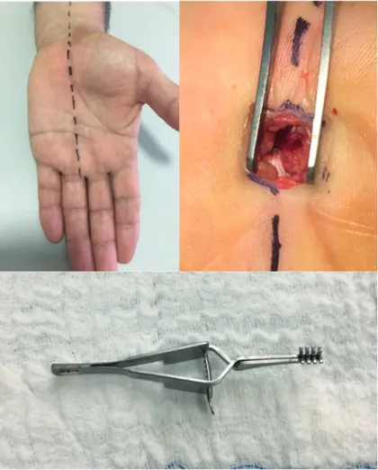
165 in the wrist and hand supine position (Figure 1). The skin was opened with mini-retractor, subcutaneous
tissue was passed through blunt dissection and palmar aponeurosis was opened and blunt dissection was performed. With the help of a mini-clamp with a curved tip, the ligament was suspended and a transverse carpal ligament was cut with a 15 number scalpel. The distal end of the median nerve in the tunnel was reached. The rest of the transverse carpal ligament was cut with tissue scissors (Figure 2). After the control of the tunnel opening with the help of dissecters, the skin was closed with 3.0 prolene after the control of bleeding (Figure 3). The average operation time was 15 minutes. All patients were discharged on the same day. All patients received 1 g of cefazolin sodium 30 minutes before the operation for prophylaxis. Patients hand wrapped with an elastic bandage and they were put on neck sling for 2 days. On post-operative day 3, the dressing was opened and the spray was left open. On the 10th post-post-operative day, sutures were taken and exercise with soft ball was recommended.
Figure 1. The incision line parallel to the medial of the ring finger, the mini-distal 1-1.5 cm skin incision and the
166
167
3. Results
Of 103 patients, 97 (94.1%) were female and 6 (5.9%) were male. The mean age of the patients was 49.4 years (21-81). Only one patient’s dominant hand was the left hand. Therefore, it was not possible to establish a statistical relationship between the dominance of the left or right hand in causing CTS. A total of 137 surgical procedures were performed. The mean duration of symptoms was 50.7 months (1-144 months). There were numbness in the hands of 73 patients, pain with numbness in 27 patients and weakness in 3 patients. Symptoms were more severe at night in 32 patients (31.1%). The Tinnel test was positive in 64 patients (62.1%) and the Phalen test was positive in 88 patients (85.4%). In 23 patients (22.3%), various degrees of weakness were detected in the hands. Muscle atrophy was found in 2 patients (1.9%). All patients had moderate or advanced conduction slowing in the median nerve detected by electromyelography (EMG). In patients with surgery, there was no median nerve injury, ulnar vessel-nerve injury, superficial palmar arc injury and motor branch injury. Pain and immobility occurred in the finger joints due to not using a hand in a patient. It was treated with physical therapy. No patient was reoperated. Results were statistically evaluated with preoperative and postoperative VAS and BCTQ scales. Preoperative VAS mean scores were 7.88±2.01 (6-9), while postoperative 1.78±0.93 (0-4) were found and statistically significant (p=0.002, p <0.05). Preoperative BCTQ scale mean score was 28.01±10.33 (22-37) while it was 16.68±5.76 (12-23) postoperatively and was statistically significant (p=0.003, p <0.05) (Table 1).
Table 1. Comparison of the preoperative and postoperative results according to the VAS and BCTQ scales.
Preoperative Postoperative p
VAS 7.88± 2.01 (6-9) 1.78± 0.93 (0-4) 0.002 BCTQ 28.01± 10.33 (22-37) 16.68± 5.76 (12-23) 0.003
4. Discussion
The compression of the median nerve, which is considered to be the prototype of entrapment neuropathies, is seen common between 40 and 60 years of age. It is seen 3-10 times more in women. It is reported that it develops more frequently on the right hand (Gök and Yalçın, 1997; Gündüz et al., 2003; Padua et al., 1997; Phalen, 1966; Szabo and Madison, 1992). Similar to the data in the literature, the mean age of the patients in our study was 49.4 years. However, the female/male ratio (16.1) was higher than the literature. In accordance with the literature, more operations were performed on the right hand side than the left side. Only one of 103 patients had a dominant left hand. There was no correlation between the dominance of left or right hand to cause CTS. Therefore, it was not possible to establish a statistical relationship between the dominance of the left or right hand in causing CTS.
The Tinnel and Phalen tests from the provocative tests are widely used. However, conflicting results have been reported about the sensitivity and specificity of these two tests. In the literature, the sensitivity of the Tinnel test is reported to be between 9-89% and the Phalen test is between 10-74.5% (Gündüz et al., 2003; Kuhlman and Hennessey, 1997; MacDermid and Wessel, 2004; Szabo et al., 1984). Tinnel test was
168
62.1% and Phalen test was 85.4% in our patients. In the light of these values, we think that the positive Phalen test is more meaningful in patients undergoing surgery.
The purpose of surgery is to eliminate the pressure in the carpal tunnel by cutting the transverse carpal ligament (Greenberg, 1994; Rengachary, 1996). Surgical outcome is worse in patients presenting with loss of strength and atrophy. Surgery can be performed with classical open, endoscopic and minimally invasive techniques. Classical open surgery is the standard method for surgical decompression of the median nerve in the carpal canal. It is more effective and safer than other surgical methods. However, with this method, the rate of hypertrophic or painful scar can be higher and duration of returning to work and daily activities may be longer. Endoscopic and minimally invasive methods are used to reduce these problems. With these surgical procedures, faster return to daily life or work and less scar formation have been reported. However, complications such as incomplete decompression and tendon or neurovascular injury are higher (Cokluk et al., 2003; Schmelzer et al., 2006).
Numerous studies have been conducted to investigate whether minimally invasive procedures are performed proximally or distally of the transverse carpal ligament. Revealing the palmar cutaneous branches of the median and ulnar nerve with detailed anatomical studies, the negative aspects of the standard incision, the long learning process and cost of endoscopic surgery; directed surgeons to mini incisions (Lee et al., 1992; Martin et al., 1996).
In their anatomical study, Martin et al. identified the palmar cutaneous branch of the median nerve in the proximal palmar region in all 25 hands, and the palmar cutaneous branch of the ulnar nerve in 24 hands. They reported that there was no safe area for the standard incision in the proximal palmar region. They reported that the incisions made from the distal palmar region were safer due to the high density of nerve in the proximal palmar region (Martin et al., 1996; Özcanlı et al., 2010).
Abouzahr et al. found a superficial palmar arch injury in one of the 28 hands in a cadaveric study with a mini-incision through the wrist flexion line (Abouzahr et al., 1995). In another study, it was reported that there was no vessel-nerve injury in the loosening of a 1-cm incision made by TCL distal in 104 of the 104 patients (Klein et al., 2003).
Bromley reported that the technique applied with a mini-incision distal to the TCL described reduced the scar formation and sensitivity and shortened the process of return to work. Lee and Strickland reported a successful result with a tool-assisted distal palmar incision at a rate of 92.2% (Lee and Stickland, 1998; Schmelzer et al., 2006). Bagatur reported that the most common finding in the revision of unsuccessful CTS surgeries was the inability to adequately release distal to the TCL. Specifically, it is difficult to see and release all of the TCL for the incisions beginning and ending at the proximal of the wrist flexion line; suggested that these incisions should not be used in CTS surgery (Bagatur, 2002). Bal et al. Compared the distal and proximal mini incisions in their study. Reported a lower incidence of morbidity in the distal incision group, although there was no difference in long-term results (Bal et al., 2008).
169 About 1 cm skin incision increased patient satisfaction. The absence of postoperative pilar pain in patients
is also an important advantage of this technique. It contributed to patient satisfaction. The absence of significant complications such as median nerve injury, ulnar vascular-nerve injury, superficial palmar arc injury and motor branch injury is the result of careful surgery, although the surgical technique has the advantage. One of the advantages of this technique is that the patients can use their hands day after the surgery and return to work in a short time.
Conclusion
We think that distal mini-open surgery is a safe and successful method in CTS surgery because of less scar tissue and tenderness on the incision, returning to daily activities in a short time after the surgery, low rate of complications and successful clinical results. We believe that minimally invasive procedures should be performed distally from the transverse carpal ligament.
Conflict of Interest
The author declares that there is no conflict of interest.
Ethical Approval
All procedures performed in studies involving human participants were in accordance with the ethical standards of the institutional and/or national research committee (Institutional Review Board of Medipol University 28.05.2020/423) and with the 1964 Helsinki Declaration and its later amendments or comparable ethical standards.
Informed Consent
Informed consent was obtained from all individual participants included in the study.
Funding
No funding was received for this research.
References
Abouzahr, M. K., Pastis, M. C., Chiu, D. T. W. (1995). Carpal tunnel release using limited direct vision. Plast Reconstr Surg., 95(3), 534-538. Doi: 10.1097/00006534-199503000-00015
Atroshi, I., Gummesson, C., Johnsson, R., Ornstein, E., Ranstam, J., Rosen, I. (1999). Prevalence of carpal tunnel syndrome in a general population. JAMA, 282, 153-158.
Bagatur, A. E. (2002). Causes of failure and revision results in carpal tunnel syndrome surgery. Acta Orthop Traumatol Turc., 36, 346-353
170
Bal, E., Pişkin, A., Ada, S., Ademoglu, Y., Toros, T., Kayalar, M. (2008). Comparison between two mini incision techniques utilized in carpal tunnel release. Acta Orthop Traumatol Turc., 42, 234-237. Doi: 10.3944/ aott.2008.234
Buchberger, W., Schon, G., Strasser, K. Jungwirth, W. (1991). High-resolution ultrasonography of carpal tunnel. J Ultrasound., 10, 531-537. Doi: 10.7863/jum.1991.10.10.531
Cokluk, C., Senel, A., Iyigun, O., Celik, F., Rakunt, C. (2003). Open median nerve release using double mini skin incision in patients with carpal tunnel syndrome: technique and clinical results. Neurol Med Chir (Tokyo), 43, 465-468. Doi: 10.2176/nmc.43.465
de Krom, M. C., Knipschild, P. G., Kester, A. D., Thijs, C.T., Boekkooi, P. F., Spaans, F. (1992). Carpal tunnel syndrome: prevalence in the general population. J Clin Epidemiol, 45, 373-376.
Deryani, E., Aki, S., Muslumanoglu, L., Rozanes, I. (2003). MR imaging and electrophysiological evaluation in carpal tunnel syndrome. Yonsei Med J, 44(1), 27-32. Doi: 10.3349/ymj.2003.44.1.27
Frank, H. N. (1997). Atlas of Human Anatomy, Paperback 2nd ed.
Gök, H., Yalçın, P. (1997). The importance of clinical tests in the diagnosis of carpal tunnel syndrome. Fiziksel Tıp Rehabil Derg., 21(1), 31-36
Greenberg, M. S. (1994). Handbook of Neurosurgery; 3rd ed. Florida: Greenberg Graphics.
Gündüz, O. H., Borman, P., Bodur, H., Uçan, H. (2003). Wrist size, clinical and electrodiagnostic features in patients with carpal tunnel syndrome. Fiziksel Tıp Rehabil Derg., 49(1), 22-26
Klein, D. C., Kotsis, S. V., Chung, K. C. (2003). Open carpal tunnel release using a 1-centimeter incision: technique and outcomes for 104 patients. Plast Reconstr Surg., 111, 1616-1622. Doi: 10.1097/01. PRS.0000057970.87632.7e
Kotevoglu, N., Gülbahce-Saglam, S. (2005). Ultrasound imaging in the diagnosis of carpal tunnel syndrome and its relevance to clinical evaluation. Joint Bone Spine, 72(2), 142-145. Doi: 10.1016/j.jbspin.2004.03.012 Kuhlman, K. A., Hennessey, W. J. (1997). Sensitivity and specificity of carpal tunnel syndrome signs. Am J Phys Med Rehabil., 76(6), 451-457. Doi: 10.1097/00002060-199711000-00004
Lee, D. H., Masear, V. R., Meyer, R. D., Stevens, D. M., Colgin, S. (1992). Endoscopic carpal tunnel release: a cadaveric study. J Hand Surg Am., 17, 1003-1008. Doi: 10.1016/s0363-5023(09)91046-2
Lee, W. P., Strickland, J. W. (1998). Safe carpal tunnel release via a limited palmar incision. Plast Reconstr Surg., 101, 418-424. Doi: 10.1097/00006534-199802000-00025
171 Martin, C. H., Seiler, J. G., Lesesne, J. S. (1996). The cutaneous innervation of the palm: an anatomic study
of the ulnar and median nerve. J Hand Surg Am., 21, 634-638. Doi: 10.1016/S0363-5023(96)80017-7 Ozcanlı, H., Coskun, N. K., Cengiz, M., Oguz, N., Sindel, M. (2010). Defination of a safe zone in open carpal tunnel surgery: a cadaver study. Surg Radiol Anat., 32, 203-206. Doi: 10.1007/s00276-009-0498-7 Padua, L., LoMonaco, M., Gregori, B., Valente, E. M., Padua, R., Tonali, P. (1997). Neurophysiological classification and sensitivity in 500 carpal tunnel syndrome hands. Acta Neurol Scand., 96(5), 211-217. Doi: 10.1111/ j.1600-0404.1997.tb00271.x
Paget, J. (1854). Lectures on surgical pathology. Lindsay any Blakiston, Philadelphia.
Phalen, G. S. (1966). The carpal tunnel syndrome: experince in diagnosis and treatment of six hundred fifty-four hands. J Bone Joint Surg., 48(2), 211-228
Rengachary, S. S. (1996). Entrapment neuropathies. In: Wilkins RH, Rengachary SS, editors. Neurosurgery. International ed. The McGraw-HillCompanies p. 3073-3098
Schmelzer, R. E., Della Rocca, G. J., Caplin, D. A. (2006). Endoscopic carpal tunnel release: a review of 753 cases in 486 patients. Plast Reconstr Surg., 117, 177-185. Doi: 10.1097/01.prs.0000194910.30455.16 Szabo, R. M., Gelberman, R. H., Dimick, R. P. (1984). Sensibility testing in patients with carpal tunnel syndrome. J Bone Joint Surg., 66(1), 60-64