R E S E A R C H A R T I C L E
Open Access
Assessment of the kinesiotherapy
’s efficacy
in male athletes with calcaneal apophysitis
Ersin Kuyucu
1, Bar
ış Gülenç
1*, Hüseyin Biçer
2and Mehmet Erdil
1Abstract
Background: The aim of the present study was to assess the efficacy of kinesiotherapy used for treating various disorders in athletes on pain and pedal functions in patients with calcaneal apophysitis.
Methods: This prospective randomized controlled study included 22 patients with calcaneal apophysitis aged 8 to 16 years presenting with heel pain among junior athletes of a professional football club. The patients were randomly grouped into two groups, with one group receiving sham tape only and the other kinesio tape. American Orthopedic Foot & Ankle Society (AOFAS) and visual analog scale (VAS) scores were recorded before and after the treatment. Results: The preoperative VAS score of the kinesio tape was 7, and AOFAS score was 62.4; the corresponding figures of the sham group were 6.81 and 70.5, respectively. The kinesio-tape group had a better AOFAS scores at 1st and 3rd month (p < 0.05). Posttreatment AOFAS score was 99.7 ± 0.9 for the kinesio-tape group and 97.4 ± 3.9 for the sham-tape group. Posttreatment VAS score was 0.1 ± 0.3 for the kinesio-tape group and 0.4 ± 0.5 for the sham-tape group (p > 0.05). Discussion: Conservative treatment modalities are preferentially used for its treatment. Kinesiotherapy is one of the treatment methods for the apophysitis. In the literature, our study is the first prospective randomized trial on the efficacy of kinesio taping in calcaneal apophysitis.
Conclusions: Although kinesio taping can be effectively used for the restoration of ankle functions of athletes with calcaneal apophysitis, its role in pain is limited. Since it lacks serious side effects, it can be used in combination with or as an alternative to pharmacological treatment in this patient group.
Background
In adolescent athletes, heel pain is a common condition that affects work force [1]. In children belonging to this age group, calcaneal apophysitis is a major cause of heel pain. Calcaneal apophysitis is a self-limiting condition and affects persons of 8–15 years of age who are overweight and physically active. It is rare after the age of 13–15 years when the calcaneal apophysis is ossified [2, 3].
Kinesiotherapy was first invented and developed by Kenzo Kase. Its principle mechanism of action is to regu-late regional blood flow and thus eliminating inflamma-tory cytokines in the region and allowing reparative cells to arrive the region, thereby providing a relief and redu-cing symptoms. We aimed to study athletes with calcaneal apophysitis to assess the efficacy of kinesiotherapy, which
we considered to be a potential alternative to the previ-ously described arch taping approach [4].
Methods
This prospective study included 22 junior football players who were diagnosed with calcaneal apophysitis with clin-ical examination and radiography between 2016 and 2017, and all were actively engaged in football playing. Radio-logically lateral calcaneal X-rays showed increased scler-osis and fragmentation of calcaneal apophysis. Those with radiologically closed calcaneal apophysis, a body mass index (BMI) greater than 25, comorbidities in addition to apophysitis, and a history of heel trauma or conservative treatment were excluded from the study.
All athletes provided informed consent before study entry. All participants were fully randomized, with one group undergoing kinesio taping and the other form of taping that mimicked kinesio taping but devoid of kine-sio taping properties (sham). Both groups were applied stretching exercises, topical analgesic treatment, and
* Correspondence:[email protected]
1Orthopaedics and Traumatology Department, Medipol University, Tem
Avrupa Göztepe çıkışı/Bağcılar, Istanbul, Turkey
Full list of author information is available at the end of the article
© The Author(s). 2017 Open Access This article is distributed under the terms of the Creative Commons Attribution 4.0 International License (http://creativecommons.org/licenses/by/4.0/), which permits unrestricted use, distribution, and reproduction in any medium, provided you give appropriate credit to the original author(s) and the source, provide a link to the Creative Commons license, and indicate if changes were made. The Creative Commons Public Domain Dedication waiver (http://creativecommons.org/publicdomain/zero/1.0/) applies to the data made available in this article, unless otherwise stated.
massage therapy aimed at heel and plantar fascia. Ages, weight, and height of all patients were recorded.
Pretreatment and posttreatment 1st week and 1st, 3rd, and 6th month visual analog scale (VAS) scores as well as pretreatment, posttreatment 1st, 3rd, and, finally, 6th month American Orthopedic Foot-Ankle Society (AOFAS) scores were recorded in all patients. We used the AOFAS for the ankle and hindfoot. All patients were monitored for recurrences from the onset of treatment to the end of the 6th month. X-rays evaluation was done before taping.
Tape application
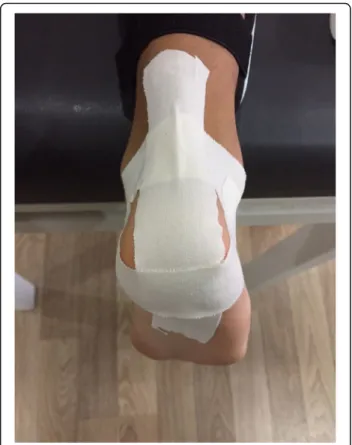
Kinesio taping was applied using the mechanical correc-tion method recommended by Kenzo Kase for calcaneal apophysitis; as such, a 4–6-in. tape portion was cut and pulled from both ends to apply moderate to severe ten-sion (50–75%) in a way to intersect the insertion point of the Achilles tendon to the calcaneus perpendicularly. The knee was placed in extension and the ankle in max-imum dorsiflexion during the application (Fig. 1) [4].
In contrast, sham taping was applied to the same region in a similar manner using Hypafix® (BSN Medical Guill-laime Kroll, Luxembourg) hypoallergenic tape, without ap-plying tension, and with the knee and ankle in extension and dorsiflexion positions as in the kinesio taping (Fig. 2).
Taping was repeated by an expert physiotherapist every 3 days, with 1 day intervals between two consecu-tive sessions in order to rest the skin. By this way, a total of 12 kinesio taping procedures were carried out and the therapy was terminated.
Statistical analysis
The descriptive statistics included mean, standard de-viation, median, minimum, maximum, frequency, and percentage. The distribution of the study variables was tested using Kolomogorov-Smirnov test. Inde-pendent quantitative variables were compared using independent samples t test and Mann Whitney U test. Dependent quantitative data were analyzed with Wil-coxon test. SPSS 22.0 software package was used for all statistical analyses.
Results
In this study, we included 27 patients with calcaneal apophysitis with radiologic and clinical diagnosis. Three patients whose BMI > 25 and two patients who have trauma history were excluded. After excluding the above patients, the study was finished with 22 pa-tients. All patients were male. The study population had an age range of 10–16 years, the mean age of the study participants was 13.18 (10–16) years, and their mean BMI was 19.6 (17–22).
Fig. 1 Kinesio taping treatment applied to the Achilles tendon and heel region
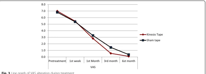
When we compared the kinesio and sham taping groups, they did not show significant differences in terms of 1st week, 1st month, 3rd month, and 6th month VAS scores (p > 0.05) (Table 1). But in itself, Kinesio group showed a sig-nificantly decreased 1st week, 1st month, 3rd month, and 6th month VAS scores compared to the pretreatment VAS score (p < 0.05). Likewise, in the sham group, the 1st week, 1st month, 3rd month, and 6th month VAS scores showed significant reductions compared to the pretreatment VAS score (p < 0.05) (Table 1). Both groups were comparable with respect to the 1st week, 1st month, 3rd month, and 6th month VAS score reductions compared to the pretreat-ment VAS score (p > 0.05) (Table 2, Fig. 3).
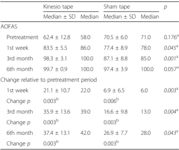
The kinesio and sham groups did not show significant dif-ferences between pretreatment and 6th month AOFAS scores (p > 0.05). The Sham group showed significant in-creases in 1st month, 3rd month, and 6th month AOFAS scores compared to the pretreatment AOFAS score (p < 0.05) (Table 2). The kinesio group had significantly higher than the 1st month and 3rd month AOFAS scores compared to the sham group (p < 0.05) (Table 2, Figs. 3 and 4). Posttreatment AOFAS score was 99.7 ± 0.9 for the kinesio-tape group and 97.4 ± 3.9 for the sham-tape group. Posttreatment VAS score was 0.1 ± 0.3 for the kinesio-tape group and 0.4 ± 0.5 for the sham-tape group (p > 0.05).
Discussion
In this prospective study, we explored no significant dif-ferences between both groups with regard to age, weight,
height, and body mass index (Table 3). No significant difference was found between the VAS scores of both groups. The results of the present study showed that the AOFAS score used for assessing ankle functions after kinesiotherapy was significantly increased in the kine-siotherapy group compared to the sham group. On the other hand, no significant difference was found between the two groups with respect to score increase.
Calcaneal apophysitis pathogenesis is thought to in-volve inflammation resulting from calcaneal apophysis being subjected to shear forces due to its constant trac-tion by Achilles tendon and plantar fascia, as well as the excessive increase in the intensity of the traction force on apophysis as a result of the Achilles tendon being un-able to keep pace with the tibial and fibular growth peak and remaining relatively “short” [5, 6]. The disease is characterized by heel pain, antalgic gait, limited ankle dorsiflexion, and performance loss in young athletes en-gaged in football and is diagnosed when a patient feels pain in the compression test. Direct films may show in-creased sclerosis and fragmentation of calcaneal apophy-sis [3] (Fig. 5). Advanced imaging techniques such as CT, MRI can be used to exclude conditions such as osteomyelitis or malignancy which are included in the differential diagnosis.
Conservative treatment modalities are preferentially used for its treatment. These include non-steroidal anti-inflammatory drugs, massage therapy, cold application, foot orthosis, and arch taping [7].
Direct forces acting on calcaneus apophysis are the traction forces that the plantar fascia and Achilles ten-don apply in opposite directions. In normal walking cycle,
Table 1 VAS alteration of both groups before and during treatment
Kinesio tape Sham tape p
Median ± SD Median Median ± SD Median VAS Pretreatment 7.0 ± 0.9 7.0 6.8 ± 1.2 7.0 0.783a 1st week 5.5 ± 0.9 6.0 5.4 ± 2.1 6.0 0.893a 1st month 2.8 ± 0.6 3.0 3.3 ± 2.0 3.0 0.812a 3rd month 0.5 ± 0.5 1.0 1.5 ± 1.2 1.0 0.069a 6th month 0.1 ± 0.3 0.0 0.4 ± 0.5 0.0 0.136a
Change relative to pretreatment period
1st week 1.5 ± 0.5 2.0 1.5 ± 1.2 1.0 0.782a Changep 0.003b 0.011b 1st month 4.2 ± 0.9 4.0 3.5 ± 1.5 4.0 0.199a Changep 0.003b 0.003b 3rd month 6.5 ± 1.1 6.0 5.4 ± 1.4 5.0 0.057a Change p 0.003b 0.003b 6th month 6.9 ± 1.0 7.0 6.5 ± 1.1 7.0 0.341a Changep 0.003b 0.003b a Mann-Whitney U test/X2 2
chi square test
Table 2 AOFAS alteration of both groups before and after treatment
Kinesio tape Sham tape p
Median ± SD Median Median ± SD Median AOFAS
Pretreatment 62.4 ± 12.8 58.0 70.5 ± 6.0 71.0 0.176a 1st week 83.5 ± 5.5 86.0 77.4 ± 8.9 78.0 0.045a 3rd month 98.3 ± 3.1 100.0 87.1 ± 8.8 85.0 0.001a 6th month 99.7 ± 0.9 100.0 97.4 ± 3.9 100.0 0.057a Change relative to pretreatment period
1st week 21.1 ± 10.7 22.0 6.9 ± 6.5 6.0 0.003a Changep 0.003b 0.006b 3rd month 35.9 ± 13.6 39.0 16.6 ± 9.8 13.0 0.004a Changep 0.003b 0.003b 6th month 37.4 ± 13.1 42.0 26.9 ± 7.7 28.0 0.043a Changep 0.003b 0.003b a Mann-Whitney U test/X2 2
chi square test
in the initial contact phase, when the foot contacts the floor, the talonavicular joint displaces distally, the ankle makes internal rotation and pronation, and the plantar fascia becomes tenser. Tense plantar fascia causes a pres-sure increase in calcaneus apophysis [8–10].
With the continuation of the walking cycle, shortening of the Achilles tendon increases its tension, further in-creasing load transfer onto calcaneal apophysis [11–13]. The plantar fascia starts in the medial tubercle and at-taches to the base of toes. In the late phase of the cycle, when heel elevation takes place, the plantar fascia is folded beneath the convex surface of the metatarsal heads with the dorsiflexion of the metatarsophalangeal joints (the windlass mechanism) [8, 9].
Again, during this phase of walking, the length of the Achilles tendon is significantly shortened and the trac-tion forces on the calcaneus’ center of growth become maximum, causing microavulsions [14, 15].
In our study, we also observed that the improvement of some patients’ complaints occurred too late in the course. The literature data shows an average healing time of several weeks to 2 months [16]. We consider
that in persons intensely performing the late swinging phase activity of walking (during shooting or sprinting), such as football players, calcaneal apophysitis is caused by microtraumas in plantar fascia due to repetitive trauma healing with fibrosis, resulting in shortening that slightly further increase the load on calcaneal apophysis. Hence, we think that patients’ complaints are not fully eliminated without this shortening being corrected. What matters here is to determine in which patients fi-brosis develops and to predict which athletes would re-turn to sports and when.
Rachel et al. sought to answer the question whether all patients have to undergo radiography to make diagnosis. They reported that pathologies other than calcaneal apo-physitis potentially causing severe heel pain were present in 5.1% of 96 patients and that a lateral film should be taken in every patient with heel pain and a positive com-pression test [17]. We also took lateral foot radiograms for every patient. No athlete was found to have any path-ology other than calcaneal apophysitis causing heel pain. Some patients had fragmentation, and some others had increased sclerosis. But we did not classify the X-rays Fig. 3 Line graph of VAS alteration during treatment
taken for the elimination of other pathologies to exclude or include the patients. Our hypothesis related to this finding is that fragmentation occurs as a result of an ex-cessively increased load on heel apophysis in patients with shortened plantar fascia secondary to sclerosis.
Hundreds of studies have been conducted on the effi-cacy of kinesio taping since its first introduction; it has been reported to have favorable effects in a considerable portion of these studies [18–25].
We are of the opinion that one of the major reasons of the absence of any significant difference between kinesio taping and sham taping is that the former not altering plantar fascia tension. The force applied by the plantar fascia with the winding wheel mechanism is as effective as the traction force applied by the Achilles tendon on the development of apophysitis [16]. Hence, we suggest that kinesiotherapy on the Achilles tendon alone with-out taping the plantar fascia would not provide ad-equate efficacy. Although it appears technically simple to tape the plantar fascia, the efficacy of kinesiotherapy wanes when applied in constantly perspiring feet of ath-letes with long training hours. We did not apply arch taping or kinesio taping as described in the literature due to difficulties in application.
The major strength of the present study is being the first prospective randomized trial on the efficacy of kine-sio taping in calcaneal apophysitis. The others include studying the procedure’s efficacy on professional foot-ballers and assessing not only pain but also providing a functional assessment.
The limitations of the study include a small sample size and the absence of an assessment of arch banding which has been performed in previous studies and has proven efficacy.
Conclusions
Kinesiotherapy was an effective treatment modality to reduce pain compared to placebo in patients with calca-neal apophysitis. Kinesio taping was significantly more effective than placebo when ankle and foot functional scores were considered.
In addition to taping, treatment modalities such as massage and manual therapy aiming at relieving plantar fascia tension can still be utilized in this age group as the most effective treatment modalities as in the past. Abbreviations
AOFAS:American Orthopedic Foot and Ankle Society; BMI: Body mass index; CT: Computed tomography; MRI: Magnetic resonance imaging; VAS: Visual analog scale
Acknowledgements
There are no funding resources or acknowledgements. Availability data and materials
Data sharing is not applicable to this article, as no data sets were generated or analyzed during the study.
Funding
There are no funding resources for this paper. Authors’ contributions
BG and EK planned the study and writing. HB coordinated the application of physiotherapy and kinesio taping. EK and ME moderated and analyzed the study. All authors read and approved the final manuscript.
Ethics approval and consent to participate
The medical ethics committee of the Istanbul Medipol University (Istanbul, Turkey) approved this study with the decision number 10840098-604. Consent for publication
Written informed consents were obtained from the patients for publication of these cases and accompanying images.
Competing interests
The authors declare that they have no competing interests.
Publisher’s Note
Springer Nature remains neutral with regard to jurisdictional claims in published maps and institutional affiliations.
Author details
1Orthopaedics and Traumatology Department, Medipol University, Tem
Avrupa Göztepe çıkışı/Bağcılar, Istanbul, Turkey.2Physiotherapy Department,
Medipol University, Tem Avrupa Göztepe çıkışı/Bağcılar, Istanbul, Turkey.
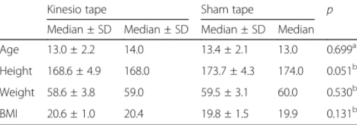
Table 3 Baseline characteristics of the study population with kinesio and sham taping
Kinesio tape Sham tape p
Median ± SD Median ± SD Median ± SD Median Age 13.0 ± 2.2 14.0 13.4 ± 2.1 13.0 0.699a Height 168.6 ± 4.9 168.0 173.7 ± 4.3 174.0 0.051b Weight 58.6 ± 3.8 59.0 59.5 ± 3.1 60.0 0.530b BMI 20.6 ± 1.0 20.4 19.8 ± 1.5 19.9 0.131b a t test b Mann-Whitney U test/X2
chi square test
Fig. 5 A foot radiogram of a 13-year-old male patient with heel pain. The radiogram shows increased sclerosis of the apophysis of calcaneus
Received: 20 June 2017 Accepted: 18 September 2017
References
1. Bailey CW, Cannon ML. Sever disease (calcaneal apophysitis). J Am Osteopath Assoc. 2014;114(5):411.
2. Wiegerinck JI, Yntema C, Brouwer HJ, Struijs PA. Incidence of calcaneal apohysitis in the general population. Eur J Pediatr. 2014;173(5):677–9. 3. Arbab D, Wingenfeld C, Rath B, Lüring C, Quack V, Tingart M.
Osteochondrosis of the pediatric foot. Orthopade. 2013;42(1):20–9. 4. Kenzo K, Jim W, Tsuyoshi K. Health & Personal Care. Clinical Therapeutic
Applications of the Kinesio Taping Method. 2rd Ed. 2003. p. 54–67. 5. Lewin P. Apophysitis of the os calcis. Surg Gynecol Obstet. 1926;41:578. 6. Madden CC, Mellion MB. Sever’s disease and other causes of heel pain in
adolescents. Am Fam Physician. 1996;54(6):1995–2000.
7. James AM, Williams CM, Haines TP. Effectiveness of interventions in reducing pain and maintaining physical activity in children and adolescents with calcaneal apophysitis. Prosthetics Orthot Int. 2013;37(6):495–8. 8. Bolgla LA, Malone TR. Plantar fasciitis and the windlass mechanism: a
biomechanical link to clinical practice. J Athl Train. 2004;39(1):77–82. 9. Hicks JH. The mechanics of the foot, II: the plantar aponeurosis and the
arch. J Anat. 1954;88:25–30.
10. Sarrafian SK. Functional characteristics of the foot and plantar aponeurosis under tibiotalar loading. Foot Ankle. 1987;8:4–18.
11. Donatelli RA. Abnormal biomechanics. In: Donatelli RA, editor. Biomechanics of the Foot and Ankle. 2nd ed. Philadelphia: FA Davis; 1996. p. 34–72. 12. Backstrom KM, Moore A. Plantar fasciitis. Phys Ther Case Rep. 2000;3:154–62. 13. Schwab SA. Apophyseal injuries in the growing athlete. Can Med Assoc J.
1977;117:626–30.
14. Kvist MH, Heinonen OJ. Calcaneal apophysitis (Sever’s disease)––a common cause of heel pain in young athletes. Scand J Med Sci Sports. 1991;1:235–8. 15. Orava S, Saarela S. Exertional injuries to young athletes. Am J Sports Med.
1978;6:68–74.
16. Micheli LJ, Ireland ML. Prevention and management of calcaneal apophysitis in children: an overuse syndrome. J Pediatr Orthop. 1987;7(1):34–8. 17. Rachel JN, Williams JB, Sawyer JR, Warner WC, Kelly DM. Is radiographic
evaluation necessary in children with a clinical diagnosis of calcaneal apophysitis (sever disease)? J Pediatr Orthop. 2011;31(5):548–50. 18. Ho KY, Epstein R, Garcia R, Riley N, Lee SP. Effects of patellofemoral taping
on patellofemoral joint alignment and contact area during weight bearing. J Orthop Sports Phys Ther. 2017;47(2):115–23.
19. Tsai FH, Chu IH, Huang CH, Liang JM, Wu JH, Wu WL. Effects of taping on Achilles tendon protection and Kendo performance. J Sport Rehabil Mar. 2017;2:1–26.
20. Huang YC, Chang KH, Liou TH, Cheng CW, Lin LF, Huang SW. Effects of Kinesio taping for stroke patients with hemiplegic shoulder pain: a double-blind, randomized, placebo-controlled study. J Rehabil Med. 2017;49(3):208–15.
21. Ortiz-Ramirez J, Perez-De la Cruz S. Efficacy of the application of kinesio tape in patients with stroke. Rev Neurol. 2017;64(4):175–79.
22. Boobphachart D, Manimmanakorn N, Manimmanakorn A, Thuwakum W, Hamlin MJ. Effects of elastic taping, non-elastic taping and static stretching on recovery after intensive eccentric exercise. Res Sports Med. 2017;25(2):1–10. 23. Keles BY, Yalcinkaya EY, Gunduz B, Bardak AN, Erhan B. Kinesio Taping in
patients with lumbar disc herniation: a randomised, controlled, double-blind study. J Back Musculoskelet Rehabil. 2016;30(3):543–50.
24. Coskun Benlidayi I, Salimov F, Kurkcu M, Guzel R. Kinesio Taping for temporomandibular disorders: single-blind, randomized, controlled trial of effectiveness. J Back Musculoskelet Rehabil. 2016;29(2):373–80.
25. Balki S, Göktaş HE, Öztemur Z. Kinesio taping as a treatment method in the acute phase of ACL reconstruction: a double-blind, placebo-controlled
study. Acta Orthop Traumatol Turc. 2016;50(6):628–34. • We accept pre-submission inquiries
• Our selector tool helps you to find the most relevant journal
• We provide round the clock customer support
• Convenient online submission
• Thorough peer review
• Inclusion in PubMed and all major indexing services
• Maximum visibility for your research Submit your manuscript at
www.biomedcentral.com/submit