Assessment of Cerebellar Dimensions Using
Magnetic Resonance Imaging in
Children Aged 0-4 Years
AABBSS TTRRAACCTT OObbjjeeccttiivvee:: Several diseases progress with cerebellar atrophy and hypoplasia. Cerebel-lum dimension measurements facilitating the assessment of decreases in cerebelCerebel-lum size on radio-logical images between the ages of 0 and 4 years in particular, when the cerebellum is still growing, will be of considerable use in clinical practice and appropriate for measurement value standardiza-tion. This will allow easier interpretetation of findings, together with more accurate diagnosis or a decrease in delays caused by false diagnosis. This study, the purpose of which was to obtain a data-base suitable for clinical use by performing cerebellar development dimension measurements in normal individuals aged 0-4 years, investigated the effect on cerebellar dimensions of parameters such as height, weight, head circumference and birth week. MMaatteerriiaall aanndd MMeetthhooddss:: One hundred fourty children aged 0-4 years, 56 girls and 84 boys, undergoing magnetic resonance imaging of the brain at the Karadeniz Technical University Farabi Hospital Radiology Department with no struc-tural cerebral or cerebellar abnormalities were included in the study. RReessuullttss:: In this study of cere-bellar dimensions in healthy children, these were positively correlated with weight, head circumference and height in both genders. Preterm birth had a significant effect on cerebellar di-mensions. CCoonncclluussiioonn:: In conclusion, this scale developed by measuring the cerebellar dimensions of healthy children aged 0-4 years will be of considerable practical assistance in terms of assessing measurements performed in a clinical environment.
KKeeyy WWoorrddss:: Magnetic resonance imaging; child Ö
ÖZZEETT AAmmaaçç:: Serebellumun atrofisi ve hipoplazisi ile giden birçok hastalık mevcuttur. Özellikle se-rebellumun büyümeye devam ettiği 0-4 yaş arasında radyolojik görüntülerde serebellum boyut-larındaki azalmanın değerlendirilebilmesi için serebellum boyutlarının ölçümleri, klinikte pratik olarak yararlı olacak ve ölçüm değerlerinin standardizasyonu için uygun olacaktır. Böylece bulgu-ların daha kolay tanımlanabileceği ve doğru tanı konulamaması ya da tanı konulmasındaki gecik-melerin azalacağı öngörülmektedir. Bu nedenle 0-4 yaş arası normal bireylerde serebellum gelişimi boyut ölçümleri ile yaparak klinik kullanıma uygun bir veri dökümü elde etmeyi amaçladığımız bu çalışmada boy, kilo, baş çevresi, doğum haftası gibi parametrelerin serebellum boyutları üzerine et-kilerini de değerlendirdik. GGeerreeçç vvee YYöönntteemmlleerr:: Araştırmaya Karadeniz Teknik Üniversitesi Rad-yoloji Bölümünde beyin manyetik rezonans görüntülemesi yapılan ve serebral ve serebellum anomalisi olmayan 0-4 yaş arasındaki 56 kız, 84 erkek çocuk dahil edilmiştir. BBuullgguullaarr:: Sağlıklı ço-cuklarda serebellum boyutlarını saptadığımız bu çalışmada, serebellum boyutlarının her iki cinsi-yette boy, kilo ve baş çevresi ile pozitif korelasyon olduğu ve erken doğumun serebellum boyutları üzerine önemli etkileri olduğu belirlendi. SSoonnuuçç:: Sonuç olarak 0-4 yaş arası sağlıklı çocukların se-rebellum boyutlarını ölçerek geliştirdiğimiz bu skala klinik ortamda yapılan ölçümlerin değerlen-dirilmesinin yapılabilmesi açısından oldukça pratik bir kolaylık sağlayacaktır.
AAnnaahh ttaarr KKee llii mmee lleerr:: Manyetik rezonans görüntüleme; çocuk
TTuurrkkiiyyee KKlliinniikklleerrii JJ MMeedd SSccii 22001155;;3355((44))::225566--6622
Fatma KÜÇÜKSÜMBÜL AYHAN,
aGülay YEĞİNOĞLU
bSibel KUL
caKaramanoglu Mehmetbey University,
Health College, Karaman Departments of
bAnatomy, cRadiology,
Karadeniz Technical University Faculty of Medicine, Trabzon Ge liş Ta ri hi/Re ce i ved: 10.09.2015 Ka bul Ta ri hi/Ac cep ted: 04.12.2015 Ya zış ma Ad re si/Cor res pon den ce: Fatma KÜÇÜKSÜMBÜL AYHAN Karamanoglu Mehmetbey University, Health College, Karaman,
TÜRKİYE/TURKEY [email protected]
doi: 10.5336/medsci.2015-47320 Cop yright © 2015 by Tür ki ye Kli nik le ri
n the embryological period, the central
nerv-ous system that develops from the ectoderm
begins to form at the start of the 3rd week.
Neural tube development is completed toward the
middle of the 4th embryological week. This neural
tube subsequently differentiates in the rostral area
to form the cerebrum and cerebellum.
Develop-ment of the cerebellum begins in days 40-45, and
the fissurae cerebelli and folia cerebelli appear in
the cerebellum at the end of the 4th month. The
cerebellum is part of the hindbrain, located below
the temporal and occipital lobes. Despite being a
structure where many impulses from sensory
re-ceptors terminate, it is regarded as one of the motor
components of the central nervous system (CNS).
Its volume represents approximately 10% of the
entire brain, but it consists of more than 50% of all
neurons.
1The basic functions of the cerebellum are the
motor control of coordination, balance and speech
functions. Recent neuroanatomical and clinical
studies and imaging of the CNS have proved that
the cerebellum also has cognitive, affective and
ex-ecutive functions.
2-4New studies are revealing
fur-ther cerebellar functions, and this is increasing the
amount of research into the cerebellum. One of
these studies reported a relation between higher
cognitive function, speech memory, consecutive
learning and language functions and the cerebellar
hemispheres and vermal lobules IV-VII.
5One
study involving functional magnetic imaging
showed that activity in the anterior lobe of the
cerebellum increased with speed of motor activity.
6Another study evaluating cerebellar size
deter-mined a significant relation between a reduced
in-telligence function and decreased cerebellar
dimension.
7These studies all reveal the importance
of measuring cerebellar dimensions.
Several previous studies have measured
cere-bellar volume, but none of these developed an
av-erage volume scale for age groups between 0 and 4
years.
3, 8-16One study that measured cerebellar
vol-ume between 0 and 2 years identified a 253%
in-crease in cerebellar volume between birth and 2
years.
17Another study measured cerebral and
cere-bellar volume using magnetic resonance imaging
(MRI) images in children between 4 and 18 years
and reported that cerebral and cerebellar volumes
did not change in this age range.
18We therefore
in-cluded children aged between 0 and 4 years in this
study, hypothesizing a potential change in
cere-bellar dimensions.
Several diseases progress with cerebellar
atro-phy and hypoplasia, including attention deficit
hy-peractivity disorder, autism, macrocephaly, cleft lip
and palate, Down syndrome, schizophrenia,
bipo-lar disorder, cerebral palsy and epilepsy.
19-31The
cerebellar dimensions obtained in the study will be
useful in clinical practice by making it possible to
assess a decrease in cerebellum dimensions on
ra-diological images between the ages of 0 and 4 years,
when the cerebellum is still growing, and will also
be applicable for the standardization of
measure-ment values. The purpose of this study was
there-fore to obtain a database suitable for clinical use by
performing cerebellar development dimension
measurements in normal individuals aged 0-4
years. We also assessed the effect of parameters
such as height, weight, head circumference and
weeks of gestation on cerebellar dimensions.
MATERIAL AND METHODS
One hundred forty children aged 0-4 years, 56
girls and 84 boys, undergoing MRI of the brain at
the Karadeniz Technical University Farabi
Hos-pital Radiology Department for reasons such as
headache, sleeplessness, delayed speech and
walk-ing, preliminary diagnoses of meningitis and
hy-poxic-ischemic encephalopathy and seizures and
with no structural cerebral or cerebellar
abnor-mality, as confirmed by a specialist radiologist,
were included. Families were told not to permit
their children to sleep during the preceding night,
thus ensuring they would sleep and remain
immo-bile during MRI. Images from children who woke
or moved during MRI were excluded from the
re-search for lacking sufficient clarity. MRIs were
taken of all sections requested by radiology
spe-cialists, but only midsagittal and axial images were
used in this study. Local ethical approval was
granted and written consent was obtained from
parents. Children’s height, weight and head
cir-cumferences at MRI were measured in order to
in-vestigate correlations with cerebellar dimensions.
Families were administered a questionnaire
con-taining questions about factors that might affect
cerebellar development, such as children’s weeks
of gestation, whether they cried at birth, cyanosis
during birth, history of intubation and maternal
ex-posure to viral infection during pregnancy.
Children were divided into premature (<37
weeks of gestation) and term (≥37 weeks of
gesta-tion).
MEASUREMENTS
Measurements were performed using a 1.5T MRI
system (Simens Magnetom Symphony, Germany).
Two-dimensional T
1weighted sagittal images (TR:
400 ms, TE: 8.6 ms, slice thickness: 5 mm, FOV:
230x230 mm, Dist factor: 40%, Average: 1, Matrix:
256x192) and two-dimensional T
2axial images
(TR: 6800 ms, TE: 121 ms, slice thickness: 5 mm,
FOV: 230x168 mm, Dist factor: 30%, Average:1,
Matrix: 256x156) were recorded. Cerebellar
di-mensions were measured personally by one of the
authors using automatic calibrations on a work
sta-tion (Leonardo, Siemens).
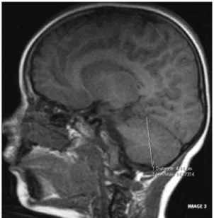
Vermis height (VH) was measured on the
ver-tical axis, and vermis anterior-posterior dimension
(VAPD) on the transverse axis by accurate
deter-mination of cerebellar vermis margins on T
1weighted midsagittal images (Figure 1).
Right cerebellar hemisphere height (HH
RIGHT)
was measured on the vertical axis on that section
where the right cerebellar hemisphere was largest
and left hemisphere height (HH
LEFT) was
meas-ured on the section where that hemisphere was
largest by accurate determination of hemisphere
margins on T
1weighted midsagittal images (Figures
2, 3).
Transcerebellar diameter (TCD) was measured
between the two lateral margins of the cerebellum
from the section in which the cerebellum appeared
largest on T
2weighted axial images (Figure 4) and
cerebellar hemisphere anterior-posterior
dimen-sions (HAPD
RIGHT-and HAPD
LEFT) were
meas-ured perpendicular to that length (Figure 5).
STATISTICAL ANALYSIS
Data were analyzed and compared on SPSS 13.0
soft-ware. Median, arithmetic mean, standard deviation
and standard error values were determined. The
para-metric Student’s t test and paired t test were used to
compare means of normally distributed data and the
Mann Whitney U test for non-normally distributed
data. P<0.05 was considered statistically significant.
STUDY LIMITATIONS AND RECOMMENDATIONS
Thirty-two of the children in the study were
pre-mature, and 108 were term. Due to the small case
FIGURE 1: VH measurement (1), VAPD measurement (2).
number in this study, no comparison for preterms
on the basis of weeks of gestation was possible, and
we were unable to determine whether or not
cere-bellar dimensions of these babies after the 37
thweek were comparable to those of normal
new-borns. In addition, cerebellar dimensions increased
rapidly in the 0-1 years age range. We therefore
think that it will be useful for change in cerebellar
dimensions in preterms and rate of cerebellar
growth in the 0-1 age range to be evaluated in
greater detail by increasing the numbers of children
aged 0-1 years in future studies on the subject.
RESULTS
Children’s mean age was 48±15.39 months, mean
height 70.25 ± 19.78 cm, mean weight 8563.86 ±
5283.15 g and mean head circumference
42.50±6.87 cm. Correlation analysis revealed that
cerebellar dimensions increased with height,
weight, head circumference and age (Table 1).
Children born on term and aged 0-4 years
were divided into 7 groups 0-3, 4-6, 7-9, 10-12,
13-24, 25-36 and 37-48 months. Mean cerebellar
di-mensions (cm) of these 7 groups are shown in Table
2.
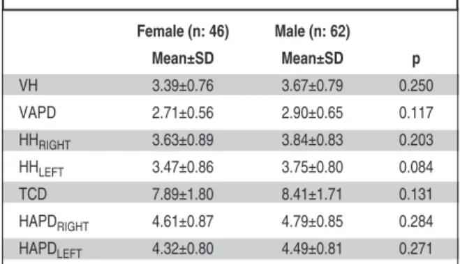
No statistically significant difference was
de-termined between male and female term children
in terms of cerebellar dimensions (Table 3).
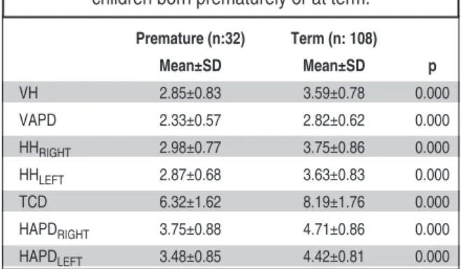
Comparison of cerebellar dimensions between
premature and term children revealed statistically
significantly lower VH, VAPD, HH
RIGHT, HH
LEFT,
TCD, HAPD
RIGHTand HAPD
LEFTparameters in
prematurely born children compared to those born
on term (p< 0.001) (Table 4).
DISCUSSION
Several studies in the literature have assessed
cerebellar dimension with volume measurement.
FIGURE 3: HHLEFTmeasurement FIGURE 4: TCD measurement.
One study measuring cerebellar vermis
dimen-sions assessed vermis size in individuals aged 0-60
years, but no mean vermis dimensions were given
for the 0-4 age group.
32Another study measured
cerebral and cerebellar volumes at MRI in
chil-dren aged 4-18 years and determined no change
in volumes in this age group.
18Our research
therefore involved the 0-4 age group, in which
we predicted a possible change in cerebellar
di-mensions, and these were observed to increase up
to the age of 4 years.
Analysis of the rate of increase in cerebellar
dimensions between 0 and 4 years determined
quite rapid growth in the first year of life.
Simi-larly, a study in which dimensions of the
cerebel-lar vermis were measured reported a marked
increase in cerebellar vermis size between the ages
of 0 and 1.
32One study performed with volume
measurement paralleled our own research finding
by determining rapid growth in cerebellar volume
in the first year of life.
17The difference in cerebellar size between the
sexes is unclear in the literature, and conflicting
opinions exist. Some authors have suggested that
gender variations in cerebellar size may be attributed
to sex hormones.
32No difference in cerebellar
di-mensions was determined between the sexes in this
study. Various studies involving individuals in
dif-ferent age groups (newborn-86 years) have reported,
VH VAPD TCD HAPDRIGHT HAPDLEFT HHRIGHT HHLEFT
Height r 0.872 0.797 0.927 0.881 0.856 0.877 0.889 p 0.000 0.000 0.000 0.000 0.000 0.000 0.000 Weight r 0.842 0.775 0.916 0.864 0.848 0.848 0.863 p 0.000 0.000 0.000 0.000 0.000 0.000 0.000 Head circumference r 0.897 0.852 0.956 0.921 0.898 0.897 0.915 p 0.000 0.000 0.000 0.000 0.000 0.000 0.000 Age r 0.751 0.715 0.819 0.759 0.748 0.761 0.780 p 0.000 0.000 0.000 0.000 0.000 0.000 0.000
TABLE 1: Cerebellar dimension correlations with height, weight, head circumference and age.
VH VAPD HHRIGHT HHLEFT TCD HAPDRIGHT HAPDLEFT
Age Interval n Mean±SD Mean±SD Mean±SD Mean±SD Mean±SD Mean±SD Mean± SD
0-3 months 32 2.57±0.34 2.10±0.27 2.64±0.40 2.55±0.31 5.85±0.66 3.62±0.39 3.46±0.38 4-6 months 10 3.40±0.25 2.57±0.24 3.49±0.37 3.42±0.30 7.59±0.43 4.28±0.33 4.12±0.37 7-9 months 7 3.85±0.51 2.94±0.47 3.87±0.34 3.66±0.48 8.06±0.94 4.62±0.40 4.10±0.46 10-12 months 9 3.99±0.31 3.13±0.43 4.35±0.30 4.10±0.32 8.92±0.39 5.23±0.37 4.84±0.40 13-24 months 12 4.08±0.40 3.13±0.28 4.15±0.40 4.07±0.36 9.28±0.47 5.32±0.40 4.92±0.45 25-36 months 14 4.20±0.32 3.23±0.30 4.51±0.37 4.38±0.39 9.77±0.41 5.48±0.23 5.14±0.30 37-48 months 24 4.22±0.36 3.34±0.46 4.45±0.34 4.34±0.30 9.85±0.45 5.41±0.31 5.09±0.43
TABLE 2: Mean cerebellar dimensions at 0-3, 4-6, 7-9, 10-12, 13-24, 25-36 and 37-48 months.
n: Specimen number. All values are given in centimeters.
Female (n: 46) Male (n: 62) Mean±SD Mean±SD p VH 3.39±0.76 3.67±0.79 0.250 VAPD 2.71±0.56 2.90±0.65 0.117 HHRIGHT 3.63±0.89 3.84±0.83 0.203 HHLEFT 3.47±0.86 3.75±0.80 0.084 TCD 7.89±1.80 8.41±1.71 0.131 HAPDRIGHT 4.61±0.87 4.79±0.85 0.284 HAPDLEFT 4.32±0.80 4.49±0.81 0.271
TABLE 3: Comparison of male and female children in
terms of cerebellar dimensions.
in agreement with our research, no difference
be-tween the genders in terms of cerebellar
dimen-sion.
8,11,16,32However, studies involving individuals
from different age groups across a broad range of ages
(4-77 years) have reported a greater cerebellar
vol-ume in men compared to women.
9,10,18,33,34Volumet-ric analysis of organ and tissue dimensions is without
doubt more reliable that dimensional measurements.
However, analysis of the findings of various studies
that have compared gender and volume size
8-11,16,18,32-34reveals no definitive conclusion regarding whether
cerebellar size differs between the sexes.
Several previous studies have reported that
fetal cerebellar dimensions increase with weeks of
gestation.
14,35-42Premature births can interrupt
cerebellum growth. Investigation of whether the
adverse effect or prematurity on cerebellar
di-mensions persists after birth has shown smaller
cerebellar dimensions in children born
prema-turely compared to those born at term.
One study in which fetal cerebellar
dimen-sions were evaluated using ultrasound reported
that head circumference, TCD, cerebellar
circum-ference, vermis surface area, VAPD and VH
in-creased in a linear manner with week of gestation.
Another study that employed ultrasound to
eval-uate the fetal cerebellum also revealed a parallel
increase in TCD and head circumference.
36At
analysis of increases in cerebellar dimensions with
head circumference, height, weight and age, this
increase was reported to persist in a parallel
man-ner also after birth. Another similar study
deter-mined that cerebellar circumference and area were
positively correlated with head circumference,
humerus length and weight.
41One study
com-paring cerebellar volume and head circumference
supports this research by reporting a positive
cor-relation.
39Varying degrees of cerebellar atrophy have
been determined in studies involving individuals
with attention deficit hyperactivity disorder,
29,43autism,
23, 30cleft palate or cleft lip,
31Down
syn-drome,
20cerebral palsy,
28and epilepsy
26. In the
con-text of the findings in the literature, this study
provides a database suitable for clinical use by
measuring cerebellar dimension development in
healthy individuals aged 0-4 years in order to for it
to be possible for such anomalies to be identified in
the early period.
CONCLUSIONS
The findings from this study reveal a rapid increase
in cerebellar dimensions between the ages of 0 and
4 years. We think that a scale drawn up for these
ages when the cerebellum is growing will be of
considerable use in the assessment of cerebellar size
in clinical practice.
Premature (n:32) Term (n: 108) Mean±SD Mean±SD p VH 2.85±0.83 3.59±0.78 0.000 VAPD 2.33±0.57 2.82±0.62 0.000 HHRIGHT 2.98±0.77 3.75±0.86 0.000 HHLEFT 2.87±0.68 3.63±0.83 0.000 TCD 6.32±1.62 8.19±1.76 0.000 HAPDRIGHT 3.75±0.88 4.71±0.86 0.000 HAPDLEFT 3.48±0.85 4.42±0.81 0.000TABLE 4: Comparison of cerebellar dimensions in
children born prematurely or at term.
All values are given in centimeters.
1. Sadler TW. Medical Embriology-Langmann. Başaklar C. (Çeviri Ed.). 11. Baskı. Ankara: Palme Yayın; 2011. p.358-94.
2. Baillieux H, De Smet HJ, Paquier PF, De Deyn PP, Mariën P. Cerebellar neurocognition: in-sights into the bottom of the brain. Clin Neurol Neurosurg 2008;110(8):763-73.
3. Shi L, Wang D, Hui SC, Tong MC, Cheng JC,
Chu WC. Volumetric changes in cerebellar re-gions in adolescent idiopathic scoliosis com-pared with healthy controls. Spine J 2013;13(12):1904-11.
4. Stoodley CJ, Schmahmann JD. Evidence for topographic organization in the cerebel-lum of motor control versus cognitive and af-fective processing. Cortex 2010;46(7): 831-44.
5. MacLullich AM, Edmond CL, Ferguson KJ, Wardlaw JM, Starr JM, Seckl JR, et al. Size of the neocerebellar vermis is associated with cognition in healthly elderly men. Brain Cogn 2004;56(3):344-8.
6. Wenzel U, Taubert M, Ragert P, Krug J, Vill-ringer A. Functional and structural correlates of motor speed in the cerebellar anterior lobe. PLoS One 2014;9(5):e96871.
7. Fu CH, Morgan K, Suckling J, Williams SC, Andrew C, Vythelingum GN, et al. A functional magnetic resonance imaging study of overt letter verbal fluency using a clustered acqui-sition sequence: greater anterior cingulate ac-tivation with increased task demand. Neuroimage 2002;17(2):871-9.
8. Luft AR, Skalej M, Welte D, Voigt K, Klock-gether T. Age and sex do not affect cerebellar volume in humans. AJNR Am J Neuroradiol 1997;18(3):593-6.
9. Chung SC, Lee BY, Tack GR, Lee SY, Eom JS, Sohn JH. Effects of age, gender, and weight on cerebellar volume of Korean peo-ple. Brain Res 2005;1042(2):233-5. 10. Acer N, Sahin B, Usanmaz M, Tatoğlu H,
Irmak Z. Comparison of point counting and planimetry methods for the assessment of cerebellar volume in human using magnetic resonance imaging: a stereological study. Surg Radiol Anat 2008;30(4):335-9. 11. Gocmen-Mas N, Pelin C, Canan S, Yazici AC,
Zagyapan R, Senan S, et al. Stereological evaluation of volumetric asymmetry in healthy human cerebellum. Surg Radiol Anat 2009;31(3):177-81.
12. Kosar MI, Karacan I, Otag K, Isleyen M, Gültürk S, Cimen M. Determination of cere-bellar volume in children and adolescents with magnetic resonance images. Folia Morphol (Warsz) 2012;71(2):65-70.
13. van Soelen IL, Brouwer RM, van Baal GC, Schnack HG, Peper JS, Chen L, et al. Heri-tability of volumetric brain changes and height in children entering puberty. Hum Brain Mapp 2013;34(3):713-25.
14. Holland D, Chang L, Ernst TM, Curran M, Buchthal SD, Alicata D, et al. Structural growth trajectories and rates of change in the first 3 months of ınfant brain development. JAMA Neurol 2014;71(10):1266-74.
15. Orasanu E, Melbourne A, Cardoso MJ, Modat M, Taylor AM, Thayyil S, et al. Brain volume estimation from post-mortem newborn and fetal MRI. Neuroimage Clin 2014;6:438-44. 16. Wierenga L, Langen M, Ambrosino S, van Dijk
S, Oranje B, Durston S. Typical development of basal ganglia, hippocampus, amygdala and cerebellum from age 7 to 24. Neuroimage 2014;96:67-72.
17. Knickmeyer RC, Gouttard S, Kang C, Evans D, Wilber K, Smith JK, et al. Structural MRI study of human brain development from birth to 2 years. J Neurosci 2008;28(47):12176-82. 18. Giedd JN, Snell JW, Lange N, Rajapakse JC, Casey BJ, Kozuch PL, et al. Quantitative mag-netic resonance imaging of human brain
de-velopment: ages 4-18. Cereb Cortex 1996; 6(4):551-60.
19. Ney GC, Lantos G, Barr WB, Schaul N. Cere-bellar atrophy in patients with long-term phenytoin exposure and epilepsy. Arch Neurol 1994;51(8):767-71.
20. Aylward EH, Habbak R, Warren AC, Pulsifer MB, Barta PE, Jerram M, et al. Cerebellar vol-ume in adults with Down syndrome. Arch Neu-rol 1997;54(2):209-12.
21. Specht U, Mayıs T, Schulz R, Rohde M, Ebner A, Schmidt RC, et al. Cerebellar atrophy and prognosis after temporal lobe resection. J Neurol Neurosurg Psychiatry 1997;62(5):501-6.
22. DelBello MP, Strakowski SM, Zimmerman ME, Hawkins JM, Sax KW. MRI analysis of the cerebellum in bipolar disorder: a pilot study. Neuropsychopharmacology 1999;21(1):63-8. 23. Levitt JG, Blanton R, Capetillo-Cunliffe L, Guthrie D, Toga A, McCracken JT. Cerebellar vermis lobules VIII-X in autism. Prog Neu-ropsychopharmacol Biol Psychiatry 1999;23(4):625-33.
24. Carper RA, Courchesne E. Inverse correlation between frontal lobe and cerebellum sizes in children with autism. Brain 2000;123(Pt 4):836-44.
25. Crooks R, Mitchell T, Thom M. Pattern of cere-bellar atrophy in patients with chronic epilepsy: a quantitative neuropathological study. Epilepsy Res 2000;41(1):63-73.
26. Sandok EK, O’brien TJ, Jackc CR, So EL. Significance of cerebellar atrophy in in-tractable temporal lobe epilepsy a quantitative MRI study. Epilepsia 2000;41(10):1315-20. 27. Keller A, Castellanos FX, Vaituzis AC, Jeffries
NO, Giedd JN, Rapoport RJ. Progressive loss of cerebellar volume in childhood-onset schiz-ophrenia. Am J Psychiatry 2003;160(1):128-33.
28. Kulak W, Sobaniec W. Magnetic resonance imaging of the cerebellum and brain stem in children with cerebral palsy. Adv Med Sci 2007;52(Suppl 1):180-2.
29. Mackie S, Shaw P, Lenroot R, Pierson R, Greenstein DK, Nugent TF 3rd, et al. Cere-bellar development and clinical outcome in at-tention deficit hyperactivity disorder. Am J Psychiatry 2007;164(4):647-55.
30. Bigler ED, Johnson JL, Lu J, McMahon W, Lainhart JE. Quantitative magnetic resonance imaging analysis of the cerebellum in macro-cephalic and normomacro-cephalic children and adults with autism. J Int Neuropsychol Soc 2008;14(3):401-13.
31. Nopoulos P, Langbehn DR, Canady J,
Mag-notta V, Richman L. Abnormal brain structure in children with isolated clefts of the lip or plate. Arch Pediatr Adolec Med 2008;161(8): 753-8.
32. Hayakawa K, Konishi Y, Matsuda T, Kuriyama M, Konishi K, Yamashita K, et al. Develop-ment and aging of brain midline structures: as-sesment with MR imaging. Radiology 1989;172(1):171-7.
33. Raz N, Dupuis JH, Briggs SD, McGavran C, Acker JD. Differential effect of age and sex on the cerebellar hemispheres and the vermis: a prospective MR study. AJNR Am J Neurora-diol 1998;19(1):65-71.
34. Carne RP, Vogrin S, Litewka L, Cook MJ. Cerebral cortex: an MRI-based study of vol-ume and variance with age and sex. J Clin Neurosci 2006;13(1):60-72.
35. Co E, Raju TN, Aldana O. Cerebellar dimen-sions in assessment of gestational age in neonates. Radiology 1991;181(2):581-5. 36. Vinkesteijn AS, Mulder PG, Wladimiroff JW.
Fetal transverse cerebellar diameter meas-urements in normal and reduced fetal growth. Ultrasound Obstet Gynecol 2000;15(1):47-51. 37. Malinger G, Ginath S, Lerman-Sagie T, Wa-temberg N, Lev D, Glezerman M. The fetal cerebellar vermis: normal development as shown by transvaginal ultrasound. Prenat Diagn 2001;21(8):687-92.
38. Zalel Y, Seidman DS, Brand N, Lipitz S, Ach-iron R. The development of the fetal vermis: an in-uterosonographic evaluation. Ultrasound Obstet Gynecol 2002;19(2):136-9. 39. Limperopoulus C, Soul JS, Gauvreau K, Huppi
PS, Walfield SK, Bassan H, et al. Late gesta-tion cerebellar growth is rapid and impeded by premature birth. Pediatrics 2005;115(3):688-95.
40. Messerchmidt A, Brugger PC, Boltshauser E, Zoder G, Sterniste W, Birnbacher R, et al. Distruption of cerebellar development: poten-tial complication of extreme prematurity. AJNR Am J Neuroradiol 2005;26(7):1659-67. 41. Sherer DM, Sokolovski M, Dalloul M, Pezzullo
JC, Osho JA, Abulafia O. Nomograms of the axial fetal cerebellar hemisphere circumfer-ence and area throught gestation. Ultrasound Obstet Gynecol 2007;29(1):32-7.
42. Bednarek N, Akhavi A, Pietrement C, Mesmin F, Loron G, Morville P. Outcome of cerebellar injury in very low birth-weight infants: 6 case raports. J Child Neurol 2008;23(8):906-11. 43. Valera EM, Faraone SV, Murray KE, Seidman
LJ. Meta-analysis of structural imaging finding in attention-deficit/hyperactivity disorder. Biol Psychiatry 2006;61(12):1361-9.