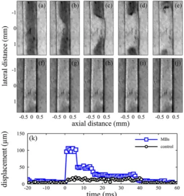
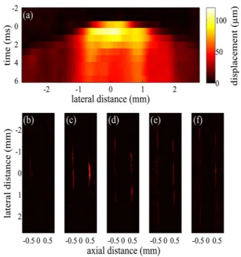
Acoustic particle palpation for measuring tissue elasticity
Tam metin
Şekil
Benzer Belgeler
the present study demon- strates that office HS and concurrent endometrial biopsy performed in the luteal phase, on the day of GnrH ago- nist initiation, improves implantation in
In the present study we aimed to investigate the anti-inflammatory and hypoglycemic activities of limonene, one of the major compounds of volatile fennel oil extract and
Department of Infectious Diseases and Clinical Microbiology, Gazi Yasargil Training and Research Hospital, Diyarbakir; 38 Department of Clinical Microbiology, Sakarya University
Previous optical implementations of the two-dimensional fractional Fourier transform have assumed identical transform orders in both dimensions.. We let the orders
We emphasize that public awareness is required to enable people to make an informed decision to share their genomic data publicly because such sharing compro- mises their own and
In this research, it was aimed to determine the effects of five different modi fied atmosphere packaging gas compositions with different concentrations of O 2 /CO 2 /N 2 on the
Indeed, three main mechanisms have been described so far by which neutrophils can contribute to thrombo- inflammation in either inflammatory or neoplastic conditions: ( 1 ) by
We achieved a good outcome in this case because of rapid resus- citation and aggressive treatment with monitoring and supportive care, including mechanical ventilation..
