88
ABSTRACT
Objective: Chewing includes the rhythmic movement of the jaw muscles. In this study, we investigated volumetric changes in the masticatory muscles and morphometric changes in the mandibular condyle due to unilateral extraction of the teeth in the sixth and twelfth weeks of life.
Materials and Methods: Eighteen rats were used. The rats were divided into three groups. In the experimental groups (Groups I and II), all of the teeth on one side of the upper and lower jaws of the rats were extracted. In the control group all the teeth were intact. Volumetric changes of the masseter and temporal muscles were evaluated. In addition, morphometric changes of the mandibular condyle were investigated.
Results: The measurements performed six weeks after tooth extraction did not reveal any difference in the temporal muscles between the two sides, however the volume of the masseter muscle on the side of the extraction was significantly reduced (Group I). Twelve weeks after the tooth extraction, the volumes of the masseter and temporal muscles showed significant reduction on the side of the extraction (Group II). A morphometric reduction in the mandibular condyle was observed in the longer period of time. No asymmetry was observed either for muscular volumes or for mandibular condyle dimensions in the control group.
Conclusion: Unilateral tooth loss may cause volumetric reduction in the masseter and temporal muscles ipsilaterally. However, the cause of the morphometric reduction in the mandibular condyle on the contralateral side in the longer period of time is surprising.
Keywords: Masseter muscle, Temporal muscle, Mandibular condyle, Muscle volume, Dental extraction
ÖZ
Amaç: Çiğneme, çiğneme kaslarının ritmik bir hareketidir. Bu çalışmada, tek taraflı diş çekimi sonrasında, altıncı ve onikinci haftalarda, çiğneme kaslarındaki volumetrik ve condylus mandibula’da morfometrik değişikliklerin değerlendirilmesi amaçlanmıştır.
Gereç ve Yöntem: Çalışmamızda 18 sıçan kullanılmıştır. Sıçanlar üç gruba ayrılmıştır. Deney grubunda (Grup I ve Grup II) sıçanların alt ve üst çene yarımındaki dişler çekilmiştir. Kontrol grubunda ise tüm dişler eksiksiz olarak bulunmaktaydı. M. masseter ve m. temporalis’deki volumetric değişiklikler değerlendirilmiştir. Ayrıca, condylus mandibula’daki morfometrik değişiklikler incelenmiştir.
Bulgular: Diş çekimi sonrası altıncı haftada m. temporalis volümünde her iki taraf arasında bir farklılık gözlenmezken, diş çekimi yapılan tarafta m. masseter volümü diğer tarafa göre anlamlı derecede azalmıştır (Grup I). Diş çekimi sonrası onikinci haftada, diş çekimi yapılan tarafta her iki kas volümünde azalma gözlenmiştir (Grup II). Ayrıca, Grup II’de tek taraflı diş kaybının, karşı tarafta condylus mandibula boyutlarında azalmaya neden olduğu gözlenmiştir. Kontrol grubunda ise her iki kas volümlerinde ve condylus mandibula boyutlarında iki taraf arasında fark gözlenmemiştir.
Sonuç: Tek taraflı diş kaybı, uzun zaman süreci içerisinde aynı tarafta m. masseter ve m. temporalis volumünde azalmaya neden olur. Ayrıca, aynı grupta, tek taraflı diş kaybının nedeni karşı tarafta condylus mandibula boyutlarında azalmaya neden olması şaşırtıcıdır.
Anahtar kelimeler: M. masseter, M temporalis, Condylus mandibula, Kas volümü, Diş çekimi
Introduction
Chewing includes the rhythmic movement of the jaw muscles. The components in charge of the movements are collectively referred to as the masticatory system, including the teeth, temporomandibular joint (TMJ), masticatory muscles, and the nerves controlling them. The teeth may be damaged due to different causes, and if this situation persists for a long
Volumetric and morphologic changes due to effect of unilateral
extraction of teeth
Tek taraflı diş çekim etkisine bağlı olarak çiğneme kaslarındaki volumetrik ve morfolojik
değişiklikler
Ayla Kurkcuoglu ( ), Can Pelin
Department of Anatomy, School of Medicine, Başkent University, Ankara, Turkey
e-mail: [email protected]
Submitted/Gönderme: 04.12.2015 Accepted/Kabul: 24.02.2016 Ayla KURKCUOGLU, Can PELİN
time, the muscles and jaw joints may also be affected [1,2]. In previous studies, it has been proposed that a reaction may occur after the occlusal alterations in response to the new distribution of loading, and bone tissues in the mandibular condyle may be altered [3,4]. Studies in humans and rats showed disturbance of the local microcirculation, irregular fibers in the disc, smaller and condensed chondrocytes in the condylar cartilage, and destruction of articular cartilage of the TMJ [5,6]. Thickening of TMJ articular cartilage was reported in rats after unilateral or bilateral removal of the teeth [7], and in monkeys with occlusal splints. The same finding was also observed by light microscopy [8] although extractions, removal of muscle and denervation were performed in these experiments, these altered the masticatory system or caused functional stresses [7,8]. Some experiments led to a modification in the morphology of the jaw. Osteosclerotic changes or a thinning of cartilage on the mandibular condyle were also observed [3,6,7]. Some other animal studies, reported several types of occlusal changes with effects on the masticatory muscles [6,9]. When occlusal changes and the loss of posterior teeth occurred, the electromyogram showed changes also associated with fatigue, discomfort, and pain of the masticatory muscles [10]. Tooth loss also influences anatomical features of the jaw muscles. A computed tomographic analysis revealed a reduction in radiodensity due to an increased proportion of fat tissue and cross-sectional area of the masseter and temporal muscles in subjects who had been edentulous for a long time [10,11]. Many studies, proposed that masticatory muscles and mandibular condyles may be altered due to loss of teeth. However, none of these studies showed how these changes alter over time. The present study was conducted to evaluate the effects of unilateral loss of teeth on the mandibular condyles and the masseter and the temporal muscles, after six weeks and twelve weeks.
Material and Methods
The project was approved by the Committee for the Use of Living Animals in Teaching and Research on project number: DA11/28 Başkent University. A total of 18 adult male Sprague Dawley rats (body weight, 350 - 400 g) were used in the present study. The rats were fed with unlimited rat pellet food and water during the experiment. All animals were evaluated weekly for their weights and physical activities. The rats were divided into three groups as the control and the two experimental groups (Groups I and II). In the experimental groups teeth on the right side of the upper and lower jaws were extracted unilaterally under general
anesthesia.General anesthesia was induced by intramuscular injection of 60 mg/kg ketamine hydrochloride IP, and 7 mg/ kg xylazine hydrochloride IP. After extraction, the rats were injected with intramuscular antibiotics (baytril %5, 0.05 mg/ kg) for 6 days to prevent infection of the wound. Oral 0.02 mg/kg fentanyl was given once a day, as an analgesic. Group I (six rats) were killed at the end of the 6th week and Group
II (six rats) were sacrificed at the end of 12th week. Control
group (six rats) had no teeth extracted, they were fed with unlimited rat food and water during the experiment and were killed at the end of the 12th week. In the control group and the
experimental groups the mandibles were removed from their surrounding tissues. The mandibular condyles, the masseter and the temporal muscles were harvested at 6 weeks (Group I) and 12 weeks (Group II and Control Group) after the tooth extractions. The extending front teeth of the animals were clipped 2 mm below the gingiva during the experiment.
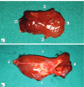
The superficial and deep parts of the masseter and temporal muscles were dissected (Fig.1a, b). The muscles were removed together with their fascia to prevent fluid influx into the tissues. Muscle volumes were measured using a water displacement method. The muscles were directly put into 10% formaldehyde for a few seconds, and the rise of the liquid was measured to record the muscle volume. Since the specimens were held in the formaldehyde solution for a very short time the effect of osmosis was disregarded. All measurements were done for both sides.
Fig. 1. External view of the temporal muscle (a), the superficial and deep parts of the masseter muscle (b) separated from the surrounding tissue (I inferior,S superior, M medial, L lateral)
a.
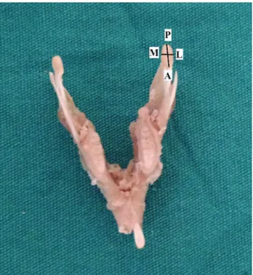
After dissection of the masseter and temporal muscles, the mandible was completely cleared from the surrounding tissues. Right and left mandibular condyles were used to investigate the changes in size. Osteometric measurements were obtained using four different points on the mandibular condyle. One of these measurements was performed between the protruding front and rear points of mandibular condyle (anteroposterior, A-P), and the other was performed at the ends of the protruding lateral sides of the condyle (mediolateral, M-L) (Fig. 2). Morphometric measurements were performed with 1 mm precision, using a digital caliber.
Fig. 2. Morphometric measurements were performed between the protruding front and rear points of mandible condyle (anteroposterior, A-P), and the other was performed at the ends of the protruding lateral sides of the mandibular condyle (M-L mediolateral) using a digital caliber.
After descriptive statistics were obtained for discrete and continuous variables, we determined whether the prerequisite of parametric tests (normality and homogeneity of the variances) was procured. Since, it was determined that the prerequisite could not be reached for parametric tests, the Wilcoxon test was used to compare dependent two groups. The Dunn multiple comparisons test was used to determine which groups caused the significant difference. The data were analyzed with SPSS 17.0 package program. Approximately 15% of all measurements were randomly selected to be measured again, and consistency testing was
performed using these two measurements. Consistency varied between 0.928 and 0.997, showing a remarkable reliability of the measurements (Table I).
Table I. The evaluation of consistency between the measurements
ICC p
The volume of the masseter muscles 0.983 <0.001 The volume of the temporal muscles 0.997 <0.00 Antero-posterior length of the mandibular
condyle 0.996 <0.00
Medio-lateral length of the mandibular condyle 0.928 <0.00 ICC: Intraclass correlation coefficient
Wilcoxon Signed Ranks Test (p<0.05)
Results
The rats were weighed every week. In the experimental groups, rats lost weight rapidly after extraction of their teeth. In the following weeks the nutrition of the rats improved and they started to gain weight. However, weight gain reached to a plateau at the end of the sixth week .In Group II extracted incisors of the rats grew slightly in the eighth week. These teeth were filled down in order to facilitate one-sided use. In this time period, first there was weight gain, and then weight loss following filling down the teeth, but later there was no change in the weights of the animals. The weights of the control rats did not change during the study. All rats were also examined for their regular physical activities and defecation habits, and all were regarded as normal.
Evaluation of the temporal and masseter muscles
In all rats, volumes of the masseter and the temporal muscles were evaluated on both sides (Fig. 1a, b). Concerning the volume of the muscles, no differences were found between the two sides in the control group (p≥ 0.05) for the masseter and temporal muscles. In group I, the volume of the masseter muscle was 2.08 ± 0.20 mL and the volume of temporal muscle was 0.55 ± 0.13 mL 6 weeks after extraction of the teeth, on the extraction side. On the non-extracted side, the volumes of the masseter and the temporal muscles were 2.67 ± 0.26 mL and 0.57 ± 0.12 mL, respectively in Group I. No difference was found between the extracted and non-extracted sides for the volumes of the temporal muscles in group I (p>0.05). However, the volumes of the masseter muscles were significantly different (p<0.05). The comparison of the extracted and non-extracted sides for the volumes of the masseter and temporal muscles yielded statistically significant differences in Group II (p<0.05) (Table II).
Table II. Volumes of the masseter and the temporal muscles
Masseter muscle (mL) Temporal muscle (mL)
Extraction side Mean ± std Non-extrac-tion side Mean ± std p Extraction side Mean ± std Non-extrac-tion side Mean ± std p CONTROL GROUP 2.50 ± 0.45 2.33 ± 0.41 p > 0.05 0.60 ± 0.13 0.58 ± 0.12 p > 0.05 GROUP I 2.08 ± 0.20 2.67 ± 0.26 p < 0.05 0.55 ± 0.13 0.57 ± 0.12 p > 0.05 GROUP II 1.91 ± 0.38 3.08 ± 0.38 p < 0.05 0.42 ± 0.08 0.63 ± 0.10 p < 0.05 Std: Standard deviation
Wilcoxon Signed Ranks Test (p<0.05)
Evaluation of the morphometric measurements of the mandibular condyle
We measured the mandibular condyles in all the rats. The extracted and non-extracted sides were not different in the A-P measurements in the control group. But the measured A-P lengths were greater on the extracted side for Group I, when compared to the non-extracted side (p<0.05) in Group I. Additionally, the values for Group II were greater than for Group I in the A-P measurements. M-L measurements revealed no differences between extracted and non-extracted sides in the control group. The values of the M-L measurements for the extracted and non-extracted sides were significantly different in Groups I and II (p<0.05). In Group I the M-L values in the extraction side were smaller when compared to the non-extraction side, however in Group II, M-L values of the mandibular condyle were greater in the extraction side when compared to the non-extraction side (Table III).
Table III. Morphometric measurement on the mandibular condyle in all rats
Anteroposterior length (A-P, mm) Mandibular condyle Mediolateral length (M-L, mm) Mandibular condyle Extraction side Mean ± std Non-extraction side Mean ± std p Extraction side Mean ± std Non-extrac-tion side Mean ± std p CONTROL GROUP 3.49 ± 0.36 3.52 ± 0.29 p > 0.05 1.57 ± 0.05 1.59 ± 0.04 p > 0.05 GROUP I 3.31 ± 0.26 3.26 ± 0.28 p < 0.05 1.47 ± 0,16 1.52 ± 0.06 p < 0.05 GROUP II 3.83 ± 0.23 3.41 ± 0.41 p < 0.05 1.53 ± 0.09 1.46 ± 0,10 p < 0.05
Std: Standard deviation Wilcoxon Signed Ranks Test (p<0.05)
Discussion
In this study, we primarily examined the volumetric changes in the masseter and temporal muscles at six and twelve weeks after unilateral tooth extraction in rats. Secondarily, we investigated morphometric changes in the mandibular condyle resulting from asymmetrical use of the TMJ. We fed and watered the rats well throughout the study. The rats continued to use one side of their jaws to chew their food. We compared the extracted and non-extracted sides. We filled down the lengthened teeth in Group II rats in order to perform an accurate comparison between the two sides.
Various methods were used to investigate the activity changes of the skeletal muscles in earlier studies [3,7,8,12]. These studies examined the changes in the muscle activity due to the tooth extraction, the functional disorders of the masticatory muscles after facial and trigeminal nerves had been severed, and muscle alterations due to the changes in the occlusal plane and a soft food diet. The volumes of the skeletal muscles can be measured directly and macroscopically in rats after dissecting them completely from the neighboring structures. The most frequently used method for this purpose is to place the tissue into a graduated cylinder filled with fluid, and to measure the difference of level of the fluid representing the volume of the tissue [13]. We used this simple method in our study to measure the volumes of the muscles. We were careful not to detach the fascia of the muscles in order to keep the amount of fluid entering the tissues to a minimum. That is, fluid may fill the tissue spaces and may result in a too small volume measurement [13-15]. At the end of the experiment, the masseter muscle volumes were found greater than the temporal muscle volumes on the same side in control group, Group I and Group II. In Group I rats, we analyzed the volumetric changes in the masticatory muscles after six weeks of unilateral chewing due to extraction of the teeth at one side of the jaws. Muscle volume measurements showed a significant difference in the masseter muscle volume at the teeth extracted side (p<0.05). There was a volumetric reduction, in other words, an atrophy on the extracted side. In contrast, there was muscle hypertrophy and volume increase in the non-extracted side. The volumes of the right and left temporal muscles were not found to be different after extraction of the teeth in Group I (p>0.05).
The strongest masticatory muscle is the masseter muscle in rats, and it is twice as strong as the temporal muscle and ten times stronger than the pterygoid muscles [16]. Like other skeletal muscles, masticatory muscles can adapt
themselves to anatomical, physiological, histochemical, and biochemical changes. Typically, relaxation of a skeletal muscle causes thinning of the muscle and its contraction causes thickening [17]. Takashima et al., found similar results to those of Staron et al., and explained the result as follows: the work performed by a muscle is related to the product of the transverse cross-section of the muscle and its fiber length. In addition, the work load of a muscle is directly proportional to its volume [18]. Our measurements did not show a difference between the extracted and non-extracted sides in Group I rats’ temporal muscle volumes, and this may be related to relatively small muscle volume and low functional load bearing by this muscle.
Masseter and temporal muscle volumes were smaller in the extraction side of Group II rats. Increased volume was noted on the non-extracted side, where chewing functions continued (Table III). As a result, significant volumetric differences were found on the extracted and non-extracted sides for both muscles. The volume of the temporal muscle was not different between the extraction and non-extraction sides in Group I rats, but the difference was significant in Group II rats (p<0.05). We suggest that the reason for this was 6 weeks longer period of feeding in Group II rats (12 weeks in total) compared to the Group I rats. Generally speaking, the common cause of the volumetric changes is disuse of the masticatory muscles on the extracted side, and a lot more use of the muscles on the non-extraction side. Masseter volumes were smaller in Group I rats compared to the control group on the extraction side, however it was greater on the non-extraction side. We compared our results with the results of the previous studies. However, there were no earlier reports of volumetric measurements of the masticatory muscles at different time points. Stal et al., reported that muscle deformation resulting from tooth extraction primarily involved ischemic changes [19]. The reason given for this was the high activity of the masseter muscle due to its high functional needs, and its intensive capillary network. It was reported that this was the reason for the atrophy of the masseter muscle at the side of tooth extraction [19]. Bani et al., performed muscle analyses 14 and 26 days after molar amputations [9]. They reported that there were no significant changes in the ipsilateral and contralateral muscle fibers 14 days after the amputation. However, the percentage of vacuolated fibers was significantly increased when compared to the controls. In addition, the muscle calcium content was significantly increased when compared to the controls, but it was not significantly different from that of the control counterparts in muscles contralateral to
amputated molars. The volume of the muscles ipsilateral to amputated molars were significantly increased as compared to the control rats, and were even more elevated than in the rats with molar amputation after 14 days. The calcium content of the muscles contralateral to amputated molars was not significantly different from that of the controls. The results of this study show that, in rats subjected to occlusal alteration by unilateral amputation of molar cusps, clear-cut morphologic and biochemical changes take place in the masseter muscle ipsilateral to the amputated teeth. By contrast, the contralateral masseter muscle appears to be only slightly affected or even unaffected [9]. Skeletal muscles are capable of changing a variety of their anatomical, physiological, histochemical, and biochemical characteristics to match altered functional demands [20,21]. Changes in anatomical characteristics, such as size and fiber types, are often associated with changes in the intensity, duration, and frequency of muscle activation by the central nervous system. Notwithstanding the marked differences between human jaw muscles and other skeletal muscles, their adaptational processes follow the same general concepts [21]. Typically, resistance training of a skeletal muscle, for example by means of repeated isometric contraction and relaxation, causes an increase in the thickness of the muscle and enhances muscular strength [17].
At the end of this study, the muscles were removed and measurements were made for the mandibular condyles. Mandibular condyle measurements showed significant changes either in A-P or M-L lengths between the extracted and non-extracted sides in both Group I and Group II. A-P lengths were longer in the extraction side in both groups. In contrast to this, there was an increase in M-L length of the Group II rats on the side of extraction when compared to the other side. Huang et al., found a significant increase of the histological glycosaminoglycan levels in the tissues of the mandibular condyle and articular disc in rabbits after tooth extraction. As a result of this, growth of the mandibular condyle and articular disc were seen [4]. We used only measurements in our study. However, the results of Huang et al.’s study can be seen to support the changes that occurred on the right side of our Group II rats. The authors reported that this was due to growth of the chondrocyte nuclei on the side of the extraction. Mao et al., reported hyperplasia at the proliferative and fibrocartilaginous region of the condyle cartilage on the side of the extraction in rabbits [21]. However, Ramfjord et al., did not report any difference between the right and left sides of the temporomandibular joint cartilages due to the effect of unilateral tooth extraction
in monkeys and sheep [22]. Furstman reported thickening of the condyle, joint cartilage and disc, Im et al., reported a significant histological decline in the osteoclast levels on the side of the extraction, 6 and 12 weeks after tooth extraction [7,23].
Morphological analyses performed by Lifshitz revealed mandibular shortening 3 months after excision of the masseter muscle in growing rats, and reduction of the angular process and asymmetrical growth of the maxilla were especially noted [24]. We did not study the body of the mandible, however we studied the mandibular condyle, a structure taking part in the TMJ, and noted an increase in the condylar measurements on the extraction side. The reason for this may be: the medial pterygoid muscle may partially compensate for the functions of the masseter and temporal muscles on the side of extraction. The condylar head then grows due to the excessive pulling force of the lateral pterygoid muscle at its insertion point on the condylar head.
In conclusion, our study showed that unilateral loss of teeth causes a decrease in the volumes of the masticatory muscles on the same side over a long period of time. These changes were more apparent in the masseter muscle when compared with the volumetric changes observed in the temporal muscle. In addition, we found that morphometric measurements of mandibular condyle showed differences on the extraction side both in the 6th and 12th weeks after
the extraction.
Further studies need to be done on this subject with a larger sample size with a longer period of time. However, we assume that the results of this study might be able to help with orthodontic treatment, jaw surgery and plastic and reconstructive surgery after long term unilateral loss of teeth.
Acknowledgments
We thank to Dr. Ragıba Zagyapan and Dr. Didem Bacanlı, the veterinary physician for teeth extraction and to Dr. Ersin Ogus for the statistical analysis and to Aslı Genç, Ayşe Duygu Tufan, Cem Önder, Volkan Ezici, Deniz Bulduk, the term 4 students for all their help during the study.
References
1. Huja SS, Rummel AM, Beck FM. Changes in mechanical properties of bone within the mandibular condyle with age. J Morphol 2009; 269:138-43. doi: 10.1002/jmor.10582 2. Parker MW. The significance of occlusion in restorative
fentistary. Dent Clin North Am 1993; 37: 341-51.
3. Endo Y, Mizutani H, Yasue K, Senga K, Ueda M. Influence of food consistency and dental extractions on the rat mandibular condyle: a morphological, histological and immunohistochemical study. J Craniomaxillofacial Surg 1998; 26: 185-90.
4. Huang Q, Opstelten D, Samman N, Tideman H. Experimentally induced unilateral tooth loss: histochemical studies of the temporomandibular joint. J Dent Res 2002; 81:209-13.doi: 10.1177/154405910208100313
5. Fujita S, Hoshino K. Histochemical and immunohistochemical studies on the articular disk of the temporomandibular joint in rats. Acta Anat 1989; 134: 26-30.
6. Granados JI. The ınfluence of the loss of teeth and attrition eminence. J Prosthet Dent 1979; 42: 78-85.
7. Frustman L. The effect of loss of occlusion upon the mandibular joint. Am J Orthodont 1965; 51: 245-61.
8. Christensen LV, Ziebert GJ. Effects of experimental loss of teeth on the temporomandibular joint. J Oral Rehabil 1986; 13: 587-98.
9. Bani D, Bani T, Bergamini M. Morphologic and biochemical changes of the masseter muscles induced by occlusal wear: studies in a rat model. J Dent Res 1999; 78:1735-44. doi: 10.1177/00220345990780111101
10. Raustia A M, Salonen M M, Pyhtinen J. Evaluation of masticatory muscles of edentulous patients by computed tomography and electromyography. J Oral Rehabil 1996; 23: 11-6
11. Newton JP, Yemm R , Abel RW, Menhinick S. Changes in human jaw muscles with age and dental state . Gerodontology 1993; 10: 16-22.
12. Bamaç B, Colak T, Ozbek A, Yenigün E, Çolak S, Bamaç Y. Volumetric determination of medial epicondyle and lateral epicondyle of humerus in male and female volleyball players. Okajimas Folia Anat Jpn 2003; 80:63-9.
13. Canan S, Sahin B, Odacı E. Unal B, Aslan H, Bilgiç S. Estimation of the reference volume. volume density and volume ratios by a stereological method: Cavalieri’s Principle. T Klin J Med Sci 2002; 22:7-14.
14. Cruz-Orive LM, Weibel ER. Recent stereological methods for cell biology: a brief survey. Lung. Cell Mol Physiol 1990; 2: 148-56.
15. Gundersen HJG, Bendtsen TF, Korbo L, et al. Some new simple and efficient stereological methods and their use in pathological research and diagnosis. APMIS 1989; 96: 379-94.
16. Kabak M, Gültiken ME, Onuk B. Yeni Zelanda tavşanında (oryctolagus cuniculus l.) articulatio temporomandibularis ve çiğneme kaslarının anatomisi. Ankara Üniv Vet Fak Derg 2007; 54: 149-54.
17. Staron RS, Karapondo DL, Kraemer WJ. Skeletal muscle adaptations during early phase of heavy-resistance training in men and women. J Applied Physio 1994; 76:1247–55. 18. Takashima M, Kitai N, Murakami S, Furukawa S, Kreiborg
S, Takada K. Volume and shape of masticatory muscles in patients with hemifacial microsomia. Cleft Palate Craniofac J 2003; 40:6-12.doi: 20.1177/00220985990780178101 19. Stal P, Eriksson PO, Thornell LE. Differences in capillary
supply between human orofacial, masticatory and limb muscles. J Musc Res Cell Motil 1996; 17: 183-97.
20. Kiliaridis S, Thilander B, Kjellberg H, Topouzelis N, Zafiriadis A. Effect of low masticatory function on condylar
growth: a morphometric study in the rat. Am J Orthod
Dentofacial Orthop 1999; 116:121-5. doi: 10.1016/S0889-5406(99)70207-6
21. Mao JJ, Rahemtulla F, Scott PG. Proteoglycan expression in the rat temporomandibular joint in response to unilateral bite raise. J Dent Res 1998; 77:1520-8.
22. Ramfjord SP, Walden JM, Enlow RD. Unilateral function and the temporomandibular joint in rhesus monkeys. Oral Surg
1971; 32:236-47.
23. Im JH, Kim SG, Oh JS, Lim SC, Ha JM. Influence of unilateral tooth loss in the temporomandibular joint and masseter muscle of rabbits. Oral Surg Oral Med Oral Pathol Oral Radiol 2012; 114:9-16. doi: 10.1016/j.tripleo.2011.05.022
24. Lifshitz J. Comparative anatomic study of mandibular growth in rats after bilateral resections of superficial masseter, posterior temporal and anterior digastric muscles. J Dent Res 1976; 55:854-8.