Morphological investigation of the pecten oculi in quail (Coturnix
coturnix japonica)
İ. Önder ORHAN1, Okan EKİM1, Alev Gürol BAYRAKTAROĞLU2
1 Ankara University, Faculty of Veterinary Medicine, Department of Anatomy, 2 Ankara University, Faculty of Veterinary Medicine,
Department of Histology and Embryology ANKARA.
Summary: In this study pecten oculi was investigated morphologically in quail (Coturnix coturnix japonica). It was detected
that the pecten oculi was attached to the retina and consisted of 19 folds on average. It was observed that quail pecten was in the pleated group and had a shell like rhomboid shape. The width of the folds was 259 µm on average. The width of the each fold was constant on every region of the fold. In light microscopic investigation, pecten was consisted of large number of capillaries and pigment cells (melanocytes) and the basal edge of pecten lined from the ventral part of the optic disc through the retina ventrally and projected into the vitreous body.
Key words: Anatomy, histology, pecten oculi, quail
Bıldırcında (Coturnix coturnix japonica) pecten oculi’nin morfolojik olarak incelenmesi
Özet: Bu çalışmada bıldırcında (Coturnix coturnix japonica) pecten oculi, morfolojik olarak incelendi. Ortalama 19 adet
kıvrıma sahip olan pecten oculi’nin, retina’ya tutunduğu tespit edildi. Bıldırcında pecten oculi’nin deniz kabuğu biçimli, yamuk benzeri bir şekle sahip olduğu ve bu nedenle kıvrımlı tip pecten grubuna dahil olduğu gözlendi. Ortalama olarak her bir kıvrım kalınlığı 259 µm olup, bu kalınlık kıvrımlar üzerinde her noktada sabit bulundu. Işık mikroskobik olarak, çok sayıda kapillar ve pigment hücreleri (melanositler) içeren pecten oculi’nin tabanı, discus nervi optici’nin ventral’inden retina’ya doğru yayılım göstermekteydi.
Anahtar Sözcükler: Anatomi, bıldırcın, histoloji, pecten oculi.
Introduction
The quail which belongs to the Phasianidae family is widely used as an experimental model particularly in physiology, pathology, toxicology and anatomy disciplines because of its lower feed consumption, rapid growth, early sexual maturity and also short generation intervals. The meat and egg consumption of the quail has also been increased in recent years (16, 25).
The pecten oculi which is specific to the birds is a unique vascular and pigmented structure (19, 22, 28, 29). The pecten projects from the optic disc into the vitreous body (7, 13, 14, 15, 28) and contains a large number of blood vessels and pigment cells (4, 8, 19, 28).
The studies conducted on the capillaries of the pecten demonstrated that it has a function as a nutritive tissue (7, 23, 28). The eye of the birds is characterized by an avascular retina (17, 26). Pecten oculi can be considered as an alternative structure comparing to the falciform process of some teleosts, the supraretinal vessels of amphibians (7), the conus papillaris of the reptiles (5, 11, 26) and intraretinal vessels of the mammals for vascularisation and nutrition of inner retina (7, 10, 11). Also light is absorbed by the pigments found on the
pigment cells of the pecten which provides a thermoregulation (2) and reduces penetration of light into the eye (27).
The pecten oculi was classified into three groups morphologically as conical, vaned and pleated types. It was reported that kiwi (Apteryx mantelli) has a conical type, ostrich (Struthio camelus) and rhea (Rhea
americana) have vaned type and the other bird species
commonly have pleated type pecten (21). However, bird species with pleated type pecten have similar structures; some considerable differences can be observed in thickness, number of the folds, shape and dimensions of the pecten (3, 4, 7, 8, 12, 18, 24). It was reported that these differences are directly proportional to the dissimilarities between the species and their daily activities. While day active (diurnal) birds have wide and more complicated pecten structure, night active (nocturnal) birds have narrow and basic formed pecten (4, 11, 21). As nighthawk (Chordeiles minor) has a narrow pecten with few folds (4-5 folds) (3), the daylight active bird, red-tailed hawk (Buteo jamaicensis) has fan-like pecten which consists of 17-18 accordion folds (6).
Histological structure of these three types of pecten in birds is basically composed of large number of capillaries, pericytes, melanocytes, hyalocytes and connective tissue (3, 20, 28).
In the present study it was aimed to define the anatomical and histological details oriented to the pecten oculi of the quail.
Materials and Methods
The present study was carried out in the Ankara University, Faculty of Veterinary Medicine, Department of Anatomy. The study was performed with 14 adult quails slaughtered in the abattoir. The ethical approval was also obtained for the study. Right and left eyeballs of 9 quails (5 male, 4 female) were dissected together with optic nerve apart from the orbit. The eyeballs were cut from the equatorial region. The posterior region including the pecten oculi was investigated as a fresh cadaver. The materials were photographed using Canon Powershot S70 digital camera fitted to Leica M16 stereomicroscope (Leica, Wetzlar, Germany).
Additional to the detection of the number of the folds, all morphological features of pecten were measured by Leica Application Suite software. The measurements were analyzed statistically considering male-female differences. The data was evaluated by descriptive statistics. As the differences between genders in terms of variables did not demonstrate a normal range, Mann-Whitney U test was used for the intergroup analyses. Besides, instead of mean values, median values were calculated. The significance level was considered as p<0.05.
Regardless of gender, right and left eyeballs of the 5 quails were examined histologically. Samples were washed with physiological saline and fixed in 10% neutral buffered formalin solution. Afterwards, they were processed by the standard paraffin sectioning, blocked in paraplast and finally trimmed a 5 µm sections cut. Mounted sections were stained with Mallory’s modified triple stained technique and examined under a light microscope (9).
The anatomical nomenclature of the structures was confirmed by using Nomina Anatomica Avium (1).
Results
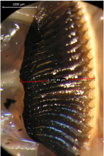
It was observed that the pecten oculi in quail was attached to the retina and the long axis of the pecten directed ventrally. Quail had a pleated type pecten and it displayed a folded structure. Beside this pleated type, pecten had short, thick and vertical coursed folds and due to that the quail pecten had a typical shell like aspect (Figure 1). Pecten was attached to the eyeball with its basal border and each fold spired and connected into the retina through this basal border (Figure 1, 2). The opposite side of the attached border, the apical border,
was projected into vitreous body and directed to the lens. Contrary to the basal border, the apical border had a flat surface (Figure 1).
Figure 1. The left pecten oculi of the quail. Şekil 1. Bıldırcının sol pecten oculi’si.
Figure 2. The basal border that folds are attached to the retina. Şekil 2. Kıvrımların retina’ya tutunduğu basal kenar.
It was determined that the pecten was composed of 19 folds on average. In all quails, fold numbers of both left and right pectens were equal for each sample inspected. It was observed that the folds at the middle of the pecten were longer than the ones at the lateral sides (Figure 1). The width of the each fold was measured from both basal and apical border and there was no variability in the width of each fold at basal or apical side. In this study the width of the folds was measured as 259 µm on average.
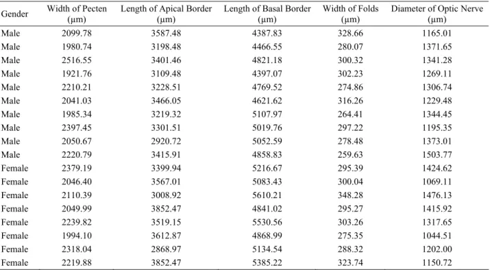
The numerical values about the pecten oculi and the optic nerve measurements were presented in Table 1. Descriptive statistical analyses were performed in 18 eyes about the width of the pecten in the thickest region, the length of both the basal and the apical border of the pecten, the width of the folds and the diameter of the optic nerve where exiting out sclera (Table 2).
The data of the intergroup statistical analyses were presented in Table 3. Due to the results of the intergroup
statistical analyses, the differences between genders in terms of the length of the basal border and the diameter of the optic nerve were found significant. There was no significant difference with regards to the other variables (p>0.05).
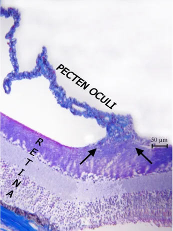
Histological examinations demonstrated that the localization of the basal border of pecten was started from the ventral part of the optic disc and extended on the retina. Therefore, histological sections of pecten were obtained from two different regions. In first region which pecten located on the ventral part of the optic disc, optic nerve (arrowhead) dispersed to the retina (arrow) but there was no direct connection existed between the pecten and the optic nerve (Figure 3). Second region in which the pecten was situated on the retina, it was determined that the pecten was only attached to the retina (Figure 4). In parallel with the first region, a direct continuation wasn’t observed from retina (arrows) to pecten (Figure 4). Pecten displayed a quite pleated
Table 1. The measurements of pecten oculi and optic nerve Tablo1. Pecten oculi ve nervus opticus’a ait ölçümler
Gender Width of Pecten (µm) Length of Apical Border(µm) Length of Basal Border(µm) Width of Folds (µm) Diameter of Optic Nerve(µm)
Male 2099.78 3587.48 4387.83 328.66 1165.01 Male 1980.74 3198.48 4466.55 280.07 1371.65 Male 2516.55 3401.46 4821.18 300.32 1341.28 Male 1921.76 3109.48 4397.07 302.23 1269.11 Male 2210.21 3228.51 4769.52 274.86 1306.74 Male 2041.03 3466.05 4621.62 316.26 1229.48 Male 1985.34 3219.32 5107.97 264.41 1344.45 Male 2397.45 3301.51 5019.76 297.22 1195.35 Male 2050.67 2920.72 5052.59 278.48 1373.01 Male 2220.79 3415.91 4858.83 259.63 1503.77 Female 2379.19 3399.94 5216.67 295.39 1424.62 Female 2046.40 3567.01 5083.43 300.04 1069.11 Female 2110.39 3008.92 5610.21 348.28 1476.13 Female 2049.99 3852.47 4841.02 295.27 1415.92 Female 2239.82 3519.15 5530.56 303.26 1317.65 Female 1994.10 3612.87 4868.99 275.35 1044.51 Female 2318.04 2868.97 5134.54 288.32 1202.00 Female 2219.88 3852.47 5385.22 323.74 1150.72
Table 2. The descriptive statistical analyses of pecten oculi and optic nerve Tablo 2. Pecten oculi ve nervus opticus’a ait tanımlayıcı istatistiksel analizler
Min. - Max. (n=18) Mean±SD (n=18) SE (n=18) Width of Pecten (µm) 1921.76 – 2516.55 2154.5628±167.38394 39.45277 Length of Apical Border (µm) 2868.97 – 3852.47 3362.8178±284.20731 66.9883 Length of Basal Border (µm) 4387.83 – 5610.21 4954.0867±356.83272 84.10628
Width of Folds (µm) 259.63 – 348.28 296.2106±22.90861 5.39961
Diameter of Optic Nerve (µm) 1044.51 - 1503.77 1288.9172±132.70416 31.27867 Min: Minimum; Max: Maximum; SD: Standard deviation; SE: Standard error
structure and a large number of capillaries (asterisks) were found in this structure (Figure 3). The vascularisation was more intensive at the apical border comparing to the basal border. The localization of pigment cells (melanocytes) (arrowheads) around the capillaries (arrows) was noticed (Figure 5).
Table 3. Intergroup analyses of the pecten oculi Tablo 3. Pecten oculi’ye ait gruplar arası analizler
Median Minimum - Maximum p
Width of Pecten (µm)
Male (n=10) 2075.2250 1921.76 – 2516.55
Female (n=8) 2165.1350 1994.10 – 2379.19 p>0.05 Length of Apical Border (µm)
Male (n=10) 3265.0100 2920.72 – 3587.48
Female (n=8) 3543.0800 2868.97 – 3852.47 p>0.05 Length of Basal Border (µm)
Male (n=10) 4795.3500 4387.83 – 5107.97
Female (n=8) 5175.6050 4841.02 – 5610.21 p<0.001 Width of Folds (µm)
Male (n=10) 288.6450 259.63 – 328.66
Female (n=8) 297.7150 275.35 – 348.28 p>0.05 Diameter of Optic Nerve (µm)
Male (n=10) 1324.0100 1165.01 – 1503.77
Female (n=8) 1259.8250 1044.51 – 1476.13 p<0.05
Figure 3. The section taken from which pecten located on the ventral part of the optic disc. Optic nerve (arrowhead) dispersed to the retina (arrow); the capillaries in the pecten (asterisks). Şekil 3. Pecten’in discus nervi optici’nin ventral kısmına yerleşen bölümünden alınan kesit. Retina’ya (ok) dağılmış n. opticus (okbaşı); pecten’deki kapillarlar (asteriskler).
Figure 4. The section taken from which pecten located on the retina (arrow).
Şekil 4. Pecten’in retina (ok) üzerine yerleşen kısmından alınan kesit.
Figure 5. Light micrographic appearance of pecten. Melanocytes (arrowheads); capillaries (arrows).
Şekil 5. Pecten’in ışık mikroskobik görünümü. Melanositler (okbaşları); kapillarlar (oklar).
Discussion and Conclusion
According to Meyer’s (21) classification it was determined that the pecten oculi of the quail was in the pleated type group. As the quail which is a daily active bird, had 19 folds on average, the other daily active species, ostrich had 16-19 (19), black kite had 12 (18) and chick had approximately 18 folds in the pecten (13). It could be noticed that there was a correlation between the pecten complexity and the activity of the species in general.
Due to the results of the intergroup statistical analyses, the differences between the genders in terms of the length of the basal border and the diameter of the optic nerve were found significant however a satisfying research hasn’t been published yet about the relationship between the diameters of the pecten and genders.
However it was stated that the pecten of the chick was located on the optic disc (13), quail pecten lined from the ventral part of the optic disc through the retina ventrally and projected into the vitreous body. It is known that the pecten oculi in birds which is thought to provide the blood supply of the avascular retina, contains large number of capillaries (3, 20). Histological findings obtained in this study were similar to those defined in other researches before. Pigment cells (melanocytes) were especially found between blood vessels. Similar to literature (2, 7) it was thought that pecten might play a role in thermoregulation and absorption of light.
References
1. Baumel JJ, King SA, Breasile JE, Evans HE, Berge JCV (1993): Nomina Anatomica Avium. 2nd edition,
prepared by the international committee on avian anatomical nomenclature, A Committee of The World Association of Veterinary Anatomists. Nuttall Ornithological Club. Cambridge, Massachusetts.
2. Bawa SR, YashRoy RC (1974): Structure and function of
vulture pecten. Acta Anat, 89, 473-480.
3. Braekevelt CR (1984): Electron microscopic observations on the pecten of the nighthawk (Chordeiles minor). Ophthalmologica, 189, 211-220.
4. Braekevelt CR (1988): Fine structure of the pecten of the
pigeon (Columba livia). Ophthalmologica, 196, 151-159.
5. Braekevelt CR (1989). Fine structure of the conus
papillaris in the bobtail goanna (Tiliqua rugosa). Histol
Histopathol, 4,287-293.
6. Braekevelt CR (1991): Fine structure of the pecten oculi
of the red-tailed hawk (Buteo jamaicensis). Anat Histol
Embryol, 20, 354-362.
7. Braekevelt CR (1994): Fine structure of the pecten oculi
in the American crow (Corvus brachyrhyncho). Anat
Histol Embryol, 23, 357-366.
8. Braekevelt CR (1998): Fine structure of the pecten oculi
of the emu (Bromaius novaehollandiae). Tissue cell, 30,
157-165.
9. Culling CFA, Allison RT, Barr WD (1985): Cellular
Pathology Technique. 4th ed. Butterworths, London;
214-255.
10. De Schaepdrijver L, Simoens P, Lauwers H, De Geest JP (1989): Retinal vascular patterns in domestic animals. Res Vet Sci, 47, 34-42.
11. Duke-Elder S (1958): System of Ophthalmology. Henry Kimpton, London.
12. Fielding M (1972): The ultrastructure of the pecten oculi
in the domestic fowl. J Anat, 113, 295-297.
13. Fischlschweiger W, O'Rahilly R (1966): The
ultrastructure of the pecten oculi in the chick. Acta Anat
(Basel), 65, 561-78.
14. Gerhardt H, Liebner S, Wolburg H (1996): The pecten
oculi of the chicken as a new in vivo model of the blood-brain barrier. Cell Tissue Res, 285, 91-100.
15. Gerhardt H, Schuck J, Wolburg H (1999):
Differentiation of a unique macroglial cell type in the pecten oculi of the chicken. Glia, 28, 201–214.
16. İde T (2003): Laboratuvar Hayvanları Biliminin Temel
İlkeleri. Medipres Yayınları – Özkan Matbaacılık, Ankara
17. Jeon GS, Kang TC, Park SW, Kim DW, Seo JH, Cho SS (2004): Microglial responses in the avascular quail
retina following transection of the optic nerve. Brain Res,
1023, 15-23.
18. Kiama SG, Bhattacharjee J, Maina JN, Weyrauch KD (1994): A scanning electron microscope study of the pecten
oculi of the black kite (Milvus migrans): possible involvement of melanosomes in protecting the pecten against damage by ultraviolet light. J Anat, 185, 637-642.
19. Kiama SG, Maina JN, Bhattacharjee J, Mwangi DK, Macharia RG, Weyrauch KD (2006): The morphology of
the pecten oculi of the ostrich, Struthio camelus. Ann Anat,
188, 519-28.
20. Llombart C, Nacher V, Ramos D, Luppo M, Carretero A, Navarro M, Melgarejo V, Armengol C, Rodríguez-Baeza A, Mendes-Jorge L, Ruberte J (2009):
Morphological characterization of pecteneal hyalocytes in the developing quail retina. J Anat, 215, 280-91.
21. Meyer DB (1977): The avian eye and its adaptations: in
crescitelli, Handbook of sensory physiology, Vol.VII/5.
The visual system in vertebrates. 549-612. Springer, Berlin.
22. Nickel R, Schummer A, Seiferle E (1977): Anatomy of
The Domestic Birds. Verlag Paul Parey Publishing
Berlin&Hamburg 2nd Ed., Berlin.
23. O’Rahilly R, Meyer DB (1968): The development and
histochemistry of the pecten oculi. The Structure of the Eye. Academic Press. New York, 207-219.
24. Porte A, Stoeckel ME, Brini A (1968): Structure du
pecten oculi chez le poulet. Archs Ophthal, 28, 7-26.
25. Sarıca M, Camcı Ö, Selçuk E (2003): Bıldırcın, Sülün,
Keklik, Etçi Güvercin, Beç Tavuğu ve Devekuşu Yetiştiriciliği. O.M.Ü. Ziraat Fakültesi Ders Kitabı No: 4,
3. baskı, Samsun.
26. Schuck J, Gerhardt H, Wolburg H (2000): The
peripapillary glia of the optic nerve head in the chicken retina. Anat Record, 259, 263–275.
27. Tucker R (1975): The surface of the pecten oculi in the
pigeon. Cell Tissue Res, 157, 457-465.
28. Yew DT (1978): The origin and initial development of the
pecten oculi. Anat Anz, 143, 383-387.
29. Walls GL (1942): The vertebrate eye and its adaptive
radiation. Cranbook Institute of Science, Bloomfield Hills.
Geliş tarihi: 15.01.2010 / Kabul tarihi: 05.07.2010
Corresponding Author
Asc.Prof.Dr. İsmail Önder Orhan
Department of Anatomy, Faculty of Veterinary Medicine, University of Ankara 06110 Altındağ – ANKARA Telephone: +90 312 317 03 15 – 395