PROTECTIVE EFFECTS OF N-ACETYL CYSTEINE ON OLIGOSACCHARIDE RESIDUES IN THE EXPERIMENTAL LIVER INTOXICATION FORMED BY CARBONTETRACHLORIDE (CCL4) IN RATS
DILEK AKSIT1, YASEMIN ATICI2, HASAN AKSIT3, HATIBE KARA3, AYSEGUL BILDIK4, KAMIL SEYREK5
1Pharmacology and Toxicology Department, Faculty of Veterinary, Balikesir University, Balikesir, Turkey- 2Medical Biochemistry Department, Faculty of Medicine, Mustafa Kemal University, Hatay, Turkey- 3Biochemistry Department, Faculty of Veterinary, Balikesir University, Balikesir, Turkey- 4Biochemistry Department, Faculty of Veterinary, Adnan Menderes University, Aydin, Turkey- 5Medical Biochemistry Department, Faculty of Medicine, Balikesir University, Balikesir, Turkey
Introduction
Liver is an organ with various physiological and biochemical roles including detoxification of toxic substances. Because of its physiological and biochemical roles liver is exposed to several toxic agents and drugs. There are more than 600 chemi-cal substances causing pathologichemi-cal alterations in liver. Carbon tetrachloride (CCl4) is one of these
substances inducing toxic alterations in liver(1). Table
To induce liver toxicity CCl4is used common-ly in experimental studies. It stimulates free radical
production and is metabolized by mitochondrial monooxygenase enzyme system (P 450 2 E 1). First product of CCl4metabolites is a not stable free
rad-ical called trichlormethyl (CCl3). Then this radical
binds lipids and proteins covalently and forms per-oxyl radicals. Perper-oxyl radicals lost their hydrogens and alter into the last product called chloroform(2,3). Peroxyl radicals damage cell membranes by peroxi-dation of membrane lipids(4). Pathological alter-ations such as cirrhosis caused by high doses of CCl4in liver cells induce numerous clinical
symp-toms such as edema, weakness, sickness etc.(5,6). N-acetyl cysteine (NAC), a sulfur-based
Received May 30, 2015; Accepted January 02, 2016 ABSTRACT
Aim: In this study, we aimed to investigate the protective effects of N-acetyl cysteine (NAC) that is known playing an important
role in detoxification reactions of biomolecules, on rat liver tissue exposed to carbon tetrachloride (CCl4).
Materials and methods: Twenty-eight rats were divided into four groups each containing seven rats: Control groups (Group 1:
Olive oil group, Group 2: Olive oil+NAC group) and experimental groups (Group 3: CCl4 group, Group 4: CCl4+NAC group). CCl4 was given in 1 ml/kg doses intraperitoneally (i.p.), NAC was given in 50 mg/kg doses. Oligosaccharide units were detected histoche-mically using biotin labeled lectins.
Results: Histochemically, NAC showed no remarkable effect on the staining intensity of biotin labeled lectins. Liver enzyme
activities and plasma protein levels were measured using commercial available kits. Compared to the control group in CCl4given rats AST, GGT and ALP activities increased significantly (P<0.001), while NAC administration alleviated the toxic effects of CCl4. CCl4decreased serum albumin concentration significantly (P<0.05), however NAC annihilated negative effect of CCl4on albumin concentration. The alterations in total protein levels and ALT activities remained insignificant (P>0.05).
Conclusion: Our findings indicate that cells may increase their oligosaccharide units to protect themselves from the toxic
effects of CCl4and NAC may support cells to scavenge free radicals induced by CCl4.
amino acid, has a role in glutathione synthesis, which is a very significant antioxidant for the immune system(7-9). NAC, the precursor of L-cys-teine and glutathione (GSH), is used in the treat-ment of liver injury caused by paracetamol as well as by acetaminophen. Likewise, to impede the harmful effects of free radicals NAC is used follow-ing liver transplantation, alcoholism, metal toxicity and fibrosis(10, 11). NAC regulates the activities of several proteins and inhibits apoptosis in liver cells(12).
Lectins are carbohydrate binding proteins with are at least two sugar binding sites(13). They fulfill their physiological and biochemical roles by decod-ing the biological code encoded in oligosaccharides of tissues. Plant lectins can be used as a tool to detect specific carbohydrate structures in animal tissues(14,15).
In the present study, we intended to detect the alterations in oligosaccharide units (N-acetylgalac-tosamine, α–D-mannose and α-2,3 bound sialic acid) as well as some liver enzymes in rat liver exposed to high doses of CCl4. We also intended to
find out the putative protective effect of NAC on liver tissue of rats given CCl4.
Materials and methods
Institutional ethics committee approval for animal studies was obtained prior to the study. All animals used in the study received care in compli-ance with the guidelines established by the commit-tee. All studies with animals described herein were reviewed and approved by the university of Balikesir Animal Ethics Committee (approval date-number: 28 November 2011-2011/12).
Animals
Twenty-eight healthy adult male Sprague Dawley rats, 15-17 weeks old and weighing 170-210 gram were used. All the animals were kept at 22±2 ºC in standard rat cages with controlled light-ning (12 h light/dark). All rats were fed ad libitum standard pelleted diet and tap water. Rats were ran-domly divided into four equal groups each contain-ing seven rats:
Control groups:
Group 1: Olive oil group,
Group 2: Olive oil+NAC group) and experi-mental group,
Group 3: CCl4 group,
Group 4: CCl4+NAC group). I
In the first control group (Group 1), animals were given three times every other day 1 mg/kg olive oil only i.p. In addition to olive oil in the sec-ond control group (Group 2), rats were given NAC (50 mg/kg/day) three days prior to the olive oil administration and continued to the end of the experiment. In the first experimental group (Group 3), rats were given CCl4 (1 ml/kg), dissolved in olive oil (1/1 proportion) i.p. Administration of CCl4 was performed three times every other day. To the rats in the second experimental group (Group 4), NAC was administrated three days prior to the CCl4 injection and NAC administration
con-tinued to the end of the experiment in 50 mg/kg/day doses i.p. At the end of the experimental period bloods were taken under ether anesthesia from the heart. Thereafter blood samples were centrifuged at 1500×g for ten minutes and serum samples stored at -80°C. Liver samples were removed for histo-chemical analyses fixed in 10% neutral buffered formalin and embedded in paraffin blocks. All ani-mals were then sacrificed.
Biochemical analysis
In serum samples AST (Aspartate aminotrans-ferase), ALT (alanine aminotransaminotrans-ferase), GGT (Gamma glutamyl transferase), ALP (Alkaline phosphatase) activities as well as albumin and total protein levels were measured using commercial available test kits (Archem, Istanbul/Turkey) at an autoanalyzer (Sinnowa D280, China).
Immunohistochemistry for Lectins
Tissues in paraffin blocks were randomly cut in 5 µm sections by a microtome (Leica RM 2135). After 2 h incubation at 40 °C, sections were deparaffinized in xylene, hydrated through graded ethanol and endogenous peroxidase blocked with 3% H2O2 in 70% methanol. The sections were washed as in step 3 for 10 min in phosphate-buffered saline (PBS, pH 7.3), and non-specific binding sites were blocked with 2% bovine serum albumin (Sigma, UK) to reduce background staining.
The sections were processed to detect the localization of oligosaccharide units using biotin labeled lectins such as Griffonia simplicifolia lectin (GSL-1) for N-acetylgalactosamine, Pisum sativum lectin (PSA) for α–D-mannose and Maackia amurensis lectin (MAA) for α- 2,3 bound sialic. Then the samples were processed with 0.05% (w/v) 3,30-diaminobenzidine and 0.010% (v/v) hydrogen
peroxide in PBS (10 mM, pH 7.4).
These sections were counter-stained with hematoxylin and mounted in entellan. Screen shots were taken with Camedia digital camera (C5050 zoom) at Olympus BX51 microscope.
Quantitative immunohistochemistry
All the slides were examined by the same observer who was blind to the tissue sections between groups. To evaluate the staining intensity of lectins (GSL-1, MAA, PSA), 8-10 different areas (per visual fields) from groups were randomly defined for experimental groups. The mean of reac-tivity intensity calculated by using Image J software at high power fields.
Statistical analysis
The analysis of the data was performed by using the Statistical Package for the Social Sciences (SPSS) 13.0 (SPSS, Inc., Chicago, Illinois, USA) statistical software. The one-way ANOVA and Duncan tests were used to compare the values of the different groups. Data were shown as mean ± standard deviation. P values <0.05 were considered significant.
Results
As shown in table 1 CCl4administration to the
rats elevated AST, GGT and ALP activities signifi-cantly (P<0.001), while the levels of these enzymes significantly dropped by applying NAC (P<0.05). Carbon tetrachloride decreased serum albumin con-centration significantly (P<0.05), however NAC annihilated negative effect of CCl4 on albumin con-centration.
The alterations in total protein levels and ALT activities remained insignificant (P>0.05).
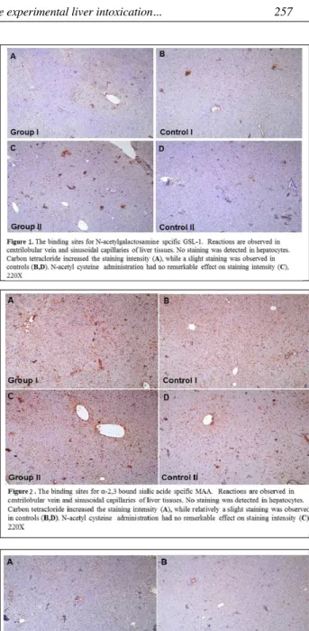
Histochemically, reactions are observed in centrilobular vein and sinusoidal capillaries of liver tissues, while no staining was detected in hepato-cytes. A slight staining for GSL-1 was visualized in centrilobular vein and sinusoidal capillaries of the liver. The staining pattern for GSL-1 was similar in all groups. However, the staining intensity in CCl4
-treated rats was stronger than the other groups
Parameters CCl4 (n = 7) CCl4+NAC (n = 7) Control (Olive Oil) (n = 7) Control (OliveOil + NAC) (n = 7) P AST (U/L) 232.42 ± 25.82a 197 ± 29.52a 72 ± 5.17b 108.23 ± 13.35ab * ALT (U/L) 79.64 ± 5.28 72.25 ± 6.73 49.52 ± 5.62 47.19 ± 5.23 -GGT (U/L) 13.28 ± 0.68a 9.12 ± 0.29b 8.66 ± 0.49b 8.57 ± 0.42b *** ALP (U/L) 34.42 ± 1.97a 32.62 ± 1.71a 21.83 ± 1.04b 23.14 ± 0.50b *** Albumin (g/dl) 3.21 ± 0.06b 3.26 ± 0.06b 3.50 ± 0.09a 3.50 ± 0.07a * Total Protein (g/dl) 6.35 ± 0.18 6.13 ± 0.12 6.49 ± 0.22 6.50 ± 0.20
-Table 1: Effect of NAC and CCl4treatments on
bioche-mical parameters
Statistical significance: *p < 0.05; ***p < 0.001. a, b: Between groups with different letters in the same row mean dif-ference is significant. CCl4: Carbontetrachloride; NAC: N-acetyl cysteine; AST: Aspartate aminotransferase; ALT: Alanine aminotransferase; GGT: Gamma glutamyl transferase; ALP: Alkaline phosphatase.
(Figure 1). A dense staining for MAA was observed in both experimental groups, while relatively mod-erate staining was detected in the liver tissue of controls (Figure 2). Compared to the GSL-1 and MAA a slighter staining was observed for PSA in all groups (Figure 3).
Discussion
Detoxification of all toxic substances takes place in liver. Therefore, liver is the central organ for removal of harmful chemical substances such as CCl4. Histopathological studies revealed that CCl4
causes acute and chronic liver damage(16,17).
Measurements the activities of some liver spe-cific enzymes (ALT, AST, ALP and GGT) can pro-vide valuable information on the condition of the liver. Increased values in the activity of these enzymes may indicate an injury in liver(18). In the case of cell membrane damage of liver cells activi-ties of ALT and AST, cytoplasmic enzymes, in serum increased drastically(19). A remarkable increase in serum activities of ALT and AST in CCl4 given rats was reported by Ustundag et
al.(20). It is revealed that within 12 hours ALT and AST activities start to increase in serum of rats exposed to 1 ml/kg CCl4 and rises to the maximum activity(21-23).
Data obtained in this study is partly in line with the statements of Ustundag et al.(20). Increased AST activities obtained in this study from the rats subjected to CCl4 were similar to the results pub-lished by Ustundag et al.(20). In spite of the eleva-tions in ALT activities in rats given CCl4 this
increase was no statistically significant. Increased levels of AST, ALP and GGT obtained in the pre-sent study may not only indicate liver injury but also heart and renal tissues may be affected nega-tively.
NAC can enter to the cells easily and it is used in vivo and in vitro studies as an antioxidant. It pro-tects the liver cells by increasing GSH levels in cells(24). Previous studies revealed that NAC protects the liver cells from the damage caused by CCl4 (24,25). Similarly, results of the present study exhibited that NAC protect the liver cell from damage caused by CCl4.
Protein and lipids in tissues are glycosylated and these sugar units attached to these molecules via glycosidic bond with tissue specificity. In the expression and localization of oligosaccharide units there are remarkable diversities based on the
physi-ological or pathphysi-ological condition the cells. There are few studies on the alteration of oligosaccharide units in liver injury(26,27).
However, to our knowledge there is no data published yet revealing the effects of CCl4 induced liver injury on the expression and localization of certain oligosaccharide units such as N-acetylgalac-tosamine, α–D-mannose and α-2,3 bound sialic acid residues. The most intensive staining in oligosaccharide units visualized in this study was for MAA lectin specific for α-2,3 bound sialic acid residues. Especially, in liver tissue exposed to CCl4
showed the strongest reaction with the biotin labeled MAA. This finding is in line with findings of previous researchers reported that damaged tis-sues as well as tistis-sues suffering from a disease with poor prognosis expresses elevated levels of sialic acid(28,29).
The intensity of the reaction for GSL-1, MAA and PSA obtained using Image J software program proved that NAC administration the CCl4 exposed
rats was stronger than that of CCl4group alone. In
the light of this findings it could be speculated that to protect themselves cells exposed to a toxic sub-stance increase the expression of N-acetylgalac-tosamine, α–D-mannose and α-2,3 bound sialic acid. In addition, NAC may protect cells from the damaging effects of free radicals not only by scav-enging free oxygen radicals but also by increasing oligosaccharide residues.
Conclusion
Cells may increase their oligosaccharide units to protect themself from the CCl4 induced liver
damage and NAC may have beneficial, protective and curative effects on hepatic cells from the toxic radicals induced by CCl4in rats.
References
1) Robbins SL, Cotran RS, Kumar V. Basic Pathology. Sixth Edition. Philadelphia: WB Saunders Company 2000.
2) Recknagel R, Glende EA, Dolak JA, Waller RL.
Mechanisms of carbon tetrachloride toxicity. Pharma
Ther 1989; 43: 139-54.
3) Cheshchevik VT, Lapshina EA, Dremza IK, Zabrodskaya SV, Reiter RJ, et al. Rat liver
mitochondr-ial damage under acute or chronic carbon tetrachlo-ride-induced intoxication: protection by melatonin and cranberry flavonoids. Toxicol Appl Pharmacol 2012;
4) Salam OM, Sleem AA, Omara EA, Hassan NS.
Hepatoprotective effects of misoprostol and silymarin on carbon tetrachloride-induced hepatic damage in rats. Fundam Clin Pharmacol 2009; 23: 179-88.
5) Al-Assaf AH. Preventive effect of corosolic acid on
lipid profile against carbon tetrachloride-induced hepatotoxic rats. Pakistan Journal of Nutrition 2013;
12: 748-52.
6) Bilgin HM, Atmaca M, Deniz Obay B, Ozekinci S, Taşdemir E, et al. Protective effects of coumarin and
coumarin derivatives against carbon tetrachloride-induced acute hepatotoxicity in rats. Exp Toxicol
Pathol 2011; 63: 325-30.
7) Kortsalioudaki C, Taylor RM, Cheeseman P, Bansal S, MieliVergani G, et al. Safety and efficacy of
n-acetyl-cysteine in children with non-acetaminophen-induced acute liver failure. Liver Transpl 2008; 14: 25-30.
8) Kumar BA, Reddy AG, Kumar PR, Reddy YR, Rao TM, et al. Protective role of n-acetyl l-cysteine against
reproductive toxicity due to interaction of lead and cadmium in male wistar rats. J Nat Sci Biol Med 2013;
4: 414-9.
9) Atkuri KR, Mantovani JJ, Herzenberg LA, Herzenberg LA. N-acetylcysteine-a safe antidote for
cysteine/glu-tathione deficiency. Curr Opin Pharmacol 2007; 7: 355-9.
10) Zafarullah M, Li WQ, Sylvester J, Ahmad M. Molecular mechanisms of n acetyl cysteine actions. Cell Mol Life Sci 2003; 60: 6-20.
11) Bernardi RM, Constantino L, Machado RA, Vuolo F, Budni P, et al. N-acetylcysteine and deferoxamine
pro-tect against acute renal failure induced by ischemia/ reperfusion in rats. Rev Bras Ter Intensiva 2012; 24: 219-23.
12) Foresti R, Sarathchandra P, Clark JE, Green CJ, Motterlini R. Peroxynitrite induces heme oxygenase-1
in vascular endothelial cells: a link to apoptosis.
Biochem J 1999; 339: 729-36.
13) Costa FH, Valença NS, Silva AR, Bezerra GA, Cavada BS, et al. Cloning and molecular modeling of
Litopenaeus vannamei (Penaeidae) C-type lectin homologswith mutated mannose binding domain-2.
Genet Mol Res 2011; 10: 650-64.
14) Bourrilon R, Aubery M. Cell surface glycoproteins in
embryonic development. Int Rev Cytol 1989; 116: 257-338.
15) Gabius HJ. Animal lectins. Eur J Biochem 1997; 243:
543-76.
16) Handa SS, Sharma A. Hepatoprotective activity of
andrographolide from Andrgraphis paniculata against carbon tetrachlorid. Indian J Med Res B 1990; 92: 276-83.
17) Rojkind M. Inhibition of liver fibrosis by-L
Azetidine-2-carboxylic acid in rats treated with carbon tetrachlo-ride. J Clin Invest 1973; 52: 2451-6.
18) Roderick P. Liver function tests: defining what’s nor-mal. Brit Med J 2004; 328: 987.
19) Lu KL, Tsai CC, Ho LK, Lin CC , Chang YS.
Preventive effect of the Taiwan folk medicine ixeris lae-vigata var. Oldhami on a-nophthyl-isothiocyarate and carbon tetrachlorideinduced acute liver injury in rats.
Phytother Res 2002; 16: 45-50.
20) Üstündağ B, Bahçecioğlu İH, Şahin K, Gülcü F, Düzgün S, et al. Soy izoflavonların karbon tetraklorüre
(CCl4) bağlı karaciğer hasarı ve plazma paraoksonaz ile arilesteraz aktivite düzeylerine olan etkileri. FÜ
Sağlık Bil Dergisi 2005; 19(4): 263-71.
21) Ariosto F, Riggio O, Cantafora A, Colucci S, Gaudoi E, et al. Carbon tetrachloride - induced experimental
cir-rhosis in the rat: A reappraisal of the model. Eur Surg
Res 1989; 21: 280-6.
22) Dashti H, Jeppson B, Hagerstrand I, Hultberg B, Srinivas U, et al. Thioacetamide and
carbontatrachlo-ride-induced liver cirrhosis. Eur Surg Res 1989; 21: 833-91.
23) Lida C, Fujii K, Koga E, Washino Y, Kitamura Y, et al.
Effect of alpha-tocopherol on carbon tetrachloride intoxication in the rat liver. Arch Toxicol 2009; 83(5): 477-83.
24) Howard RJMW, Blake DR, Pall H, Williams A, Green
ID. Allopurinol/Nacetylcysteine for carbon monoxide poisoning. Lancet 1987; 2: 628-9.
25) Kelly GS. Clinical applications of n-acetylcysteine. Altern Med Rev 1998; 3(2): 114-27.
26) Blomme B, Steenkiste CV, Vanhuysse J, Colle I, Callewaert N, et al. Impact of elevation of total
biliru-bin level and etiology of the liver disease on serum n-glycosylation patterns in mice and humans. Am J
Physiol Gastrointest Liver Physiol 2010; 298: G615-24. 27) Pepard CD, Ponard D, Colonb MG. Analysis of low
molecular weight intracellular association of a human mannan binding lectin (MBL). Mol Immunol 2004; 40: 795-801.
28) Kamigaito T, Okaneya T, Kawakubo M, Shimojo H, Nishizawa O, et al. Overexpression of O-GlcNAc by
prostate cancer cells is significantly associated with poor prognosis of patients. Prostate Cancer PD 2014;
17: 18-22.
29) Pan X, Wilson M, Mirbahai L, McConville C, Arvanitis TN., et al. In vitro metabonomic study detects increases
in UDP-GlcNAc and UDP-GalNAc, as early phase markers of cisplatin treatment response in brain tumor cells. J Proteome Res 2011; 10: 3493-500.
_______
Corresponding author
DILEK AKSIT, Assistant Professor
Faculty of Veterinary, Pharmacology and Toxicology Department, Balikesir University
Balikesir, 10145
(Turkey)
View publication stats View publication stats