* Department of Neurosurgery, School of Medicine, Dicle University, Diyarbakır
Analyzing Extradural Haematomas: A Retrospective Clinical Investigation
Ümit Özkan*, Serdar Kemaloğlu*, Mustafa Özateş**, Aslan Güzel*, Mehmet Tatlı SUMMARY
In this study, of 3462 cases admitted with cranial trauma between 1992 and 2000. The 115 cases that were diagnosed as extradural haematoma (EDH), were analyzed to obtain the outcome of extradural hematomas.
Each patient was evaluated retrospectively in terms of age, sex, and models of injury, localization of the haematoma, origins of bleeding, neurological findings and diagnostic investigations. The outcomes for each patient were retrieved and analyzed, and the chi–square test was used in the statistical analyses.
Of all the patients in this group, 88 (76%) were male and 27 (24%) were female, of the patients, 70% were between 10-40 years old. The outcomes showed a significant correlation with Glasgow Coma Scale score, pupillary sizes, and pyramidal signs (p <0.05).The radiologic findings of the the size hematoma, the degree of the brain shift, significantly correlated with outcome (p < 0.05).
Factors such as coma, pupil alterations, volume of haematoma, presence of pyramidal signs, midline shift, and lucid interval the existence of local motor findings are risk factors for the outcome in patients with EDH.
Key Words: Extradural Haematoma, Outcome, Risk Factors, Trauma.
Extradural Hematomların Analizi: Retrospektif Klinik Bir Araştırma
ÖZETBu çalışma 1992 ile 2000 yılları arasındaki yaklaşık 3462 kranial travma olgusundan 115ine extradural kanama tanısı BBT ile konuldu ve cerrahi tedavileri yapıldı.
Her bir hasta yaş, cinsiyet, yara çeşitleri, kanamanın yeri, kanamanın odağı ,nörolojik bulgular açısından retrospektif olarak değerlendirildi, her bir hasta ekstradural kanamanın yeri, kanamanın kaynağı analiz edilerek istatistiksel olarak chi –square testi ile değerlendirildi.
Bu hasta gurubunun 88( %76) i erkek ,27 (%24) si kadındı. Çeşitli yaş guruplarını kapsamaktaydı (1ile 75) ve olguların %70 i 10-40 yaşları arasındaydı. Sonuçlar; Glasgow koma skor skalası, pupil büyüklügü ve piramidal sistem bulguları, kanamanın hacmi, herniasyonun derecesi ile sonuç arasında önemli ilişkiler bulundu.( P< 0.05) .
Koma, pupil degişiklikleri, kanamanın hacmi, piramidal sistem bulguları, beyin şifti ve lusid interval, lokal motor bulguların varlığı, ekstradural kanamalı hastalarda risk faktörlerini oluşturmaktadır. Ayrıca kanamanın hacmi ve piramidal sistem bulguları ekstradural kanamalı hastalarda bağımsız risk faktörlerini oluşturmaktadır.
INTRODUCTION
Extradural haematoma, a collection of blood between the skull and the dura mater due to bleeding from extracerebral vessels is a common complication of head injury, often fatal if not treated in time (1-7).Traumatic extradural hematomas (EDHs) comprise 1 to 3% of all head trauma admissions (8-11). The availability of computed tomography (CT) has increased the diagnosis of extradural haematomas.
We analyzed 115 cases with EDH who consecutively underwent surgery for epidural hematomas over the past 8-year period in order to determine the prognostic factors, and investigating the mortality rate stemmed from different factors. Clinical characteristics, radiologic findings, and the time intervals with regard to treatment course were investigated to determine the risk factors for outcome.
MATERIAL and METHOD
This study includes 115 consecutive patients operated on for EDH in the department of Neurosurgery at the Dicle University Faculty of Medicine from January 1, 1992 to January 1, 2000. Each patient was evaluated in terms of age, sex, and models of injury, localization of the haematoma, origins of bleeding, neurological findings and diagnostic investigations. Glaskow Coma Scale (GCS) scores of patients at the time of surgery and interval between trauma and surgery were analyzed. Glasgow Coma Scale (GCS) score 8 or less were coma and those with a (GCS) score greater than 8 were considered non comatose.
In the pre-CT era, angiography and craniography was performed as the diagnostic work up in 10 patients. Of 105 patients had CT. Of 58 patients in this series had plain skull x-ray films.
The location, thickness, density, and volume of the EDHs were noted on the CT scans in 105 patients. The shift of the midline structures was measured at the level of septum pellucidium and the formula used to calculate the volume of the haematomas was as follows;
Haematoma volume (ml) = 0,5 x transverse diameter (mm) x anteroposterior diameter
...
(mm) x craniocaudal diameter (mm) (12). The outcomes for each patient, the location of EDH, the origin of bleeding and characteristics of haematoma recorded in the operative notes of the patients and were again retrieved and analysed and the chi–square test was used in the statistical analyses.
The P values those less than 0.05 were assumed statistically significant. The Odds Ratio (O.R) was evaluated in 95% confidence interval for the risk factors affecting the outcomes of patients with extradural haematoma. Multivariate analysis were performed to determine the independent risk factors with logistic regression analysis.
RESULTS
Of 3462 patients 190 (5,5%) had EDH and 115 of these patients (3,3%) were operated upon for EDH. There were 88 males (76%) and 27 females (24%). The female/male ratio was 1/3 and the ages of the patients ranged from 1 to 75 years (mean age 26.90 ± 14.74). The highest incidents (70%) of extradural haematoma were seen in the age group 10 to 40.
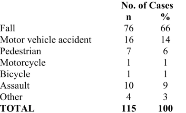
Fall was the predominant mode of injury 76 (66%). Falls comprised the majority of injuries in patients younger than 20 years old. However, motor vehicle accidents were the most common second cause of injury in the series 16 (14%) (Table1).
Table 1. Models of Injury
No. of Cases n %
Fall 76 66
Motor vehicle accident 16 14
Pedestrian 7 6 Motorcycle 1 1 Bicycle 1 1 Assault 10 9 Other 4 3 TOTAL 115 100
The origin of bleeding was detected in 68 patients (59%) at surgery;The origin of hematom was the middle meningeal artery in 44 patients, the meningeal vein in 12, the dural sinus in 5, and bone in 7 cases (Table 2).
Table 2. Origins of Bleeding
No. of Cases
LOCATION n %
Middle meningeal artery 44 39
Meningeal vein 12 10
Diploe 7 6
Dural sinus 5 4
Undetected 47 41
TOTAL 115 100
Forty-two patients (36%) had a lucid interval. Pupillary abnormalities were present in 51 (45%). Unilateral mydriasis was present in 36 patients and bilateral mydriasis in 15 patients. In addition, 21 patients (18%) GCS scores were less than 8 at the time of surgery (Fig. 1). Twelve (50%) of comatose patients had respiratory irregularities. Bradycardia and arterial hypertension were noticed in 26 patients (22%).
Figure 1. Significant operative findings in terms of outcome.
Of 58 patients who had plain skull x-rays films, 11 had a normal skull x-ray film and a fracture unrelated to EDH was seen on the skull x-ray films of 3 cases. The fractures crossing the middle meningeal artery and a dural sinus were detected in 17 and 6 patients, respectively. EDH was associated with a depressed skull fracture in 7 patients. Angiography was performed in 10 patients.
With radiological evaluation, the shape of EDHs on CT scan was biconvex in 27, monoconvex in 16 and bilenticular- biconvex in 4 patients. Temporoparietal region was the most frequent site of EDHs (35%) parietal and frontal regions were the second and third frequent locations, respectively. There were 8 patients with posterior fossa EDH (7%). The heights mortality rates were 30% in
temporoparietal region and the lowest rate was 0 % in frontoparietal region. In addition, 4 cases had bilateral EDH and the haematoma was localized in the vertex in one patient.
Among 27 patients who underwent surgery within 6 hours after injury, the mortality rate was 18,5%. The mortality rate in patients who underwent surgery between 6 to 24 hours after injury was 23,5% in our series.
In the majority of the cases a clotted hematoma was evacuated in 93 patients. It was clot and partial liquefied in 4 and pure liquefied haematoma in one case. The nature of EDH was both clod and active bleeding in 17 patients. Nineteen patients had associated intracranial lesions. Ten patients had subdural haematoma and 5 had intracerebral hematoma.
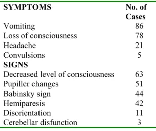
The examination on admission revealed either a scalp laceration or cephal haematoma in 92 patients (80%) and 38 (41%) had pyramidal signs such as and extensor planter response. Hemiparesis was determined in 42 (36,5%). Neurological symptoms and signs determined in patients with EDH at the time of admission are shown in (Table 3).
Table 3. Symptoms and signs on admission
Of 27 patients who had admitted in first 6 hours, 5 had died and all of these were in deep coma (GCS were 5 or less). Of 59 patients who had admitted 6-24 hours, 14 had died whose GCS were 8.
The overall mortality was 20% (23 patients). In the patients with CT and plain x-ray mortality was 23% (13 patients). Ten patients died (21%) in the only CT group.
SYMPTOMS No. of Cases Vomiting 86 Loss of consciousness 78 Headache 21 Convulsions 5 SIGNS
Decreased level of consciousness 63
Pupiller changes 51 Babinsky sign 44 Hemiparesis 42 Disorientation 11 Cerebellar disfunction 3 39 10 6 4 41 %0 %10 %20 %30 %40 %50
A midline shift greater than 10 mm (n= 88 and mortality rate 25%) and haematoma thickness greater than 30 mm (n= 75 and mortality rate 26.6%) significantly increased the mortality. Of 34 patients who have EDH greater than 90 ml in volume, 21 died (mortality rate 62%), whereas only 2 patients died (mortality rate 4%) in those with EDH of 90 ml or less ( ²=45,286 p=0,0001) O.R. 0,018 (0,004-0,086 ) (Power: 99.9%).
The presence of respiratory irregularities also worsened the outcome. The mortality rates were 70% and 9% in those with and without respiratory irregularity, respectively ( ²=28,631 p=0,0001).
Mortality was highest 45 (39%) patients in which the origin of bleeding had been the meningeal artery, 11 (10%) in venous bleeding, 11 (10%) in dural and bone oozing and 48 (41%) unidentified origin. Except 2 patients, all 23 patients who died had been comatose or had a GCS score of less than 8 at the time of surgery. There were 2 deaths among patients whose GCS scores were 8 or greater ( ²=102,766 p=0,0001) (Power:
……….
99.9%). Nine (12%) of the 73 patients without a lucid interval had died. However 14 (33%) of 42 patients who had a lucid interval died ( ²=7,352 p=0,007) O.R. 3,5 (1,4 - 9,2) (Power 77 %).
Of 38 patients with pyramidal signs, 14 died and there were 9 deaths in 77 patients not showing pyramidal signs ( ²=10,062 p=0,002) O.R. 4.4 (1,6- 11,4) (Power 89 %). Mortality rate was 40%, in the patients with pupillary changes and 2% in those with a normal pupil size and reaction ( ²=25,685 p=0,0001) (Fig. 2). In addition, all factors influenced the outcomes of the patients had statistical significance were emphasized in table 4.
Figure 2. Symptoms and signs were found to be significant in predicting the outcome
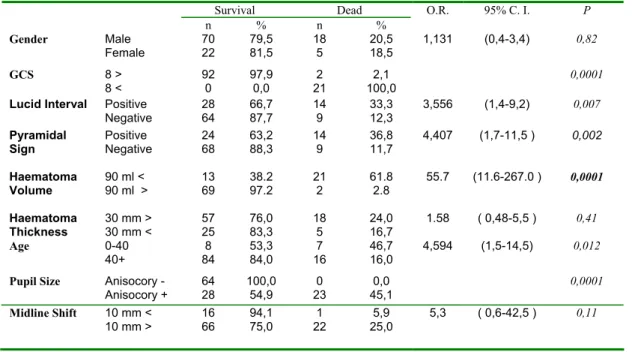
Table 4. Risk factors influencing the outcome of the patients with extradural haematoma by univariate statistical analysis.
20 31 3 61 14 24 9 68 0% 20% 40% 60% 80% 100% Abnormal pupil Normal pupil Pyramidal signs No pyramida signsl Death Survival Survival Dead n % n % O.R. 95% C. I. P Gender Male Female 70 22 79,5 81,5 18 5 20,5 18,5 1,131 (0,4-3,4) 0,82 GCS 8 > 8 < 92 0 97,9 0,0 2 21 2,1 100,0 0,0001
Lucid Interval Positive
Negative 28 64 66,7 87,7 14 9 33,3 12,3 3,556 (1,4-9,2) 0,007 Pyramidal Sign Positive Negative 24 68 63,2 88,3 14 9 36,8 11,7 4,407 (1,7-11,5 ) 0,002 Haematoma Volume 90 ml < 90 ml > 13 69 38.2 97.2 21 2 61.8 2.8 55.7 (11.6-267.0 ) 0,0001 Haematoma Thickness 30 mm > 30 mm < 57 25 76,0 83,3 18 5 24,0 16,7 1.58 ( 0,48-5,5 ) 0,41 Age 0-40 40+ 8 84 53,3 84,0 7 16 46,7 16,0 4,594 (1,5-14,5) 0,012
Pupil Size Anisocory -
Anisocory + 64 28 100,0 54,9 23 0 45,1 0,0 0,0001 Midline Shift 10 mm < 10 mm > 16 66 94,1 75,0 1 22 5,9 25,0 5,3 ( 0,6-42,5 ) 0,11
DISCUSSION
Traffic accidents and falls are the first and second predominant types of injury in many series (13, 14). This series, falls from height were the first type of injury in 66% of the patients and traffic accidents were the second frequent type of injury. Fall was determined as the most frequent mode of injury in this series, as also reported by Baykenar et al. (15), Ersahin et al. (1), and Kvarnes and Trumpy (9), in contrast to most other series, in which traffic accidents were predominant (8,10,13,16). Falls were predominant in younger children, while traffic accidents were more frequent in adolescents.
The overall mortality in our series is 20%. It has been reported that, mortality rates between 0% and 17% in patients with EDH (11,13,17). The introduction of CT has facilitated the series observation of the intracranial condition after head injury, and reports of enlargement of acute extradural haematoma have begun to appear (18).
GCS scores of 94 patients were greater than 8 and 21 (18%) were comatose. In some series, 35 - 40% of the patients were in the comatose group and 60–65% in non-comatose group (13,14). The mortality rates have been reported 18–44% (9,11,14,19). Seeling et al. (16) reported 41% mortality in comatose patients with EDH. There were no deaths among patients with a GCS score of 8 or better in the series of Bricolo and Pasut (13). Higher mortality rates have been reported in patients’ unconscious at the time of surgery (6,10,11,14). All of the our comatose patients died, whereas only two death occurred among non-comatose patients. The level of consciousness was significantly influenced the outcome.
Temporoparietal and temporal regions were predominant in terms of the location of EDHs, as reported in other series (8,9,10,11,13,17,20). There was no statistically correlation between the site of EDH and mortality, although high mortality has been reported in association with EDH in the temporal region (1,8,21). Although the most common site of EDHs has been reported as the temporal region (9,10,13). EDHs were
………..
localized in the temporo parietal or parietal regions in some recent papers, since CT has enabled us to localize the haematoma precisely. Temporo-parietal location was the most common site of EDHs in our patients and was consequently associated with the highest mortality rate (30%) (11,14,22,23). The difference in mortality rates according to the location of haematoma was not statistically significant.
The origin of bleeding was detected during surgery in 60% of the cases. The middle meningeal artery was the source of bleeding in most EDHs, as found in other series (1,7,8, 15,17). Hypertension, bradicardia and respiratory irregularities are rarely seen in patients with EDH. The presence of these findings was determined as a significant factor in terms of mortality.
In our series 69% of patients with EDH have a good outcome, good recovery and moderate disability was 80 % and all deaths had occurred in comatose patients except two. Bricolo stated that overall mortality was 5%, and 89% good recovery or moderate disability. Lobato also stated that overall mortality rate as 12%, and 88% good recovery or moderate disability. His series was include the comatose patients, and he stated no deaths occurred among patients reaching surgery with a GCS score of 8 or better. Inaddition, It showed that the outcomes of the patients had improved by routine CT examination and intensive care facilities for cranial trauma. In our series, the high mortality rates were attributed comatose patients and the late surgery. The outcome figure of our patients was nearly consistent with those in the literature (5,13,22).
In some series, surgery was performed within 6 hours, 6 to 24 hours and more than 24 than hours after injury in 30-57%, 2- 41% and 13-35% of the patients with EDH, respectively (5,11,13,22). The mortality rate has been 5-35% in patients operated upon within 6 hours following trauma (1,6,22). In our series, the main factor is high mortality rate and the 27 patients admitted in first six hours of whom 5 were died. These 5 cases were in deep coma whose GCS were 5 or less. However, 18 patients of the remaining 88 died but all had a
GCS score of 8 or less. We thus concluded that delay in transferring patients to our hospital is the other factor in the high mortality rate. We think that if these 18 cases would have admitted to hospital in the first hours, they had survived.
In conclusion, GCS scores less than 8 or deep coma state at the time of operation, pupillary abnormalities, midline shift, and lucid interval are risk factors for the outcome in patients with EDH. In addition, hypertension, bradicardia, respiratory irregularities and the time between trauma-to-surgery intervals are significant factors in terms of mortality. Finally volume of haematoma and presence of pyramidal signs are independent risk factors in patients with extradural haematoma.
REFERENCES
1. Erşahin Y, Mutluer S, Güzelbağ E Extradural hematoma analysis of 146 cases. Child’s Nerv Syst 1993; 9:96-99
2. Erşahin Y, Mutluer S. Posterior fossa extradural hematomas in children. Pediatr Neurosurg 1993;19:31-33
3. Ford LE, Mc-Laurin RI. Mechanism of extradural hematomas. J Neurosurg 1963; 20:760-769
4. Galbraith SL. Age distrubution of extradural haemorrhage without skull fracture. Lancet I 1973; 1217-1278
5. Jamjoom A. The influence of comcominant intradural pathology on the presentation and outcome of patients with acute traumatic extradural haematoma. Acta Neurochir 1992; 155:86-89
6. Lee EJ, Hung YC, Wang LC, Chung KC, Chen HH. Factors influencing the functional outcome of patients with acute epidural hematomas: analysis of 200 patients undergoing surgery. J Trauma 1998; 45:946-952
7. Yurt I, Bezircioglu H, Erşahin Y, ve ark. Extadural haematoma: Analysis of 190 cases. Turkish Neurosurgery 1996; 6:63-67
8. Cordobes F, Lobato RD, Rivas JJ, et all. Observation on 82 patients with extradural hematoma. J Neurosurg 1981; 54:179-186
9. Kvarnes TL, Trumpy JH. Extradural hematoma: Report of 132 cases. Acta Neurochir 1978; 41:223-231
10. Mc.Kıssock W, Taylor JC, Bloom WH, Till K. Extradural hematoma: observations on 125 cases. Lancet 1960; 23:167-172
11. Rivas JJ, lobato RD, Sarabia R, et all. Extradural hematoma: Analysis of factor influencing the courses of 161 patients. Neurosurgery 1988; 23:44-51
12. Peterson OF, Espersen JO. Extradural hematomas: Measurement of size by volume summation on CT scanning. Neuroradiology 1984; 26:363-367
13. Bricolo AP, Pasut LM. Extradural hematoma: Toward zero mortality. Neurosurgery 1984; 14:8-12
14. Cook RJ, Dorsch NWC, Fearnside MR, Chaseling R. Outcome prediction in extradural haematomas. Acta Neurochir 1988; 95:90-94
15. Baykaner K, Alp H, Ceviker N, Keskil S, Seçkin Z. Observation of 95 patients with extradural hematoma and review of the literature. Surg Neurol 1988; 30:339-341
16. Seeling JM, Marshall LF, Toutant SM, et all. Traumatic acute epidural hematoma: unrecognized high lethality in comatose patients. Neurosurgery 1984; 15:617-620
17. Ammirati M, Tomita T. Epidural hematomas in infancy and childhood. Riv Neurosci Pediatr 1985; 1-2:123-128
18. Rappaport ZH, Shadked I, Tadmor R. Delayed epidural hematoma demonstrated by computed tomography: Case report. Neurosurgery 1982; 10:487-488
19. Neubauer UJ. Extradural hematoma of the posterior fossa. Twelve years experiences with CT-scan. Acta Neurochir 1987; 87:105-111
20. Zuccarello M, Pardatcher K, Andrioli GC, Fiore DL, Lavicoli R. Epidural hematomas of the posterior cranial fossa. Neurosurgery 1981; 8:434-437
21. McLaurin RL, Ford LE. Extradural hematoma. Statistical survey of forty-seven cases. J Neurosurg 1964; 21:364-371
22. Lobato RD, Rivas JJ, Munoz MJ, et all. Observation on 82 patients with extradural hematoma. J Neurosurg 1981; 54:179-186
23. Sakai H, Takagi H, Ohtaka H, et all. Serial changes in acute extradural hematoma size and associated changes in level of consciousness and intracranial pressure. J Neurosurg 1988; 68:566-670
Correspondence Address
Ümit ÖZKAN
Department of Neurosurgery, School of Medicine, Dicle University, Diyarbakır