Acta zool. bulg., 65 (2), 2013: 251-257
*Corresponding author: * Correspondence author: Tel: +90 216 418 25 06. Fax: +90 216 418 25 05. E-mail: esoylu@marmara.edu.tr
Introduction
The monogenean gill parasites P. anguillae and P.
bini are known to occur on the gills of the Asian eel Anguilla japonica and the Australian eel Anguilla re-inharditi. P. bini was first reported in 1929 by Kikuchi
in Japan and P. bini and P. anguillae were reported in 1948 by Yin & Sproston 1948 in China. P. anguillae and P. bini are rather pathogenic to Anguilla spp. and can cause mortality in heavily infected fish (Gelnar et
al. 1996). Both these monogeneans were apparently
introduced via the Japanese eel A. japonica and were first reported in Anguilla anguilla in the Soviet Union (Golovin 1977). They have been reported on the gills
of both wild and cultured eels from Hungary (Molnar
1984), France (laMbert et al. 1985), Italy (SaroGlia
et al. 1985), Denmark (buchMann et al. 1987) and Poland (Dzika et al. 1995). cone, MarcoGlieSe (1995)
were the first to record P. anguillae on the gills of the American eel Anguilla rostrata in North America. A.
anguilla is an economically valuable fish species in
Turkey. However it is not farmed, all harvesting is done by fishing. The number of studies on the para-sites of A. anguilla in Turkey is limited; altunel
(1990) investigated the parasite fauna in A. anguilla in the Ekinli Lagoon, Genç et al. (2005) studied the occurrence of the swimbladder parasite Anguillicola
crassus in the same fish host in the Ceyhan River.
Microhabitat Distribution of Pseudodactylogyrus anguillae
(Monogenea), Ergasilus gibbus and Ergasilus lizae
(Copepoda) on the Gills of European Eels (Anguilla anguilla, L.)
Erhan Soylu
1*, Sibel Özesen Çolak
2, Fatime Erdogan
3, Mete Erdogan
3, Necla Tektas
11 Fisheries Department, Vocational School of Technical Sciences, Marmara University, Göztepe, 34722 Kadıköy-Istanbul, Turkey. 2 Fisheries Faculty, Istanbul University, Ordu Cad. No. 200, 34470 Laleli, Fatih-Istanbul, Turkey.
3 Aquaculture Programme, Ortaca Vocational School, Muğla University, 48600 Ortaca, Muğla, Turkey.
Abstract: Microhabitat distribution of Pseudodactylogyrus anguillae (Yin & Sproston, 1948) Gussev, 1965
(Mono-genea: Dactylogyridae), Ergasilus gibbus von Nordman, 1832 and Ergasilus lizae Kroyer, 1863 (Copep-oda: Ergasilidae) was studied on the gills of European eel Anguilla anguilla from Lake Köyceğiz-Dalyan, Estuarine Channel System, Turkey. A total of 69 A. anguilla were examined between December 2009 and March 2010. A total of 1421 P. anguillae, 143 E. gibbus and 63 E. lizae specimens were collected from the fish host. A prevalence of 81.15% and mean infection intensity of 25.16 for P. anguillae, 50.72%, 4.0 for
E. gibbus and 27.53%, 3.31 for E. lizae were found. Gill arches II, III, IV were preferred by P. anguillae,
gill arches I, II, III by E. gibbus and gill arches III, IV by E. lizae. In general occurrence of the parasite species, P. anguillae preferred proximal-dorsal segments, E. gibbus distal-dorsal segments, whereas E.
lizae exhibited a rather homogenous distribution. Site of attachment was the inner surface of the gill
arches for E. gibbus and the outer surface for E. lizae. P. anguillae was found mostly on the inner surface of the hemibranches. Bispecific infections of P. anguillae with E. gibbus and E. lizae were also analysed individually. Finally, single species infections of the three parasite species were analysed.
The coexistence of gill monogeneans has been studied widely (el-hafİDİ 1998, Dzika 1989,
koSkivaa et al. 1991, SiMkova et al. 2000, turGut
et al. 2006). Numerous studies have investigated the
spatial distribution and microhabitats selection of the monogenean gill parasites of Anguilla anguilla (buchMann 1989; roDriGueS, Saraiva 1996, Dzika
1999, MatejuSova et al. 2003). However, only a few studies have investigated co-occurrent copepods and monogeneans (raMaSaMy et al. 1985, baker, cone
2000, baker et al. 2005). The present study investi-gated the microhabitat distribution of the monogenean
Pseudodactylogyrus anguillae in co-occurrence with
the copepods Ergasilus gibbus and Ergasilus lizae on the gills of the European eel Anguilla anguilla.
Materials and Methods
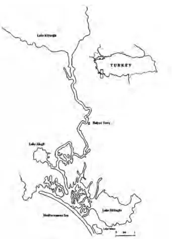
The Köyceğiz-Dalyan Nature Reserve is an impor-tant wetland area in South-western Turkey (36° 45’ and 37° 15’ N, 28° 22’ 30” and 28° 52’ 30” E). The outflow of Lake Köyceğiz, the Dalyan River enlarges into a labyrinth-like channel system, discharging into the Mediterranean Sea (Fig. 1). The estuarine area includes three lakes (Alagöl, Sülüngür and Sülüklü). The Dalyan channel system is fed by Lake Köyceğiz.
The Mediterranean Sea has many sulphu-ric thermal springs located both on the bottom and around it. The Köyceğiz-Dalyan estuarine channel system is 14 km in length and connects the meromic-tic Lake Köyceğiz and the Mediterranean Sea. The Dalyan water mass consists of a mixohaline upper layer from Lake Köyceğiz and a lower layer of the Mediterranean Sea saline water mixed with the sul-phuric thermal spring water (kazanci et al. 2003).
A total of 69 Anguilla anguilla of mean (± SD)
total length 52.5 ±10.24 cm (range 33.2-78.0 cm) and mean (± SD) weight 312.3 ±199.99 g (range
67.7-906.0 g) were examined between December 2009 and March 2010. Fish were caught by local fishermen and transferred alive to the laboratory in aerated lake water. After each eel was sacrificed all branchial arches from the left and the right sides were removed and placed in 4% formalin solution for further studies. Each gill arch was placed in a separate Petri dish filled with water, when examined. The gill arches were numbered I to IV from anterior to posterior. Each arch was divided into three gill segments: dorsal, medial and ventral; two gill areas: proximal and distal; two gill surfaces: outer and
in-ner; two gill hemibranches: anterior and posterior, as a niche for parasites (Fig. 2). All monogenean and copepod parasites on the gills were collected one by one from each sector separately under a stereo-microscope at 20X magnification and the exact loca-tion of the parasites was recorded before removal. The monogenean parasites were cleared in glycerine or lactophenol and identified on the basis of their chitinous elements according to PuGachev et al. (2010). Copepod parasites were roughly sorted into two groups on the basis of their second antenna, and preserved in 70% ethanol for further identification on the basis of the keys of bauer (1987).
Kruskal-Wallis tests were used to test the signif-icance of the differences in the number of parasites between the dorsal, medial and ventral segments. The differences in the parasite numbers between the proximal and distal parts, left and right sides, and gill arches were tested using the Mann Whitney U-test. Differences of P < 0.05 were considered significant.
Results
A total of 69 Anguilla anguilla were examined, 4 of which (5.8%) were not infected at all, 56 (81%)
Fig. 1. Map of the study area.
were infected by Pseudodactylogyrus anguillae, 34 (49%) by Ergasilus gibbus and 19 (28%) by E. lizae. In only 5 of the infected fish were there simultane-ous infections of E. gibbus and E. lizae with P.
an-guillae. A total of 1421 P. anguillae, 143 E. gibbus
and 63 E. lizae individuals were recorded in general occurrence. The overall prevalence, mean intensity and mean abundance for P. anguillae were found to be 81.2%, 25.4, 20.6, for E. gibbus 49.3%, 4.2, 2.1 and for E. lizae 27.5%, 3.3, 0.9 respectively. The
in-fection intensities (per eel) of P. anguillae, E.
gib-bus and E. lizae respectively ranged between 1-202,
1-41 and 1-14 individuals.
General occurrence of the parasites
Branchial distribution of P. anguillae, E.
gib-bus and E. lizae on the gills of Anguilla anguilla
was examined. Of the 69 dissected fish, 56 were infected with Pseudodactylogyrus anguillae (preva-lence 81.2%). The distribution of 1421 P. anguillae in general occurrence is shown (Table 1). The dif-ferences were not found to be significant between the number of P. anguillae on the left and right gill arches (P=0.861 > 0.05). Gill arches II, III and IV were preferred over I. The differences were signifi-cant between the numbers of P. anguillae found on the segments (P=0.00 < 0.05), P. anguillae preferred dorsal segments (84.1%), namely 55.9% of the P.
anguillae settled in sector 4 and then 28.2% in sector
3. There were no statistically significant differences in the number of the parasites between the proximal and distal parts (P=0.225 > 0.05), 64.9% of P.
an-guillae were recorded on the proximal part, 80.0% of P. anguillae preferred the inner surface, 52.6%
set-tled in the anterior hemibraches.
Of the 69 examined fish, 34 were infected with
Ergasilus gibbus (prevalence 49.3%). A total of 143 E. gibbus were recorded. E. gibbus did not show
preference for the left or right side of the gill (P
Fig. 2. Division of branchial arch (1-2-3 Distal part, 4-5-6
Proximal part).
Table 1. General occurrence of Pseudodactylogyrus anguillae, Ergasilus gibbus and Ergasilus lizae on the gills of
Anguilla anguilla.
P. anguillae E. gibbus E. lizae
Number of infected eels 56 34 19
Mean intensity 25.4 4.2 3.3
Number % Number % Number %
Left side 733 51.6 78 54.5 35 55.6
Right side 688 48.4 65 45.5 28 44.4
Gill arch I 224 15.8 43 30.0 2 3.2
Gill arch II 425 29.9 36 25.2 7 11.1
Gill arch III 407 28.6 40 28.0 25 39.7
Gill arch IV 365 25.7 24 16.8 29 46.0 Dorsal segment 1195 84.1 94 65.7 22 34.9 Medial segment 192 13.5 41 28.7 26 41.3 Ventral segment 34 2.4 8 5.6 15 23.8 Proximal part 922 64.9 29 20.3 31 49.2 Distal part 499 35.1 114 79.7 32 50.8 Anterior hemibranch 748 52.6 75 52.4 37 58.7 Posterior hemibranch 673 47.4 68 47.6 26 41.3 Inner surface 1137 80.0 136 95.1 3 4.8 Outer surface 284 20.0 7 4.9 60 95.2 Dorsal
=0.341 > 0.05), E. gibbus preferred gill arches I, II and III. A significantly greater number of E. gibbus occurred on the dorsal segment of the gills (65.7%) than on the medial and ventral segments (P=0.00 < 0.05). E. gibbus preferred distal part of the gill arches (79.7%) and significant differences were found with the proximal part (P=0.001 < 0.05). E. gibbus did not show preference for anterior or posterior hemi-branches, and 95.1% of the parasites occupied the inner surface of the gill hemibranches.
Of the 69 examined fish, 19 were infected with
Ergasilus lizae (prevalence 27.5%). A total of 63 E. lizae were recorded. E. lizae did not show
prefer-ence for the left or right side of the gills (P=0.283 > 0.05), gill arches IV and III were preferably infected, differences between the number of E. lizae on gill arches IV-III and I-II were found to be significant (P=0.00 < 0.05). But distribution of E. lizae both on dorsal, medial, ventral segments and proximal, dis-tal parts was found rather homogenous. E. lizae pre-ferred outer surface (95.2%) of the hemibranches. E.
lizae did not show preference for anterior or
poste-rior hemibranches and the left or right side.
Bispecific infections
Bispecific infections of A. anguilla were exam-ined with P. anguillae – E. gibbus and P. anguillae – E.
lizae combinations. Of the 69 eels examined, 24 were
infected with only P. anguillae – E. gibbus (prevalence
34.8%). In these eels, 849 P. anguillae and 133 E.
gib-bus were recorded. P. anguillae preferred gill arches
II, III and IV, dorsal segments (86.0%), and proximal part (60.7%). E. gibbus preferred gill arches I and II, dorsal segments (64.7%), distal part (77.4%) and in-ner surface (94.0%) of the hemibranches (Table 2).
Of the 69 eels examined, 10 were infected with only P. anguillae – E. lizae (prevalence 14.5%). In these eels, 175 P. anguillae and 44 E. lizae were re-corded. P. anguillae did not show a right or left side preference. P. anguillae preferred gill arches II and III, dorsal segment (84.0%), proximal part (71.4%) and inner surface (88.6%). In these bispecific in-fections, E. lizae preferred the left side (prevalence 61.4%). E. lizae preferred gill arches IV and III, dorsal (40.9%) and medial (36.4%) segments. E.
lizae were evenly distributed over both proximal and
distal parts, and they all (100%) were found on the outer surface of the hemibranches (Table 2).
Monospecific infections
A total of 17 eels (prevalence 24.6%) were in-fected with only P. anguillae, 5 eels (7.2%) with only
E. gibbus and 4 eels (5.8%) with only E. lizae.
A total of 397 P. anguillae were recorded in these monospecific infections. P. anguillae settled in gill arches II, III and IV more frequently. The spe-cies predominantly occurred on dorsal segments (79.1%), significant differences were found between
Table 2. Distribution of Pseudodactylogyrus anguillae – Ergasilus gibbus and Pseudodactylogyrus anguillae –
Ergasi-lus lizae on the gills of Anguilla anguilla in bispecific infections.
P. anguillae E. gibbus P. anguillae E.lizae
Number of infected eels 24 24 10 10
Mean intensity 35.4 5.5 17.5 4.4
Number % Number % Number % Number %
Left side 444 52.3 74 55.6 98 56.0 27 61.4
Right side 405 47.7 59 44.4 77 44.0 17 38.6
Gill arch I 127 15.0 42 31.6 33 18.9 1 2.2
Gill arch II 259 30.5 43 32.3 55 31.4 5 11.4
Gill arch III 248 29.2 28 21.1 58 33.1 18 40.9
Gill arch IV 215 25.3 20 15.0 29 16.6 20 45.5 Dorsal segment 730 86.0 86 64.7 147 84.0 18 40.9 Medial segment 106 12.5 42 31.5 22 12.6 16 36.4 Ventral segment 13 1.5 5 3.8 6 3.4 10 22.7 Proximal part 515 60.7 30 22.6 125 71.4 21 47.7 Distal part 334 39.3 103 77.4 50 28.6 23 52.3 Anterior hemibranch 473 55.7 68 51.1 92 52.6 25 56.8 Posterior hemibranch 376 44.3 65 48.9 83 47.4 19 43.2 Inner surface 712 83.9 125 94.0 155 88.6 0 0 Outer surface 137 16.1 8 6.0 20 11.4 44 100
the numbers recorded on the dorsal and the other two segments (P=0.00 < 0.05). Although there were no statistically significant differences in the number of P. anguillae between proximal and distal parts (P=0.195 > 0.05), the parasite preferred proximal parts (72.0%), inner surface of the hemibranches (75.8%), but no differences were found between the left and right side (P=0.975 > 0.05).
A total of 10 E. gibbus were recorded in these monospecific infections. E. gibbus preferred gill arch I (40.0%) and no parasite was found on arch IV. Dorsal segments were mainly preferred by E.
gib-bus (70.0%), the parasite completely occupied the
distal part (100%), inner surface (100%), and it was mainly found on the right side (70.0%).
Dorsal segments mainly preferred by E. gibbus (70.0%), parasite totally occupied distal part (100%), inner surface (100%) and of the (70.0%) found on the right side.
A total of 11 E. lizae were recorded in these monospecific infections. E. lizae preferred gill arch IV, with no parasite being found on gill arch I. E.
liz-ae mostly preferred medial segments (63.6%). There
were no significant differences in the number of E.
lizae between the proximal and distal parts (P=0.949
> 0.05), but the parasite exhibited preference for the left side (P=0.027 < 0.05); 81.8% of the E. lizae found were on the left side and 90.9% were on the outer surface of the hemibranches (Table 3).
The numbers of P. anguillae, E. gibbus and
E. lizae between bispecific and monospecific
in-fections were also examined. The numbers of P.
anguillae showed no statistically significant
differ-ence between bispecific infections with E. gibbus, bispecific infections with E. lizae and monospecific infections (Kruskal-Wallis test P=0.404; p > 0.05). There was no statistically significant difference in the numbers of E. gibbus between bispecific infec-tions with P. anguillae and monospecific infections (Mann-Whitney U test P=359 > 0.05). There were no statistically significant differences in the numbers of E. lizae between bispecific infections with P.
an-guillae and monospecific infections (Mann-Whitney U test P=0.663 > 0.05).
Table 3. Distribution of Pseudodactylogyrus anguillae, Ergasilus gibbus and Ergasilus lizae on the gills of Anguilla
anguilla in monospecific infections.
P. anguillae E. gibbus E. lizae
Number of infected eels 17 5 4
Mean intensity 23.4 2.0 2.8
Number % Number % Number %
Left side 187 47.1 3 30.0 9 81.8
Right side 210 52.9 7 70.0 2 18.2
Gill arch I 77 19.4 4 40.0 - 0
Gill arch II 112 28.2 3 30.0 3 27.3
Gill arch III 95 23.9 3 30.0 2 18.2
Gill arch IV 113 28.5 - 0 6 54.5 Dorsal segment 314 79.1 7 70.0 1 9.1 Medial segment 65 16.4 - 0 7 63.6 Ventral segment 18 4.5 3 30.0 3 27.3 Proximal part 286 72.0 - 0 5 45.5 Distal part 111 28.0 10 100.0 6 54.5 Anterior hemibranch 212 53.4 6 60.0 7 63.6 Posterior hemibranch 185 46.6 4 40.0 4 36.4 Inner surface 301 75.8 10 100.0 1 9.1 Outer surface 96 24.2 0 0 10 90.9
Table 4. Number of Pseudodactylogyrus anguillae, Ergasilus gibbus and E. lizae according to fish size. Total length (cm) P. anguillae E. gibbus E. lizae
Number % Number % Number %
33.2-44.9 322 22.7 45 31.5 6 9.5
45.0-54.9 456 32.1 72 50.3 9 14.3
55.0 ≤ 643 45.2 26 18.2 48 76.2
P. anguillae, E. gibbus and E. lizae were
re-corded in simultaneous infections in only 5 fish hosts but were not taken into account because of the low sample size.
The prevalence and the number of P.
anguil-lae increased with the size of the host and reached
45.2% and 643 individuals in the 55.0 cm and higher size classes. The highest prevalence of E. gibbus 50.3% and 72 individuals was found in the 45.0-54.9 cm size classes. The highest prevalence of E. lizae 76.2% and 48 individuals was found in the 55.0 cm and higher size classes (Table 4).
Discussion
Statistical analysis showed that P. anguillae preferred the dorsal segments; this preference was observed in the general occurrence of the three parasites, in
P. anguillae – E. gibbus and P. anguillae – E. lizae
bispecific infections and moreover in monospecific infections. P. anguillae significantly preferred dorsal segments, proximal halves and inner surfaces of the hemibranches in general occurrence, bispecific and monospecific infections. According to buchMann
(1988), P. anguillae which has large hamuli, are nev-er found embedded in a tissue reaction and thus they preferably attach more to intact gills and the more sheltered base of gill filaments. In contrast, P. bini attach more effectively to gills, as it appears that the filaments are ones with small hamuli, and it prefers distal parts (buchMann 1987). llewellyn (1956)
and SuyDaM (1971) both stated that parasite
distribu-tion over the arches is highly affected by respiratory current flow rate distribution. The greatest volume of water in the gill ventilation current pass over the second and third gill arches (PalinG 1968). The dor-sal segments, proximal halves and inner surface of the primary filaments of each hemibranch are more sheltered than the outer parts (wootten 1974). The findings mentioned above, evidence the microhabi-tat preference of P. anguillae. In the present study, the significant preference for arches II, III, and IV over arch I, dorsal segments, proximal halves, inner surface of the hemibranches and no preference for the left or right side was observed for P. anguillae. Prevalence and mean intensity of P. anguillae were higher when it coexisted with E. gibbus in bispecific infections. In general occurrence and bispecific
in-fections, E. gibbus had no significant left or right side preference, but in monospecific infections E.
gibbus preferred the right side. E. gibbus preferred
gill arches I, II and III in general occurrence, and arches I and II in bispecific infections with P.
anguil-lae. All E. gibbus were found on the inner surfaces of
the hemibranches except for 4.9% on the outer sur-face in general occurrence. There were no significant differences of E. lizae on the left or right side of the branchial apparatus in general occurrence and bis-pecific infections with P. anguillae, but E. lizae pre-ferred the left side in monospecific infections, how-ever these findings are most likely a reflection of a smaller sample size, like the right side preference of
E. gibbus in monospecific infection. But on the other
hand, E. lizae did also show left side preference in bispecific infections with P. anguillae. E. lizae pre-ferred gill arches III and IV, dorsal medial and ven-tral segments in general occurrence and in bispecific infections, but in monospecific infections E. lizae was found in arch IV, and the medial segment. In all infections, E. lizae were recorded on both proximal and distal segments, strongly preferring the outer surfaces of the hemibranches and not being found at all on the inner surface. In conclusion, our data indicate that the distribution of P. anguillae over the gill arches was not random. Even when numbers of
P. anguillae were high, vacant niches were clear, and
the parasite showed significant preference for the dorsal segment to the median and ventral segments. This aggregation of P. anguillae on the dorsal seg-ments suggests the absence of intraspecific competi-tion. From the parasite distribution observed on the gills of A. anguilla, it showed that niches of P.
an-guillae and E. gibbus overlapped each other and that
there was no interspecific competition. E. gibbus preferred anterior, E. lizae – posterior gill arches. E.
gibbus preferred inner, E. lizae – outer surface and E. gibbus preferred arch I, E. lizae – arch IV. Besides
these, a record of E. gibbus and E. lizae coexisting with P. anguillae in simultaneous infections in only 5 eels indicate negative interactions between them.
Acknowledgements: We sincerely thank Dr. Hoda El Rashidy
for confirmation of copepod specimens as Ergasilus lizae and the Dalyan Fisheries Cooperative (Muğla-Köyceğiz) for provid-ing fish specimens.
References
altunel F. N. 1990. Investigation on parasite fauna of eels
(An-guilla An(An-guilla) in Ekinli Lagoon. Prof thesis. University
of Uludag, Turkey, 55 p.
baker t. G., e. Pante, i. buron 2005. Co-occurence of
Naobran-chia lizae (Copepoda) and Metamicrocotyla macracantha
(Monogenea), gill parasites of the striped mullet Mugil
cephalus. – Parasitology Research, 97: 515-520.
barker D. e., D. k. cone 2000. Occurrence of Ergasilus celestis
(Copepoda) and Pseudodactylogyrus anguillae (Mono-genea) among wild eels (Anguilla rostrata) in relation to stream flow, pH and temperature and recommendations for controlling their transmission among captive eels. –
Aquaculture, 187: 261-274.
bauer o. n. 1987. Key for parasite determination of freshwater
fish fauna USSR. – In: Volume I3, Academy of Sciences USSR. Published by “Nauka”, Leningrad. (In Russian). BUCHMANN K., S. MELLERGAARD, M. KOIE. 1987.
Pseudodactylogyrus infections in eels: review. – Diseases of Aquatic Organisms, 3: 51-57.
buchMann K. 1988. Interaction between the gill-parasitic
Mono-geneans Pseudodactylogyrus anguillae and P. bini and the fish host Anguilla anguilla. – Bulletin of the European
Association of Fish Pathologists, 8: 98-99.
cone D. k., D. j. MarcoGlieSe 1995. Pseudodactylogyrus
anguil-lae on Anguilla rostrata in Nova Scotia: an endemic or an
introduction? – Journal of Fish Biology, 47: 177-178. Dzika e., S. SzyManSki 1989. Co-occurence and distribution of
Monogenea of the genus Dactylogyrus on gills of the bream,
Abramis brama L. – Acta Parasitologica, 34: 1-14.
Dzika E., T. Wlasow, P. Gomulka 1995. The first recorded case of the occurrence of two species of genus
Pseudodactylo-gyrus on the eel Anguilla anguilla (L.) in Poland. – Acta Parasitologica, 40: 165-167.
Dzika E. 1999. Microhabitats of Pseudodactylogyrus anguillae
and P.bini (Monogenea: Dactylogyridae) on the gills of large-size European eel Anguilla anguilla from Lake Gaj, Poland. – Folia Parasitologica, 46: 33-36.
el-hafÝDÝ f, o. berraDa-rkhaMi, t. benazzou, c. Gabrion 1998. Microhabitat distribution and coexistence of Microcotyli-dae (Monogenea) on the gills of the striped mullet Mugil
cephalus: chance or competition? – Parasitology Research,
84: 315-320.
Gelnar M, t. Scholz, i. MatejuSova, konecny r. 1996.
Oc-curence of Pseudodactylogyrus anguillae (Yin And Sproston 1948) and P. bini (Kikuchi 1929), parasites of eel, Anguilla anguilla L., in Austria. – Annalen des
Natur-historischen Museums in Wien, 98 B, 1-4.
Genç e, A. ŞAhAn, T. AlTun, I. CenGIzler, e. nevŞAT 2005.
Oc-curence of the swimbladder parasite Anguillicola crassus (Nematoda, Dracunculoidea) in European eels (Anguilla
anguilla) in Ceyhan River, Turkey. – Turkish Journal of Veterinary and Animal Sciences, 29: 661-663.
Golovin P. P. 1977. Monogeneans of eel during its culture
us-ing heated water. – In: Investigation of monogenoidea in USSR. Zoological Institute USSR. Academy of Sciences, Leningrad, 144-150.
kazanci n., D. oGuzkurt, S. GirGin, M. DüGel 2003. Distribution
of benthic macroinvertebrates in relation to
physico-chem-ical properties in the Köyceğiz-Dalyan estuarin channel system (Mediterranean Sea, Turkey). – Indian Journal of
Marine Science, 32: 141-146.
laMbert a., n. lebrun, a. PariSelle 1985. Présence en France de Pseudodactylogyrus anguillae (Yin and SProSton, 1948) Gussev, 1965 (Monogenea: Monopisthocotylea). – Annales
de Parasitologie Humaine et Comparée, 60: 91-92.
llewellyn J. 1956. The host-specificity, micro-ecology, adhesive
attitudes, and comparative morphology of some trematode gill parasites. – Journal of Marine Biology Association of
the United Kingdom, 35: 113-127.
MatejuSova i, a. SiMkova P. SaSal, M. Gelnar 2003.
Micro-habitat distribution of Pseudodactylogyrus anguillae and
Pseudodactylogyrus bini among and within gill arches of
the European eel (Anguilla anguilla L.). – Parasitology
Research, 89: 290-296.
MOLNAR K. 1984. Occurrence of new monogeneans of Far-East origin of the gills of fishes in Hungary. – Acta Veterinaria
Hungaria, 32: 153-157.
PalinG J. E. 1968. A method of estimating the relative volumes of water flow over the different gills of a freshwater fish. – Journal of Experimental Biology, 48: 533-544.
PuGachev o.n., P.i. GeraSev, a. v. GuSSev, r. erGenS, i. kho -tenowSky 2010. Guide to Monogenoideaof freshwater fish
of Palaeartic and Amur regions. – In: PuGachev, o. n., P.
Galli and D. kriStSky (Eds.): Milano (LediPublishing)
567 p.
raMaSaMy P., r. e. raMalinGaM, b. hanna, D. w. halton 1985.
Microhabitats of gill parasites (Monogenea and Copepoda) of teleost (Scomberoides spp.). – International Journal for
Parasitology, 15: 385-397.
roDriGueS a. a, a. Saraiva 1996. Spatial distribution and
seasonality of Pseudodactylogyrus anguillae and P. bini (Monogenea: Pseudodactylogyridae) on the gills of the Eu-ropean eel Anguilla anguilla from Portugal. – Bulletin of the
European Association of Fish Pathologists, 16: 85-88.
SaroGlia M. G., P. fantin, G. arlati 1985. Eel production in Italy-problems and perspectives. EIFAC (FAO). Working Party on Eel, Perpignan, France, 17.-18. Sept. 1985, pp.5. SiMkova a., y. DeSDeviSeS, M. Gelnar, S. MoranD 2000.
Co-existence of nine gill ectoparasites (Dactylogyrus: Mono-genea) parasitizing the roach (Rutilus rutilus L.) history and present ecology. – International Journal of Parasitology,
30: 1077-1088.
SuyDaM E. L. 1971. The micro-ecology of three species of mono-genetic trematodes of fishes from the Beaufort-Cape Hat-teras Area. – Proceedings of the Helminthological Society
of Washington, 38: 240-246.
wooten R. 1974. The spatial distribution of Dactylogyrus
am-phibothrium on the gills of ruffle Gymnocephalus cernua
and its relation to the relative amounts of water passing over the parts of the gills. – Journal of Helminthology,
48: 167-174.
Received: 15.07.2011 Accepted: 21.09.2012