https://doi.org/10.1007/s00586-017-5399-6
IDEAS AND TECHNICAL INNOVATIONS
Management of persistent coccydynia with transrectal manipulation:
results of a combined procedure
Ali Seker1 · Ilker Abdullah Sarikaya2 · Ozgur Korkmaz1 · Sercan Yalcin1 · Melih Malkoc1 · Ahmet Murat Bulbul1 Received: 21 April 2017 / Accepted: 14 November 2017 / Published online: 12 December 2017
© Springer-Verlag GmbH Germany, part of Springer Nature 2017 Abstract
Objective We compared the results of manual therapy combined with steroid injection with single steroid injection in the treatment of persistent coccydynia.
Patients and methods Combined therapy was performed in 21 patients (Group 1) and steroid injection in 23 patients (Group 2). We compared two groups and investigated the combined therapy group in details. Patients were classified according to the underlying cause, BMI, anatomic type of coccyx and duration of symptoms (< 6 or ≥ 6 months).
Results Mean age was 30.5 years at the time of procedures. Mean followup time was 27.8 months. VAS scores were decreased in both groups but combined therapy group had much more better results. Complete pain relief was achieved in 61.9% of patients in Group 1 whereas it was only 17.4% in Group 2. In 23.8% of Group 1, the VAS score was significantly decreased but the feeling of uncomfortability persisted. This was 73.9% in Group 2. We had no relapse in Group 1 but in Group 2 the relapse rate was 56.5%. Underlying cause, body mass index, anatomic type of coccyx and duration of symptoms had no effect on results.
Conclusion Manual therapy combined with steroid injection would be an alternative method in case of persistent coccydynia. It is a safe and easy option before surgical treatment.
Keywords Coccydynia · Manipulation · Steroid injection
Introduction
The coccydynia describes the symptoms of pain around the coccyx. It affects patients of all age groups but mostly females [1]. Pain can radiate to the genital region or lower extremities. Sitting exacerbates the pain but defecation, sex-ual intercourse, or the initial movement into a standing posi-tion may also increase the symptoms. Prolonged standing and sitting in improper positions may cause back pain [2].
The trauma, e.g. fall directly on coccyx or prolonged sit-ting on hard surfaces, is an important cause of coccydynia. Pilonidal cysts, pathologies in adjacent visceral organs, infections, bursitis, tumors or psychologic problems can be the underlying cause. In some cases, there is no explanation
for symptoms and those are accepted as idiopathic. In idi-opathic cases, local pressure over prominent coccyx or inflammation of the ligaments attached to the coccyx is asserted to be the reason [2, 3].
The treatment options are non-steroidal anti-inflamma-tory drugs (NSAIDs), cushions, ice packs, acupuncture, hot baths, physiotherapy, Extracorporeal Shock Wave Therapy (ESWT), manipulation, steroid injection, ganglion impar blocks or coccygectomy [4–6]. We performed a combined treatment (rectal manipulation and fluoroscopy-guided ster-oid injection) for persistent coccydynia. This study aimed to present the results of this combined treatment method and compare its results with single-dose steroid injection.
Patients and methods
Between 2012 and 2014, 82 patients were treated for per-sistent coccydynia in two different medical centers. Patients who had history of pain at least for 3 months and failed prior conservative treatment with NSAIDs and cushion were
* Sercan Yalcin [email protected]
1 Department of Orthopaedics and Traumatology, Istanbul
Medipol University, Istanbul, Turkey
included in the study. In Center 1, combination of steroid injection and rectal manipulation was performed. In Center 2, patients were treated with single-dose steroid injection under fluoroscopy. Patients who had followup less than 2 years were excluded. A total of 21 patients [Group 1; 18 females (85.7%) and 3 males (14.3%)] were treated in Center 1 and another 23 patients [Group 2; 15 females (65.2%) and 8 males (34.8%)] were treated in Center 2. First, we retro-spectively evaluated and compared the effectiveness of these two treatment modalities. The Visual Analog Scale (VAS) scores were recorded before the procedure, at post-procedure (10th day), first year and last control. The VAS scores were compared within subjects and between two groups.
Second, we evaluated the combined steroid injection and rectal manipulation method in details. Patients were asked for presence of previous trauma and time of symptom onset. Antero-posterior/lateral plain coccyx radiographies, and Magnetic Resonance Imaging (MRI) were obtained for pos-sible underlying pathologies. MRI was ordered to exclude possible visceral pathologies. The body mass index (BMI) was calculated for all patients. The types of coccyges were determined according to Postacchini–Massobrio classifica-tion [7] (Table 1). Patients were classified according to the underlying cause (traumatic or idiopathic), BMI (normal or overweight), anatomic type of coccyx and duration of symp-toms (< 6 or ≥ 6 months). The VAS scores of the groups were compared. Informed consent was obtained from all individual participants included in the study and the study has local ethical committee approval.
Technique
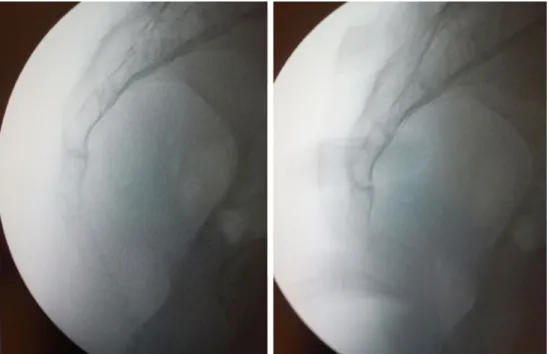
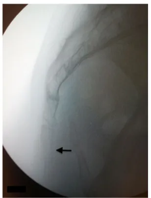
Under sedative anesthesia, patients were placed in lateral decubitus position. An external pressure was applied on coccyx under fluoroscopy before manipulation and possi-ble hypermobility was checked (Fig. 1). Angulation more than 25° was accepted as hypermobility [8]. A combined manipulation technique was performed. First, massage of the levator anus muscle, as well as anterior coccygeal sur-face, was performed as described by Thiele for 3 min [9] (Fig. 2). Second, coccyx was flexed and extended repeat-edly (stretching) for a minute and finally mobilization of the coccyx was performed as described by Maigne [10]. During last step, the coccyx was hyperextended and kept in this position for a minute (Figs. 3, 4). At the end of the session, 10 cc of a solution, which contains 1 cc (40 mg) methyl-prednisolone acetate, 3 cc (60 mg) prilocaine hydrochloride and 6 cc (30 mg) bupivacaine hydrochloride, was injected under fluoroscopy. Approximately 1 cc of the solution was injected into the sacrococcygeal joint and the remainder was injected into the soft tissues around the posterior aspect of the coccyx. Patients were advised to use cushion for 1 month and NSAII (dexketoprofen trometamol, 25 mg, twice a day) for 1 week after procedure. In Group 2, same solution was injected under fluoroscopy as in Group 1.
The statistical analysis was performed using SPSS 15 (SPSS Inc., Chicago, IL, USA). For repeated meas-ures, the Friedman test was used. Wilcoxon Signed Ranks test was used for post hoc analysis. Mean values of the
different groups were compared using Kruskal–Wallis test. Mann–Whitney U test was used for post hoc analy-sis. A p value of less than 0.05 was accepted as statistically significant.
Results
Mean age was 30.5 (range 21–48) years (31.1 and 30.2 years in Group 1 and Group 2, respectively) at the time of proce-dures. The average time between onset of symptoms and procedure was 8.1 (range 3–21) months (8.6 and 7.7 months in Group 1 and Group 2, respectively). Mean followup time was 27.8 (range 24–36) months (28.2 and 27.4 months in Group 1 and Group 2, respectively) at final control.
In Group 1, mean pre-treatment and post-procedure VAS scores were 4.6 (range 2–8) points and 1.4 (range 0–6) points, respectively. The difference between these values was statistically significant (p < 0.001). The average VAS scores were 1.5 (range 0–6) points at 1 year following the procedure and at the final followup. The difference between average post-procedure and followup VAS scores was not significant (p = 0.157). Thirteen patients (61.9%) had com-plete pain relief after treatment. In five patients (23.8%), the severity of symptoms decreased but feeling of uncomfort-ability continued. The mean pre-treatment and post-proce-dure VAS scores of those five patients were 6.2 (range 4–8) points and 3.4 (range 1–5) points, respectively. There was
Fig. 2 Index finger moves circumferentially and upward–downward directions during massage
Fig. 3 In stretching, the coccyx was flexed and extended repeatedly
a significant decrease in terms of VAS scores immediately after procedure for those patients (p = 0.034). The first year and last control VAS scores were not found to be changed for these five patients. In three patients (14.3%), VAS scores did not change after treatment. There was no relapse during followups.
In Group 2, mean pre-treatment and post-procedure VAS scores were 4.5 (range 2–7) and 1.3 (range 0–4) points, respectively. The difference between these values was statis-tically significant (p < 0.001). The average VAS scores were 2.3 (range 0–4) points at 1 year following the procedure and at the final followup. The increase in VAS scores was sig-nificant (p = 0.003). Five patients (21.7%) had complete pain relief after treatment. In 17 patients (73.9%), the severity of symptoms decreased but feeling of uncomfortability contin-ued. The mean pre-treatment and final control VAS scores of those 17 patients were 5.1 (range 3–7) points and 2.9 (range 1–4) points, respectively. The difference was statistically significant also for those patients (p < 0.001). In one patient (4.3%), VAS scores did not change. In 13 patients (56.5%), we detected significant increase in VAS scores during fol-lowups (p = 0.001). The mean post-procedure and followup VAS scores were 1.5 and 3.5 points, respectively, for these patients.
When we compared Group 1 and Group 2, we found no difference between pre- and post-procedure VAS scores (p = 0.9 and p = 0.4, respectively) But, the mean VAS score was significantly lower in Group 1 compared to Group 2 at 1 year and final controls (p = 0.038).
In Group 1, nine patients (42.9%) had history of trauma and 12 cases were idiopathic. The average BMI of the par-ticipants was 22.6 (range 18.9–27.1) kg/m2. According to the
BMI measures, there were 16 (76.2%) normal and 5 (23.8%) overweight patients (Table 2). There were 17 (80.9%) Type I, 2 (9.5%) Type II, 1 (4.8%) Type III and 1 (4.8%) Type IV patients according to Postacchini–Massobrio classifica-tion. Hypermobility was detected under fluoroscopic control in ten cases and all those patients had Type I coccyx. We compared patients who had Type 1 coccyx (group Type 1) with the other patients (group Others). Mean VAS scores in group Type 1 were 4.5 (range 2–8) points before treat-ment and 1.7 (range 0–6) points at post-procedure and fol-lowup controls. These values were 5.3 (range 3–7) points at pre-treatment, 0 point at post-procedure and 0.5 (0–1) points at followup evaluations in group Others. The differ-ences between average VAS scores of two groups before and after treatment were not significant (p = 0.388 before treatment, p = 0.101 at post-procedure, p = 0.561 at follow ups). When we checked the groups individually, we detected significant decrease in VAS scores after treatment in both groups (p = 0.001 in group Type 1 and p = 0.024 in group Others) There was no difference between post-procedure and followup scores (p = 1 in both groups).
The mean VAS score of the patients with hypermobile coccyx was 4.4 (range 2–7) points before treatment. This value was calculated as 1.6 (range 0–5) points at post-procedure and followup evaluations. When we checked the patients without hypermobility, the mean VAS scores were found to be 4.8 (range 2–8) points before treatment, 1.2 (0–6) points at post-procedure and 1.4 (range 0–6) points at followup controls. The differences between average VAS scores of two groups before and after treatment were not significant (p = 0.617 before treatment, p = 0.44 at post-procedure, p = 0.790 at followups). But in each group, there was significant decrease in VAS scores after procedure (p = 0.007 in hypermobile group and p = 0.006 in others). The difference between post-procedure and followup scores was not significant in both groups (p = 1 in hypermobile group and p = 0.157 in others).
We grouped the patients in Group 1 according to the BMI, as normal (Group A: 16 patients) or overweight (Group B: 5 patients). Mean VAS scores in Group A were 4.6 (range 2–8) points before treatment and 1.8 (range 0–6) points at post-procedure and followup controls. These val-ues were 4.6 (range 2–7) points at pre-treatment, 0 point at post-procedure and 0.2 (0–1) points at followups in Group B. The differences between average VAS scores of two groups before and after treatment were not significant (p = 0.930 before treatment, p = 0.060 at post-procedure, p = 0.108 at followups). When we checked the groups individually, we detected significant decrease in VAS scores after treat-ment in both groups (p = 0.001 in Group 1 and p = 0.042 in Group 2). There was no difference between post-procedure and followup scores (p = 0.317 in Group 1 and p = 0.330 in Group 2).
We divided the patients in Group 1 into two groups according to the underlying causes as traumatic (Group T: 9 patients) or non-traumatic (Group NT: 12 patients). Mean VAS scores in Group T were 4.4 (range 2–7) points before treatment and 1.3 (range 0–5) points at post-procedure and followup controls. These values were 4.8 (range 2–8) points before treatment, 1.4 (range 0–6) points at post-procedure and 1.6 (0–6) points at followups in Group NT. We observed significant decrease in VAS scores in both groups after pro-cedure (p = 0.007 in Group T and Group NT). The VAS scores did not change significantly during followups in both groups (p = 1 in Group T and p = 0.157 in Group NT). The differences between two groups’ mean VAS scores before and after treatment were not significant (p = 0.718 before treatment, p = 0.839 at post-procedure, p = 0.788 at followups).
We classified the patients in Group 1 according to the time between onset of the symptoms and treatment as Group X (< 6 months) and Y (≥ 6 months). There were 9 patients (42.9%) in Group X and 12 patients (57.1%) in Group Y. Mean VAS scores in Group X were 4 (range 2–6) points
before treatment and 1.1 (range 0–6) points at post-proce-dure and followup controls. These values were 5.1 (range 2–8) points before treatment, 1.6 (range 0–5) points at post-procedure and 1.8 (0–5) points at followups in Group Y. We observed significant decrease in VAS scores in both groups after procedure (p = 0.01 in Group X and p = 0.005 in Group Y). No significant change was observed during followups in both groups (p = 1 in Group X and p = 0.159 in Group Y). There was no difference between mean VAS scores of the two groups (p = 0.149 before treatment, p = 0.654 at post-procedure, p = 0.336 at followup).
Discussion
There are numerous causes of coccydynia but most of them are traumatic or idiopathic. Females are affected five times more than males. Spasm of the muscles of the pelvic floor, anomalies of the soft tissues in the mid-sacral region, chronic inflammation of an adventitious coccygeal bursa, pilonidal sinus and arachnoiditis of the lower sacral nerve roots may also cause pain in coccygeal region. Trauma is one of the leading causes of coccydynia. As well as direct hit, prolonged sitting and even pressure of the fetus during pregnancy may cause trauma to the coccyx [3, 11, 12]. In our study, there were nine patients who reported a history of trauma (42.9%) with the remaining patients unable to identify the cause of their coccydynia (idiopathic). Females consisted 75% of our study group (33 out of 44 patients).
Patel et al. pointed the importance of spasticity or other abnormalities affecting the musculature of the pelvic floor in patients with coccydynia [11]. The anterior sacrococcy-geal ligament, the gluteus maximus muscle, the coccygeus muscle, and the levator ani muscles attach to the coccyx [2]. The logic of massage is to stretch those structures to treat the tonic spasm thought to be responsible for the pain. Mobilization and stretching maneuvers of the sacrococ-cygeal and intercocsacrococ-cygeal joints are performed to enhance coccygeal mobility. Maigne and Chatellier compared these three manual treatment methods and reported 29.2, 16 and 32% success rates with massage, mobilization and stretch-ing methods, respectively, after 6 months followup [13]. We combined those three techniques with a steroid injection to further decrease the local inflammatory response. The increase in our success rate (61.9% complete pain relief) could also be related with addition of steroid injection.
There are different options for conservative treatment of coccydynia. NSAIDs and cushions are generally first choice in the treatment [5, 11]. Fogel et al. advised to use NSAIDs, cushions and stool softeners for 8 weeks after acute onset of the symptoms [1]. Kwon et al. accepted 8 weeks for acute period and named the cases as chronic coccydynia if the symptoms persist more than 8 weeks [15]. In persistent
cases, different modalities can be used. Mitra et al. claimed that steroid injection would be effective if it is applied less than 6 months after the onset of the pain [16]. Lin et al. used extracorporeal shock wave therapy (ESWT) in the treat-ment of coccydynia and reported more satisfactory results compared with physical modalities [6]. Coccygectomy can be preferred in case of refractory pain. But the complica-tion rates are high as 22% and most common complicacomplica-tion is postoperative infection [1, 11]. Good or excellent results were obtained in 60–90% of patients with excision [8]. In our series, all patients had chronic symptoms. We included only patients whose complaints persist more than 3 months. Cushions and painkillers were used during first 3 months. We performed manipulation and fluoroscopy-guided steroid injection if complaints persisted despite treatment. We inves-tigated the importance of time between onset of complaints and treatment. When we compared the patients who had coccydynia less and more than 6 months, we found no dif-ference between these two groups. Our results do not support advices of Mitra et al. and the chronicity of symptoms is not important for this treatment method.
We found no significant difference between Group 1 and Group 2 immediately after procedure but at first year and final controls the Group 1 had significantly lower VAS scores than Group 2. Our findings supported the efficacy of manipulation. Wray et al. compared the results of steroid injection with steroid injection after manipulation. Authors reported 59% success rate after injection, whereas it was 85% for injection and manipulation group [3]. The late relapse rates were 21 and 28%, respectively. In our series, the complete pain relief was achieved in 61.9% of patients in Group 1, whereas it was only 17.4% in Group 2. In 23.8% of Group 1, the VAS score was significantly decreased but the feeling of uncomfortability persisted. This was 73.9% in Group 2. We had no relapse in Group 1 but in Group 2 the relapse rate was 56.5%. We detected minor increase in aver-age VAS score during followups (1.4 points after procedure vs. 1.5 points at last followup controls) but that difference was not significant. Our success rate was lower than the Wray’s results but the author’s relapse rate was higher than ours in manipulation group. In our study, relapse rate was higher than Wray’s study in patients who were treated with single steroid injection. Since the steroid cocktails were sim-ilar in both studies (methylprednisolone and bupivacaine), the difference can be related with manipulation techniques. The authors preferred to perform only stretching maneuver but we combined three techniques. We recommended coc-cyx resection for the patients who were not pleased after treatment.
Increased body weight can be accepted as a risk fac-tor for coccydynia. Fogel et al. reported that coccydynia is three times more common in obese people [1]. In our study, there were only five overweight patients (23.8%).
We found no difference between normal and overweight patients. Maigne et al. claimed that coccygeal configuration may vary according to the BMI of the patients. Their study revealed that obese patients have mainly posterior subluxa-tion, normal-weight patients have mainly hypermobility or radiographically normal coccyges, and thin patients have mainly anterior subluxation and spicules [14]. There were ten hypermobile coccyges in our study group and all of them had normal BMI and coccygeal configuration. Our data is consistent with the theory of Maigne. There was no patient with posterior subluxation. According to our results, hyper-mobility does not affect the severity of pain and success of the treatment.
Postacchini and Massobrio classified coccygeal configu-ration and defined four types. They claimed that Types II, III, and IV were more prone to become painful than those with Type I [7]. We classified our patients according to this classification system. Most of the patients (80.9%) had Type I coccygeal configuration. Since the number of patients with Type II, III and IV coccyges was low, we compared the patients with Type 1 coccyx with all other types. We found no difference between two groups in terms of pre-treatment VAS scores and efficacy of procedure.
Our study has some limitations such as its retrospective design and low number of patients. In addition, comparison with other treatment modalities (e.g. ESWT or coccygec-tomy) could be added to determine the best option for persis-tent coccydynia. But to our knowledge there is no such study in the literature. A multicentric study would be designed to achieve enough number of patients.
In conclusion, manual therapy combined with steroid injection would be an alternative method in case of persis-tent coccydynia. It is a safe and easy option before surgical treatment.
Compliance with ethical standards
Conflict of interest The authors declare that they have no conflict of interest.
References
1. Fogel G, Cunningham P, Esses S (2004) Coccygodynia: evaluation and management. J Am Acad Orthop Surg 12:49–54
2. Emerson SS, Speece AJ 3rd (2012) Manipulation of the coccyx with anesthesia for the management of coccydynia. J Am Osteo-path Assoc 112:805–807
3. Wray CC, Easom S, Hoskinson J (1991) Coccydynia. Aetiology and treatment. J Bone Joint Surg Br 73:335–338
4. Haddad B, Prasad V, Khan W, Alam M, Tucker S (2014) Favour-able outcomes of coccygectomy for refractory coccygodynia. Ann R Coll Surg Engl 96:136–139
5. Howard PD, Dolan AN, Falco AN, Holland BM, Wilkinson CF, Zink AM (2013) A comparison of conservative interventions and their effectiveness for coccydynia: a systematic review. J Man Manip Ther 21:213–219
6. Lin SF, Chen YJ, Tu HP, Lee CL, Hsieh CL, Wu WL, Chen CH (2015) The effects of extracorporeal shock wave therapy in patients with coccydynia: a randomized controlled trial. PLoS One 10:e0142475. https://doi.org/10.1371/journal.pone.0142475
7. Postacchini F, Massobrio M (1983) Idiopathic coccygodynia. Analysis of fifty-one operative cases and a radiographic study of the normal coccyx. J Bone Joint Surg [Am] 65:1116–1124 8. Maigne JY, Lagauche D, Doursounian L (2000) Instability of the
coccyx in coccydynia. J Bone Joint Surg Br 82:1038–1041 9. Thiele GH (1937) Coccydynia and pain in the superior gluteal
region. JAMA 109:1271–1275
10. Maigne R (1996) Diagnosis and treatment of pain of vertebral origin: a manual medicine approach. Williams and Wilkins, Bal-timore, pp 339–340
11. Patel R, Appannagari A, Whang PG (2008) Coccydynia. Curr Rev Musculoskelet Med 1:223–226
12. Jaiswal A, Shetty AP, Rajasekaran S (2008) Precoccygeal epider-mal inclusion cyst presenting as coccygodynia. Singapore Med J 49:e212–e214
13. Maigne JY, Chatellier G (2001) Comparison of three manual coccydynia treatments: a pilot study. Spine (Phila Pa 1976) 26:E479–E483
14. Maigne J, Doursounian L, Chattelier G (2000) Causes and mecha-nisms of common coccydynia: role of body mass index and coc-cygeal trauma. Spine 25:3072–3079
15. Kwon HD, Schrot RJ, Kerr EE, Kim KD (2012) Coccygodynia and coccygectomy. Korean J Spine 9:326–333
16. Mitra R, Cheung L, Perry P (2007) Efficacy of fluoroscopically guided steroid injections in the management of coccydynia. Pain Physician 10:775–778