Preclinical and clinical evidence of nephro- and
cardiovascular protective effects of glycosaminoglycans
Arrigo F. Cicero
1, Sibel Ertek
2A b s t r a c t
Despite advances in pharmacological treatment, diabetic nephropathy is still the leading cause of end-stage renal disease and an important cause of morbidity and mortality in diabetics. Glycosaminoglycans are long, unbranched mucopolysaccharides that play an important role in establishing a charge-selective barrier that restricts the passage of negatively charged molecules, such as albumin and other proteins, at the level of the glomerular basal membrane. Their loss is associated with loss of selectivity and proteinuria. Extensive preclinical evidence and some clinical trials suggest that glycosaminoglycans replacement is associated with improvement of glomerular selectivity and of proteinuria. Sulodexide could also have some other effects, potentially useful to reduce the renal damage and the cardiovascular disease associated with proteinuria, such as improvement of haemorheological and blood lipid parameters, an endothelium protective effect and anti-inflammatory action. This review will discuss the evidence supporting the potential nephroprotective effects of sulodexide and other glycosaminoglycans.
Key words: glycosaminoglycans, cardiovascular diseases, proteinuria, diabetic nephropathy, sulodexide.
Introduction
Despite advances in dialysis techniques, pharmacological treatment,
and patient rehabilitation programmes, mortality and morbidity rates of
end-stage renal disease (ESRD) are still high and mainly related to
cardiovascular diseases (CVD) [1, 2]. Diabetic nephropathy is still the leading
cause of ESRD [3] and an important cause of morbidity and mortality in
patients with either type 1 or type 2 diabetes mellitus, both directly and
as a risk factor for cardiovascular disease [4]. The microangiopathic
complications of diabetes increase with longer duration of diabetes and
worse glycaemic control [5]. In fact, the presence of albuminuria or
proteinuria is a well known risk factor for coronary heart disease [6, 7].
Although recent evidence shows that an early multi-pharmacological
approach is able to slow the progression of diabetic nephropathy to ESRD
[8], the disease rarely stops and slightly regresses just in a few selected
and optimally treated patients [9]. So, a better understanding of the
pathophysiology of chronic kidney disease is needed in order to develop
new efficacious treatments for this pandemic disease.
Corresponding author: Arrigo F. Cicero, MD, PhD Internal Medicine, Aging and Kidney Diseases Department Sant’Orsola-Malpighi Hospital University of Bologna Via Albertoni 15 40138 Bologna, Italy Phone: +39 0516364920 Fax: +39 051391320 E-mail: [email protected]
1Hypertension Research Unit, Internal Medicine, Aging and Kidney Diseases Department,
Alma Mater Studiorum University of Bologna, Italy
2Endocrinology and Metabolic Diseases Department, Ufuk University, Ankara, Turkey
Submitted: 5 May 2009 Accepted: 17 October 2009 Arch Med Sci 2010; 6, 4: 469-477 DOI: 10.5114/aoms.2010.14456 Copyright © 2010 Termedia & Banach
The aim of this review is to evaluate the
available literature data supporting the possible role
of glycosaminoglycans (GAGs) in renovascular
pathology and their possible usefulness in the
treatment of patients affected by diabetic
nephropathy.
Role of glycosaminoglycans in renal physiology
The glomerular filtration barrier consists of
fenestrated glomerular endothelium, podocyte foot
processes interconnected by slit diaphragms, and
intervening glomerular basement membrane
(GBM). Its characterization as both a size and
charge-selective barrier emerged from studies
conducted decades ago. Podocyte cytoskeleton and
its connections with specific glycans and proteins
constitute the basis of slit diaphragm and
cell-extracellular matrix interactions. Anionic sites in
GBM consist of GAGs rich in heparin sulphate (HS)
and their removal by enzymatic digestion resulted
in increased permeability [10-12].
Glycosaminoglycans are long, unbranched
mucopolysaccharides that consist of repeating
disaccharide units. Apart from hyaluronan, which
is uniquely synthesized without a protein core and
is "spun out" by enzymes at cell surfaces directly
into the extracellular space, the other GAGs are
usually added to protein cores in the Golgi
apparatus to yield proteoglycans [13].
Glomerular basement membrane charge is
imparted by the sulphated GAG side chains of
proteoglycans (HS proteoglycans [HSPGs] and, less,
hyaluronic acid) and to a lesser extent by carboxyl
and sialyl groups of glycoproteins. These negatively
charged molecules play an important role in
establishing a charge-selective barrier that restricts
the passage of negatively charged molecules, such
as albumin and other proteins [14].
Role of glycosaminoglycans in renal
pathologies: experimental data
Nowadays, the charge selectivity phenomenon
has received renewed attention with the
identification of mechanisms of synthesis of
barrier-related molecules [15, 16]. In particular, GBM
HS proteoglycans (and more specifically on
perlecan, collagen XVIII, and agrin) are considered
primary charge barrier components. Segmented
loss of GBM HS has been reported in membranous
glomerulo nephritis, lupus nephritis, minimal
change disease and diabetic nephropathy
in humans [17, 18] and rat models of Adriamycin
and Heymann nephritis [19]. Van der Born
et al. observed that streptozotocin-induced
dia betic rats with diabetic nephropathy
experienced a significant decrease in glomerular
HS/4-hydroxyproline ratio (showing increased
collagen and relatively decreased GBM HS content)
compared with control rats, and that was
associated with selective proteinuria and
glomerular hyperfiltration [20]. In another
experimental model, non-diabetic mice knock-out
for the Ext1 gene encoding a subunit of HS
co-polymerase develop proteinuria that is less
impressive than that expected from the available
knowledge on renal physiology, suggesting that
the cessation of polymerisation of
podocyte-secreted HS that affects glomerular ultrafiltration
charge may not be a serious cause of albuminuria,
and there may be other roles of these molecules,
including podocyte behaviour or morphology, as
indicated in this study [21].
However, whether charge selectivity is actually
important for glomerular function for the extent
of proteinuria is still a matter of debate [22].
Recent in vivo manipulations of glomerular HS
proteoglycans put in perspective (but did not
exclude) a role for either molecules themselves
or their anionic charge, which is altered greatly
by loss of HS but caused insignificant albu
-minuria, in glomerular filtration [16]. On the other
hand, mice without HS attachment sites on
perlecan revealed normal glomerular structure
and no evidence of renal disease, except slightly
increased susceptibility to protein-overload
albuminuria [23, 24]. Collagen XVIII mutants had
mild mesangial expansion with slightly elevated
serum creatinine levels [25] and podocyte-specific
agrin knock-out mice had a significant GBM
charge defect but normal renal function [26].
Perlecan HS and perlecan-HS/agrin double mutant
rats experience significant charge reduction on
GBM, but no renal dysfunction or proteinuria [27].
Lastly, the removal of HS in rats did not result in
acute proteinuria [28].
Moreover, in renal biopsies of different human
primary proteinuric diseases, pronounced alteration
in tubulointerstitial HS proteoglycans is evident
and strongly related to the inflammatory processes
[29]. In fact, GAGs have a role in modulation of
inflammation in tissues. Heparin sulphate proteo
-glycans can bind the leukocyte adhesion molecule
L-selectin and chemokines, suggesting their role in
inflammation [29-32]. Mouse and human
glomerular endothelial cells activated by tumour
necrosis factor (TNF)-
α or interleukin 1-β showed
increased expression of inflammatory N- and
6Osulphated HS domains and these are impor
-tant in leukocyte trafficking and inflammation [33].
Heparin sulphate is an important constituent of
subendothelial extracellular matrix and basement
membrane structure and vascular HS is decreased
in atherosclerosis, diabetes and during
inflammation. It is also affected by lipoproteins,
and lipoprotein-modulated perlecan may play an
important role in vascular smooth muscle growth,
and thus in atherosclerosis [34]. Celie et al. showed
the role of microvascular BM HS in inflammatory
responses, in human renal allograft biopsies [35].
Heparin sulphate and GAGs play a major role in
adhesion of leukocytes to glomerular cells, and that
is important for proliferative glomerulopathies,
inflammation and angiogenesis. Under dynamic
flow conditions addition of HS, heparin and
tinzaparin and removal of HS on mouse glomerular
endothelial cells the number of rolling and adhering
leukocytes decreases about 2-3-fold and the rolling
velocity doubles [36]. Heparin sulphate also binds
to cell surface receptors and is involved in the
modulation of inflammation. CD44/HS actions are
well studied in inflamed synovial membrane
macrophages, and it is involved in the regulation of
growth factors during inflammation and wound
healing [37, 38].
Experimental data supporting the potential use
of glycosaminoglycans in proteinuric diseases
Besides HSPGs, heparanase may have a role in
renal diseases; together with the changes in
glomerular cell-GBM interactions and loss of HS,
increased heparanase release might cause the
release of HS-bound factors and HS fragments in
the glomeruli or changes in intracellular signalling
by binding of heparanase to glomerular cells [39].
In diabetic nephropathy, the HS content of the GBM
is decreased and that causes protein leak into the
urinary space [40]; the increased amount of
heparanase enzyme in response to hyperglycaemia
may be one explanation. On the other hand,
heparanase upregulation by high glucose is
prevented by insulin and/or heparin in endothelial
cell cultures [41]. In humans, increased heparanase
level is associated with reduced HS, as observed in
renal biopsies of diabetic patients with nephropathy
[42], whereas in renal biopsies of different human
primary proteinuric diseases pronounced alterations
in tubulointerstitial HSPGs were evident and
strongly related to inflammatory processes [29].
This is probably related to more relevant
involvement of renal glomeruli in diabetic
nephropathy than in other proteinuric diseases.
Evidence from in vitro and diabetic animal
studies reveal that the administration of heparin
increases synthesis of HS [43], and other anionic
glycoproteins can effectively prevent the bioche
-mical alterations that promote albuminuria [44].
Enoxaparin, a low-molecular weight heparin, was
also tested on patients with diabetic and
non-diabetic glomerulopathies. The
proteinuria-decreasing effect of this heparin was found not to
be related to the renin-angiotensin system, and its
glomerular filter-related effect was suggested [45].
Angiotensin II (AT-II) receptor blockers are
renin-angiotensin system (RAS) modulators with very
well known antiproteinuric activity [46].
Angiotensin II inhibits HSPG expression in human
podocytes [47] and heparins modulate AT-II
signalling in glomerular cells [48], inhibiting
aldosterone synthesis [49] and lowering proteinuria
in diabetic patients [45], but this effect is less
pronounced in other forms of proteinuric renal
diseases and its relation to haemodynamic
changes produced by RAS is not proven in clinical
trials [45]. Of course, heparins are not easily
administrable for chronic treatments.
In this context, heparinoids were considered as
potentially useful antiproteinuric drugs that could
have synergistic effects with an RAS modulator [50].
In particular, sulodexide, a soluble, highly purified
preparation of low-molecular weight GAGs
composed of fast-moving heparin (80%) and
dermatan sulphate (20%) derived from porcine
intestine, appeared to be a promising treatment for
diabetic proteinuria partially resistant to RAS
blocking agents [51]. It prevents HS degradation,
reconstruction of HS content of GBM, and in vivo
inhibition of heparanase [40].
In fact, sulodexide is concentrated in renal
parenchyma for a long time after administration
[52] and in preliminary trials it has been supposed
that it reduces albuminuria acting in vivo as
a heparanase inhibitor that reaches the glomerular
capillary wall and prevents HS degradation, thus
allowing reconstruction of HS content and
restoration of glomerular basement membrane
ionic permselectivity [40]. Its antiproteinuric effect
appears to be mainly related to the basal
proteinuria and to the treatment duration [53],
independently from its antithrombotic and
profibrinolytic activity.
Studies in mouse articular chondrocytes after
lipopolysaccharide stimulation also showed
anti-inflammatory and anti-apoptotic actions of GAGs
[54]. Moreover, sulodexide seems to have powerful
anti-inflammatory activity in experimental models
[55]. In a model of cultured human umbilical
endothelial cells exposed to high glucose
concentration, sulodexide suppresses cellular
inflammation and prevents glucose cytotoxicity
[56]; it is able to reverse the glucose-related cell
release of free oxygen radicals, monocyte
chemotactic protein-1 (MCP-1) and interleukin-6
(IL-6), and the inactivation of the cell repair
mechanism induced by exposure to glucose.
However, these anti-inflammatory effects have not
been demonstrated in humans yet. Therefore, in
rats with streptozotocin-induced diabetes,
sulodexide exerts direct endothelial protective
effects [57] that could also be involved in kidney
protection. However, the anti-inflammatory role of
GAGs and heparin was already known in humans
for decades, and especially in patients with allergy
and asthma [58].
Clinical evidence of antiproteinuric effects
of glycosaminoglycans
The antiproteinuric effects of GAGs, and
especially of sulodexide, have been known for
nearly two decades [59], and many clinical studies
confirm its potential usefulness in treating
nephropathies and especially in diabetic nephro
-pathy.
In particular, the largest and more methodo
-logically correct clinical trial was the one carried out
by Gambaro et al.: the Diabetic nephropathy and
Albuminuria Study (Di.N.A.S.) involved 223 patients
with type 1 and 2 diabetes with both macro- and
micro-albuminuria [60]. In this trial, 200 mg/day
sulodexide treatment for 4 months was associated
with 46% decrease in albumin excretion rate from
baseline in diabetics who were not receiving
concomitant angiotensin-converting enzyme (ACE)
inhibitor treatment, and urinary albumin excretion
was maintained even 2 months after drug intake
interruption. The reduction in albuminuria was
dose-dependent in this study, i.e. 100 mg/day
sulodexide caused a mean decrease in albumin
excretion rate of about 17% from baseline among
the patients who were not receiving concomitant
ACE inhibitor medication. The difference of albumin
excretion vs. placebo was 62% in all diabetics
with 200 mg/day sulodexide. The antiproteinuric
effect of sulodexide appeared to be independent
from the baseline blood pressure level and from the
use of ACE inhibitors, meaning that even in patients
already receiving ACE inhibitors, sulodexide was
able to decrease the albumin excretion rate to
approximately the same extent as in patients
without ACE inhibitors (respectively 40% and 46%
from baseline after 4 months with 200 mg/day
sulodexide) [60].
However, this supposed additive effect of
sulodexide was not confirmed either for
ACE inhibitors when used at the maximum
recommended dosage or for angiotensin receptor
blockers (ARBs) also when used at the maximum
recommended dosage. In fact, although in a pilot
study on 149 microalbuminuric type 2 diabetic
patients a trend for an increased rate of therapeutic
success (return to normoalbuminuria or a decrease
in albumin : creatinine ratio [ACR] of at least 50%
from the baseline value) was observed for
sulodexide, 200 mg/day for 6 months, compared
to placebo (33.3% vs. 15.4% of the patients
achieving the efficacy endpoint respectively;
p = 0.075) [75], in the two related subsequent large
trials respectively on microalbuminuric and
macroalbuminuric type 2 diabetic patients the
favourable trend of the preliminary pilot study for
the additive effect of sulodexide in patients already
treated with the maximum recommended dosages
of ACE inhibitors or ARBs was not confirmed, even
if the detailed results of these two studies are not
known yet [76].
Other clinical trials focused on the mechanism
of the antiproteinuric activity of sulodexide.
Sulikowska et al. studied whether the
albuminuria-lowering effect of sulodexide comes from its
renovascular or tubular effects [61]. Dopamine
infusion causes efferent arteriolar dilatation and
increases creatinine clearance in normal people [62].
They tested dopamine-induced glomerular filtration
response testing and urinary N-acetyl-
βDgluco
-saminidase measurements to test proximal tubular
integrity, besides albuminuria, on type 1 diabetic
patients. Patients were divided into placebo and
daily 100 mg sulodexide groups for 120 days and
dopamine testing was performed only on patients
taking sulodexide. Sulodexide caused a decrease in
albumin excretion from 126.1 ±15.41 to 96.3 ±13.7
mg/day in the treatment group compared with
a decrease from 106.8 ±21.4 to 126.8 ±29.6 mg/day
in controls. N-acetyl-
βDglucosaminidase measure
-ments also changed from 5.1 ±0.62 to 4.7 ±0.40
U/gCre in the sulodexide group and from 5.9 ±0.87
to 6.3 ±1.35 U/gCre in the control group with
placebo. The response increase in creatinine
clearance to dopamine infusion was from 13.2 ±2.1
to 15.44 ±1.9% (+16.9% increase) in patients taking
this drug. The conclusion was that sulodexide
affects intrarenal vascular reactivity and also
improves N-acetyl-
β-D-glucosaminidase tests,
indicating amelioration of tubular damage [61].
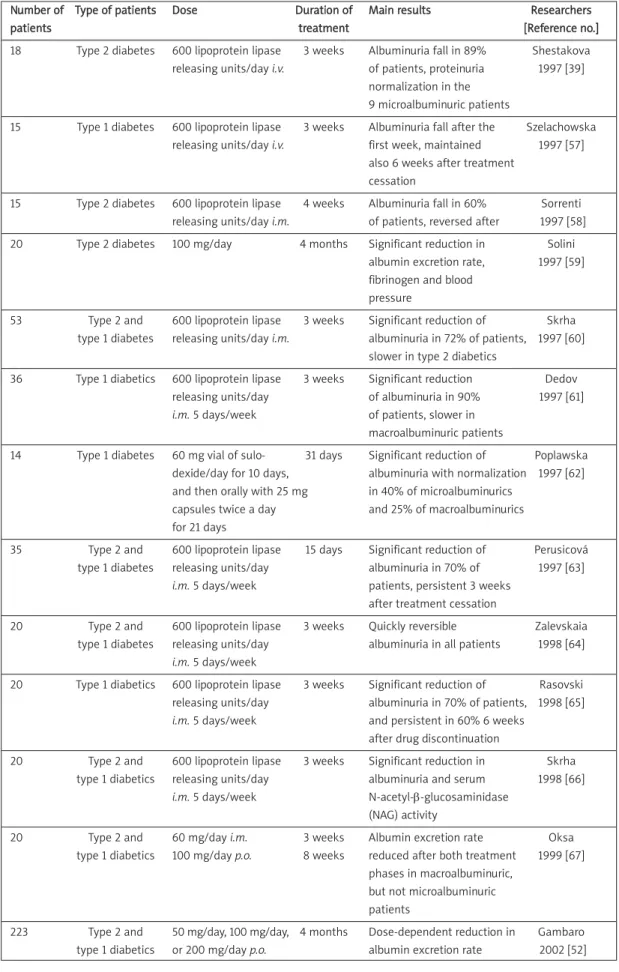
Whatever the main mechanism may be, the role
of sulodexide as an antiproteinuric agent is
suggested by the fundamental Di.N.A.S. trial and
a large number of smaller studies on both type 1
and 2 diabetic patients (Table I) [53, 60, 61, 63-75].
Although these smaller studies often had an open
design and a short duration, and did not involve
homogeneous patient categories, nevertheless they
contributed to the evidence of a clinically favourable
antiproteinuric effect of sulodexide.
Other effects of glycosaminoglycans
on cardiorenal physiology
The potential cardiovascular effects of sulodexide
and GAGs are summarized in Table II.
Interactions with HS modify and contribute to
various protein actions and intercellular signalling by
cytokines and growth factors, and some proteins
share binding sites with HS [78]. Extracellular matrix
of blood vessel walls also contain considerable
amounts of proteoglycans and systemic hypertension
may change the content of subendothelial matrix of
vessels: this may also contribute to increased
Number of Type of patients Dose Duration of Main results Researchers
patients treatment [Reference no.]
18 Type 2 diabetes 600 lipoprotein lipase 3 weeks Albuminuria fall in 89% Shestakova releasing units/day i.v. of patients, proteinuria 1997 [39]
normalization in the 9 microalbuminuric patients
15 Type 1 diabetes 600 lipoprotein lipase 3 weeks Albuminuria fall after the Szelachowska releasing units/day i.v. first week, maintained 1997 [57]
also 6 weeks after treatment cessation
15 Type 2 diabetes 600 lipoprotein lipase 4 weeks Albuminuria fall in 60% Sorrenti releasing units/day i.m. of patients, reversed after 1997 [58] 20 Type 2 diabetes 100 mg/day 4 months Significant reduction in Solini
albumin excretion rate, 1997 [59] fibrinogen and blood
pressure
53 Type 2 and 600 lipoprotein lipase 3 weeks Significant reduction of Skrha type 1 diabetes releasing units/day i.m. albuminuria in 72% of patients, 1997 [60]
slower in type 2 diabetics
36 Type 1 diabetics 600 lipoprotein lipase 3 weeks Significant reduction Dedov releasing units/day of albuminuria in 90% 1997 [61]
i.m. 5 days/week of patients, slower in
macroalbuminuric patients
14 Type 1 diabetes 60 mg vial of sulo- 31 days Significant reduction of Poplawska dexide/day for 10 days, albuminuria with normalization 1997 [62] and then orally with 25 mg in 40% of microalbuminurics
capsules twice a day and 25% of macroalbuminurics for 21 days
35 Type 2 and 600 lipoprotein lipase 15 days Significant reduction of Perusicová type 1 diabetes releasing units/day albuminuria in 70% of 1997 [63]
i.m. 5 days/week patients, persistent 3 weeks
after treatment cessation
20 Type 2 and 600 lipoprotein lipase 3 weeks Quickly reversible Zalevskaia type 1 diabetes releasing units/day albuminuria in all patients 1998 [64]
i.m. 5 days/week
20 Type 1 diabetics 600 lipoprotein lipase 3 weeks Significant reduction of Rasovski releasing units/day albuminuria in 70% of patients, 1998 [65]
i.m. 5 days/week and persistent in 60% 6 weeks
after drug discontinuation
20 Type 2 and 600 lipoprotein lipase 3 weeks Significant reduction in Skrha type 1 diabetics releasing units/day albuminuria and serum 1998 [66]
i.m. 5 days/week N-acetyl-β-glucosaminidase
(NAG) activity
20 Type 2 and 60 mg/day i.m. 3 weeks Albumin excretion rate Oksa type 1 diabetics 100 mg/day p.o. 8 weeks reduced after both treatment 1999 [67]
phases in macroalbuminuric, but not microalbuminuric patients
223 Type 2 and 50 mg/day, 100 mg/day, 4 months Dose-dependent reduction in Gambaro type 1 diabetics or 200 mg/day p.o. albumin excretion rate 2002 [52] Table I. Clinical studies evaluating the effects of sulodexide on proteinuria and albuminuria
peripheral vascular resistance, as shown in animal
models [79].
As stated above, at least a part of the renal
histological degradation observed in diabetes is
related to inflammatory processes, and sulodexide
showed anti-inflammatory activity in different
animal models [55]. A pilot study recently conducted
on 11 healthy men concluded that it may also
decrease transforming growth factor
β1 (TGF-β1)
release [80].
The haemorheological and lipid lowering actions
of GAGs have also been known for the last two
decades. In fact, sulodexide decreases triglycerides
[81], increases Apo-A1 and HDL-C levels [82] and
blood viscosity [83], decreases D-dimer and
fibrinogen levels [84, 85], and releases tissue
plasminogen activator [86, 87]. Sulodexide has a dual
effect on coagulation: antithrombin catalysis by fast
moving heparin component and heparin cofactor II
catalysis by dermatan sulphate component [88].
It causes inhibition of thrombus formation and
growth with less systemic anticoagulation than
comparable antithrombotic doses of heparins [89]
and may also reduce oxidative stress slightly
expressed by malonylaldehyde and superoxide
dismutase in diabetics [72]. Therefore, in rats with
streptozotocin-induced diabetes, sulodexide exerts
direct endothelial protective effects, improving
acetylcholine-induced relaxation of isolated aorta
and mesangial arteries and reducing the number of
circulating endothelial cells [57].
Conclusions
A relatively large body of literature supports the
antiproteinuric and nephroprotective effects of
GAGs and sulodexide, especially in diabetic
nephropathy. These could derive from their effect
on vascular permeability and inflammation, and on
endothelium protection and haemorheology
improvement. Sulodexide has the advantage of
being an oral medication with few and usually mild
side effects of intestinal type (i.e. diarrhoea, nausea,
dyspepsia). Its antiproteinuric and nephroprotective
role in therapy may be played in patients not
tolerating ARBs and ACE inhibitors or in patients
who are resistant to dosages of ARBs and ACE
inhibitors which are not being given at the
maximum recommended level, for safety or other
reasons. In fact two recent clinical trials were not
able to confirm in patients already receiving the
maximal recommended dosages of ARBs or ACE
inhibitors the previous statistically significant
additional effects of sulodexide observed by
Gambaro et al. [60] also in patients being treated
Number of Type of patients Dose Duration of Main results Researchers
patients treatment [Reference no.]
60 Type 2 and 50 mg/day p.o. 12 months Albuminuria strongly reduced Achour type 1 diabetics in all patients vs. controls 2005 [68]
and vs. baseline
45 Type 1 diabetics 120 mg/day p.o. 6 months Reduction in albuminuria and Sulikowska N-acetyl-β-D-glucosaminidase 2006 [56] (NAG) excretion, increase
in renal vascular function
149 Obese type 2 200-400 mg/day p.o. 6 months 25.3% and 33.3% of the patients Heerspink diabetics in addition to ACEI respectively in the two sulodexide 2008 [69]
or ARBs in resistant groups combined and in the patients 200 mg/day group achieved
a significant reduction or normalization of albuminuria vs. 15.4% of the patients in the control group (p = 0.26 and p = 0.075 respectively) Table I. Clinical studies evaluating the effects of sulodexide on proteinuria and albuminuria – cont.
• Antithrombotic action • Decreased oxidative stress • Antihyperlipidaemic actions • Prevention of glucose toxicity • Suppression of cellular inflammation • Cytokines and growth factors modulation • Interactions with AT-II signalling and RAS system • Reduction of peripheral vascular resistance
and improvement of vascular elasticity • Antiproteinuric effects
Table II. Potential cardiovascular beneficial effects of sulodexide and GAGs in diabetic patients
with unspecified dosages of ACE inhibitors.
Certainly, more clinical research is needed to
understand which factors influence the drug
efficacy and, consequently, which patients could
a priori obtain the best effect from this treatment.
Therefore, further clinical trials are currently ongoing
with sulodexide as an antiproteinuric agent.
R e f e r e n c e s
1. Levin A. Clinical epidemiology of cardiovascular disease in chronic kidney disease prior to dialysis. Semin Dial 2003; 16: 101-5.
2. Sarnak MJ, Levey AS. Epidemiology of cardiac disease in dialysis patients. Semin Dial 1999; 12: 69-76.
3. Radbill B, Murphy B, LeRoith D. Rationale and strategies for early detection and management of diabetic kidney disease. Mayo Clin Proc 2008; 83: 1373-81.
4. Martínez Castelao A. Advances in diabetes mellitus, diabetic nephropathy, metabolic syndrome and cardio-vascular-renal risk. Nefrologia 2008; 28 Suppl 5: 79-84. 5. Rogowicz A, Litwinowicz M, Pilacinski S, Zozulinska D,
Wierusz-Wysocka B. Does early insulin treatment decrease the risk of microangiopathy in non-obese adults with diabetes. Arch Med Sci 2007; 3: 129-35.
6. Perkovic V, Verdon C, Ninomiya T, et al. The relationship between proteinuria and coronary risk: a systematic review and meta-analysis. PLoS Med 2008; 5: e207. 7. Barylski M, Banach M, Mikhailidis DP, Pawlicki L, Kowal
-ski J. Decreased kidney function as a risk factor for cardiovascular events in subjects with metabolic syndrome – a pilot study. Arch Med Sci 2008; 4: 417-23. 8. Weir MR. Microalbuminuria and cardiovascular disease.
Clin J Am Soc Nephrol 2007; 2: 581-90.
9. Cortinovis M, Cattaneo D, Perico N, Remuzzi G. Investigational drugs for diabetic nephropathy. Expert Opin Investig Drugs 2008; 17: 1487-500.
10. Kanwar YS, Farquhar MG. Presence of heparan sulphate in the glomerular basement membranes. Proc Natl Acad Sci U S A 1979; 76: 1303-7.
11. Kanwar YS, Farquhar MG. Isolation of glycosaminoglycans (heparin sulphate) from glomerular basement membranes. Proc Natl Acad Sci U S A 1979; 76: 4493.
12. Kanwar YS, Linker A, Farquhar MG. Increased permeability of the glomerular basement membrane to ferritin after removal of glycosaminoglycans heparin sulphate) by enzyme digestion. J Cell Biol 1980; 86: 688-93.
13. Raman R, Sasisekharan V, Sasisekharan R. Structural insights into biological roles of protein-glycosaminoglycan interactions. Chem Biol 2005; 12: 267-77.
14. Deen WM, Lazzara MJ, Myers BD. Structural determinants of glomerular permeability. Am J Physiol Renal Physiol 2001; 281: F579-F596.
15. Harvey SJ, Miner JH. Revisiting the glomerular charge barrier in the molecular era. Curr Opin Nephrol Hypertens 2008; 17: 393-8.
16. Miner JH. Glomerular filtration: the charge debate charges ahead. Kidney Int 2008; 74: 259-61.
17. van der Born J, van del Havuel L, Bakker M, et al. Distribution of GBM heparin sulphate proteoglycan core protein and side chanins in human glomerular disease. Kidney Int 1993; 43: 454-63.
18. Tamsma J, van der Born J, Bruijn J, et al. Expression of glomerular extracellular matrix components in human
diabetic nephropathy: decrease of heparin sulphate in the glomerular basal membrane. Diabetologia 1994; 37: 313-20. 19. Raats C, Luca M, Bakker M, et al. Reduction in glomerular heparin sulphate correlates with complement deposition and albuminuria in active Heymann nephritis. J Am Soc Nephrol 1999; 10: 1689-99.
20. van den Born J, van Kraats AA, Bakker MA, et al. Selective proteinuria in diabetic nephropathy in the rat is associated with a relative decrease in glomerular basement membrane heparin sulphate. Diabetologia 1995; 38: 161-72. 21. Chen S, Wassenhove-McCarthy DJ, Yamaguchi Y, et al. Loss
of heparin sulfate glycosaminoglycan assembly in podocytes does not lead to proteinuria. Kidney Int 2008; 74: 289-99.
22. Comper WD, Glascow EF. Charge selectivity in kidney ultrafiltration. Kidney Int 1995; 47: 1242-51.
23. Rossi M, Morita H, Sormunen R, et al. Heparan sulphate chains of perlecan are indispensible in the lens capsule but not in the kidney. EMBO J 2003; 22: 236-45. 24. Morita H, Yoshimura A, Inui K, et al. Heparan sulphate of
perlecan is involved in glomerular filtration. J Am Soc Nephrol 2005; 16: 1703-10.
25. Utriainen A, Sormunen R, Kettunen M, et al. Structurally altered besement membranes and hydrocephalus in a type XVIII collagen deficient mouse line. Hum Mol Genet 2004; 13: 2089-99.
26. Harvey S, Burgess R, Miner J. Podocyte derived agrin is responsible for glomerular basement membrane anionic charge [Abstract]. J Am Soc Nephrol 2005; 16: 1A. 27. Goldberg S, Harvey SJ, Cunningham J, Tryggvason K, Miner
JH. Glomerular filtration is normal in the absence of both agrin and perlecan-heparan sulphate from the glomerular basement membrane. Nephrol Dial Transplant 2009; 24: 2044-51.
28. Winjhoven TJ, Lensen JF, Wismans RG, et al. In vivo degradation of heparan sulphates in the glomerular basement membrane does not result in proteinuria. J Am Soc Nephrol 2007; 18: 823-32.
29. Celie JW, Reijmers RM, Slot EM, et al. Tubulointerstitial heparan sulfate proteoglycan changes in human renal diseases correlate with leukocyte influx and proteinuria. Am J Physiol Renal Physiol 2008; 294: F253-63. 30. Kawashima H, Watanabe N, Hirose M, et al. Collagen XVIII
a basement membrane heparin sulphate proteoglycan, interacts with L-selectin and monocyte chemoattractant protein-1. J Biol Chem 2003; 278: 13069-76.
31. Parish CR. Role of heparin sulphate I in inflammation. Nat Rev Immunol 2006; 6: 633-43.
32. Wang L, Fuster M, Sriramarao P, Esko JD. Endothelial heparin sulphate deficiency impairs L-selectin – and chemokine-mediated neutrophile trafficking during inflammatory responses. Nat Immunol 2005; 6: 902-10. 33. Rops AL, van der Hoven MJ, Baselmans MM, et al.
Heparan sulphate domains on cultured activated glomerular endothelial cells mediate leukocyte trafficking. Kidney Int 2008; 73: 52-62.
34. Pillarisetti S. Lipoprotein modulation of seubendothelial heparin sulphate proteoglycans (perlecan) and athero -genicity. Trends Cardiovasc Med 2000; 10: 60-5. 35. Celie JW, Rutjes NW, Keuning ED, et al. Subendothelial
heparan sulphate proteoglycans become major L-selectin and monocyte chemoattractant protein-1 ligands upon renal ischemia/reperfusion. Am J Pathol 2007; 170: 1865-78.
36. Rops AL, Jacobs CW, Linssen PC, et al. Heparan sulphate on activated glomerular endothelial cells and exogenious heparinoids influence the rolling and adhesion of leuckocytes. Nephrol Dial Transplant 2007; 22: 1070-7.
37. Jones M, Tussey M, Athanasou N, Jackson DG. Heparan sulphate proteoglycan isoforms of the CD44 hyaluronan receptor induced in human inflammatory macrophages can function as paracrine regulators of fibroblast growth factor action. J Biol Chem 2000; 275: 7964-74.
38. Taylor KR, Gallo RL. Glycosaminoglycans and their proteoglycans; host-associated molecular aptterns for initiation and modulation of inflammation. FASEB J 2006; 20: 9-22.
39. van der Hoven MJ, Rops AL, Vladovsky I, Levidiotis V, Berden JH, van der Vlag J. Heparanase in glomerular diseases. Kidney Int 2007; 72: 543-8.
40. Lewis EJ, Xu X. Abnormal glomerular permeability characteristics in diabetic nephropathy, implications for the therapeutic use of low-molecular weight heparin. Diabetes Care 2008; 31 Suppl 2: S202-7.
41. Han J, Woytowitch AE, Mandal AK, Hiebert LM. Heparanase upregulation in high glucose-treated endothelial cells is prevented by insulin and heparin. Exp Biol Med (Maywood) 2007; 232: 927-34.
42. Wijnhoven TJ, van den Hoven MJ, Ding H, et al. Heparanase induces a differential loss of heparan sulphate domains in overt diabetic nephropathy. Diabetologia 2008; 51: 372-82.
43. Jensen T. Pathogenesis of diabetic vascular disease, evidence for the role of reduced heparin sulphate proteoglycan. Diabetes 1997; 46 Suppl 2: S98-100. 44. Wijnhoven TJ, Lensen JF, Rops AL, et al. Anti-proteinuric
effects of glycosaminoglycan-based drugs. Curr Opin Mol Ther 2007; 9: 364-77.
45. Benck U, Haeckel S, Clorius JH, van der Woude FJ. Proteinuria-lowering effect of heparin therapy in diabetic nephropathy without affecting the renin-angiotensin-aldosterone system. Clin J Am Soc Nephrol 2007; 2: 58-67. 46. Ravera M, Re M, Weiss U, Deferrari L, Deferrari G. Emerging therapeutic strategies in diabetic nephropathy. J Nephrol 2007; 20 Suppl 12: S23-32.
47. Brinkkoetter PT, Holtgrefe S, van der Woude FJ, Yard BA. Angiotensin II type-1 receptor mediated changes in heparan sulphate proteoglycans in human SV40 transformed podocytes. J Am Soc Nephrol 2004; 15: 33-40. 48. Köppel H, Yard BA, Christ M, Wehling M, van der Woude FJ. Modulation of angiotensin-II mediated signalling by heparan sulphate glycosaminoglycans. Nephrol Dial Transplant 2003; 18: 2240-7.
49. Oster JR, Singer I, Fishman LM. Heparin induced aldosterone suppression and hyperkalemia. Am J Med 1995; 98: 575-86.
50. Goh SY, Jasik M, Cooper ME. Agents in development for the treatment of diabetic nephropathy. Expert Opin Emerg Drugs 2008; 13: 447-63.
51. Weiss R, Niecestro R, Raz I. The role of sulodexide in the treatment of diabetic nephropathy. Drugs 2007; 67: 2681-96.
52. Ruggeri A, Guizzardi S, Franchi M, Morocutti M, Mastacchi R. Pharmacokinetics and distribution of a fluoresceinated glycosaminoglycan, sulodexide, in rats. Part II: Organ distribution in rats. Arzneimittelforschung 1985; 35: 1517-9.
53. Shestakova MV, Chugunova LA, Vorontsov AV, Dedov II. The efficacy of sulodexide – a low-molecular heparin – in the therapy of diabetic nephropathy. Ter Arkh 1997; 69: 34-7.
54. Campo GM, Avenoso A, Campo S, et al. Glycosami -noglycans modulate inflammation and apoptosis in LPS-treated chondrocytes. J Cell Biochem 2009; 106: 83-92. 55. Karoń J, Połubinska A, Antoniewicz AA, Sumińska-Jasińska K,
Breborowicz A. Anti-inflammatory effect of sulodexide during acute peritonitis in rats. Blood Purif 2007; 25: 510-4.
56. Ciszewicz M, Polubinska A, Antoniewicz A, Suminska-Jasinska K, Breborowicz A. Sulodexide suppresses inflammation in human endothelial cells and prevents glucose cytotoxicity. Transl Res 2009; 153: 118-23. 57. Kristová V, Lísková S, Sotníková R, Vojtko R, Kurtan ský A.
Sulodexide improves endothelial dysfunction in streptozotocin-induced diabetes in rats. Physiol Res 2008; 57: 491-4.
58. Lever R, Page C. Glycosaminoglycans, airways inflam -mation and bronchial hyperresponsiveness. Pulm Pharmacol Ther 2001; 14: 249-54.
59. Tamsma JT, van der Woude FJ, Lemkes HH. Effect of sulphated glycosaminoglycans on proteinuria in patients with overt diabetic (type 1) nephropathy. Nephrol Dial Transplant 1996; 11: 182-5.
60. Gambaro G, Kinalska Oksa A, Pont’uch P, et al. Oral sulodexide reduces albuminuria in microalbuminuric and macroalbuminuric and type 1 and type 2 diabetic patients; the Di.N.A.S. randomized trial. J Am Soc Nephr 2002; 13: 1615-25.
61. Sulikowska B, Olejniczak H, Muszynska M, et al. Effect of sulodexide on albuminuria, NAG excretion and glomerular filtration response to dopamine in diabetic patients. Am J Nephrol 2006; 26: 621-8.
62. Sulikowska B, Nieweglowski T, Manitius J, Lisiak-Szydlowska W, Rutkowski B. Effect of 12 month therapy with omega-3 polyunsaturated acids on glomerular filtration response to dopamine in IgA nephropathy. Am J Nephrol 2004; 24: 474-82.
63. Szelachowska M, Poplawska A, Topolska J, Kinalska I, Grimaldi M. A pilot study of the effect of the glyco -saminoglycan sulodexide on microalbuminuria in type I diabetic patients. Curr Med Res Opin 1997; 13: 539-45. 64. Sorrenti G, Grimaldi M, Canova N, Palazzini E, Melchionda N.
Glycosaminoglycans as a possible tool for micro- and macroalbuminuria in diabetic patients. A pilot study. J Int Med Res 1997; 25: 81-6.
65. Solini A, Vergnani L, Ricci F, Crepaldi G. Glycosamino -glycans delay the progression of nephropathy in NIDDM. Diabetes Care 1997; 20: 819-23.
66. Skrha J, Perusicová J, Pont’uch P, Oksa A. Glycosami -noglycan sulodexide decreases albuminuria in diabetic patients. Diabetes Res Clin Pract 1997; 38: 25-31. 67. Dedov I, Shestakova M, Vorontzov A, Palazzini E.
A randomized, controlled study of sulodexide therapy for the treatment of diabetic nephropathy. Nephrol Dial Transplant 1997; 12: 2295-300.
68. Poplawska A, Szelachowska M, Topolska J, Wysocka-Solowie B, Kinalska I. Effect of glycosaminoglycans on urinary albumin excretion in insulin-dependent diabetic patients with micro- or macroalbuminuria. Diabetes Res Clin Pract 1997; 38: 109-14.
69. Perusicová J, Skrha J. The effect of sulodexide, a glycosaminoglycan, on albuminuria in diabetic patients. Vnitr Lek 1997; 43: 748-52.
70. Zalevskaia AG, Astamirova KhS, Karpova IA, Popova SG. A trial of the use of the low-molecular heparin sulodexide in the therapy of diabetic nephropathy. Ter Arkh 1998; 70: 71-4.
71. Rasovskii BL, Tarasov AV, Trel’skaia NIu, Severina TI, Chernykh EF. Sulodexide in the treatment of diabetic nephropathy. Klin Med (Mosk) 1998; 76: 40-42. 72. Skrha J, Perusicová J, Kvasnicka J, Hilgertová J. The effect
of glycosaminoglycan sulodexide on oxidative stress and fibrinolysis in diabetes mellitus. Sb Lek 1998; 99: 103-9. 73. Oksa A, Pontuch P, Kratochvilova H. The effect of
glycosaminoglycan sulodexide on albuminuria in patients with diabetes mellitus. Bratisl Lek Listy 1999; 100: 486-9.
74. Achour A, Kacem M, Dibej K, Skhiri H, Bouraoui S, El May M. One year course of oral sulodexide in the management of diabetic nephropathy. J Nephrol 2005; 18: 568-74. 75. Heerspink HL, Greene T, Lewis JB, et al.; Collaborative
Study Group. Effects of sulodexide in patients with type 2 diabetes and persistent albuminuria. Nephrol Dial Transplant 2008; 23: 1946-54.
76. Lambers Heerspink HJ, Fowler MJ, Volgi J, et al.; Collaborative Study Group. Rationale for and study design of the sulodexide trials in Type 2 diabetic, hypertensive patients with microalbuminuria or overt nephropathy. Diabet Med 2007; 24: 1290-5.
77. Vilayur E, Harris DC. Emerging therapies for chronic kidney disease: what is their role? Nat Rev Nephrol 2009; 5: 375-83.
78. Kreguger J, Spillmann D, Li J, Lindahl U. Interactions between heparan sulphate and proteins: the concept of specificity. J Cell Biol 2006; 174: 323-7.
79. Reynertson RH, Parmley RT, Rodén L, Oparil S. Proteoglycans and hypertension 1. A biochemical and ultrastructural study of arta glycosaminoglycans in spontaneously hypertensive rats. Coll Relat Res 1986; 6: 77-101.
80. Borawski J, Dubowski M, Pawlak K, Mysliwiec M. Effect of sulodexide on plasma transforming growth factor-beta1 in healthy volunteers. Clin Appl Thromb Hemost 2010; 16: 60-5.
81. Pisano L, Moronesi F, Falco F, et al. The use of sulodexide in the treatment of peripheral vasculopathy accompanying metabolic diseases. Controlled study in hyperlipidemic and diabetic subjects. Thromb Res 1986; 41: 23-31. 82. Crepaldi G, Fellin R, Calabro` A, et al. Double-blind
multicenter trial on a new medium molecular weight glycosaminoglycan. Current therapeutic effects and perspectives for clinical use. Atherosclerosis 1990; 81: 233-43.
83. Calabro` A, Rossi A, Baiocchi MR, Coscetti G, Fellin R, Crepaldi G. Effect of sulodexide on hemorheological parameters in a group of patients with peripheral atherosclerotic vascular disease. Ric Clin Lab 1985; 15 Suppl 1: 455-63.
84. Kim SB, Kim SH, Lee MS, Chang JW, Lee SK, Park JS. Effects of sulodexide on hemostatic factors, lipid profile and inflammation in chronic peritoneal dialysis patients. Perit Dial Int 2007; 27: 456-60.
85. Coccheri S, Scondotto G, Agnelli G, Palazzini E, Zamboni V; Arterial Arm of the Suavis (Sulodexide Arterial Venous Italian Study Group). Sulodexide in the treatment of intermittent claudication. Results of a randomized, double-blind, multicentre, placebo-controlled study. Eur Heart J 2002; 23: 1057-65.
86. Harenberg J. Review of pharmacodynamics, pharma -cokinetics and therapeutic properties of sulodexide. Med Res Rev 1998; 18: 1-20.
87. Lauver DA, Lucchesi BR. Sulodexide a renewed interest in this glycosaminoglycan. Cardiovasc Drug Rev 2006; 24: 214-26.
88. Cosmi B, Cini M, Legnani C, Pancani C, Calanni F, Coccheri S. Additive thrombin inhibition by fast moving heparin and dermatan sulphate explains the anticoagulant effect of sulodexide, a natural mixture of glycosaminoglycans. Thromb Res 2003; 109: 333-9.
89. Buchanan MR, Liao P, Smith LJ, Ofosu FA. Prevention of thrombus formation and growth by antithrombin III and heparin cofactor-II dependent thrombin inhibitors: importance of heparin cofactor II. Thromb Res 1994; 74: 463-75.