R E S E A R C H A R T I C L E
Open Access
Comparison of four different reduction methods
for anterior dislocation of the shoulder
Olcay Guler
1*, Safak Ekinci
2, Faruk Akyildiz
3, Uzeyir Tirmik
4, Selami Cakmak
5, Akin Ugras
1, Ahmet Piskin
6and Mahir Mahirogullari
1Abstract
Background: Shoulder dislocations account for almost 50 % of all major joint dislocations and are mainly anterior. Objective: The aim is a comparative retrospective study of different reduction maneuvers without anesthesia to reduce the dislocated shoulder.
Methods: Patients were treated with different reduction maneuvers, including various forms of traction and external rotation, in the emergency departments of four training hospitals between 2009 and 2012. Each of the four hospitals had different treatment protocols for reduction and applying one of four maneuvers: Spaso, Chair, Kocher, and Matsen methods. Thirty-nine patients were treated by the Spaso method, 47 by the Chair reduction method, 40 by the Kocher method, and 27 patients by Matsen’s traction-countertraction method. All patients’ demographic data were recorded. Dislocation number, reduction time, time interval between dislocation and reduction, and associated complications, pre- and post-reduction period, were recorded prospectively. No anesthetic method was used for the reduction. Results: All of the methods used included traction and some external rotation. The Chair method had the shortest reduction time. All surgeons involved in the study agreed that the Kocher and Matsen methods needed more force for the reduction. Patients could contract their muscles because of the pain in these two methods. The Spaso method includes flexion of the shoulder and blocks muscle contraction somewhat. The Chair method was found to be the easiest because the patients could not contract their muscles while sitting on a chair with the affected arm at their side.
Conclusions: We suggest that the Chair method is an effective and fast reduction maneuver that may be an alternative for the treatment of anterior shoulder dislocations. Further prospective studies with larger sample size are needed to compare safety of different reduction techniques.
Keywords: Shoulder, Anterior shoulder dislocation, Closed reduction Background
Shoulder dislocations account for nearly 50 % of all major joint dislocations presenting to emergency departments [1]. Very often, shoulder dislocations are anterior (90–98 %) and occur due to trauma [2]. The primary anterior disloca-tion incidence is estimated to be around 12.3 per 100,000 people [3]. Many reduction methods have been described in the literature [1]. The methods include different reduc-tion maneuvers. However, few studies have compared the efficacy, reliability, and safety of the various techniques
[1–3]. As a result, deciding which technique to use is sel-dom based on objective criteria. Which method is super-ior is also unclear. An“ideal” reduction method would be effective, rapid, and as painless as possible for patients and should not cause iatrogenic complications.
The aim of this study was to compare the clinical out-come, primarily reduction time and pain, of four differ-ent reduction maneuvers to reduce the dislocated shoulder, which were all performed without anesthesia.
Patients and methods
Study design and population
In total, 162 patients who were treated with any of four reduction maneuvers, with different forms of traction
* Correspondence:[email protected]
1
Orthopedics and Traumatology Department, Medical Faculty, Medipol University, Atatürk Bulvarı. No:27 Unkapanı, 34083 Fatih, Istanbul, Turkey Full list of author information is available at the end of the article
© 2015 Guler et al.; licensee BioMed Central. This is an Open Access article distributed under the terms of the Creative Commons Attribution License (http://creativecommons.org/licenses/by/4.0), which permits unrestricted use, distribution, and reproduction in any medium, provided the original work is properly credited. The Creative Commons Public Domain Dedication waiver (http://creativecommons.org/publicdomain/zero/1.0/) applies to the data made available in this article, unless otherwise stated.
and external rotation (Spaso, Chair, Kocher, and Matsen methods) in the emergency units of four hospitals be-tween 2009 and 2012, were retrospectively included in the study. Each of the four hospitals had different treat-ment protocols for reduction and applying one of the four maneuvers.
Patients aged 18 years or over who had acute traumatic anterior shoulder dislocation, were cooperative, and could communicate were included. Patients with hemodynamic instability (two patients), polytrauma (one patient), Ideberg type 2–5 glenoid fracture associated with dislocation (one patient) [4], and recurrent dislocation with a history of re-duction under sedative/anxiolytic/analgesic/muscle relaxant (two patients) and those who wanted sedation prior to the reduction maneuver (three patients) were excluded. Thus, nine patients were excluded from the study, leaving 153 pa-tients (36 females, 127 males) for analysis.
Anterior shoulder dislocation was diagnosed by phys-ical examination and radiography. Conventional antero-posterior and trans-scapular view plain radiographs were taken pre- and post-reduction. Each of the four reduc-tion maneuvers was performed by one of four physicians who were in the third year of residencies in orthopedic surgery.
This retrospective study was approved by the Institu-tional Ethics Committee of Medipol University and was conducted in accordance with the latest version of the Helsinki Declaration. All patients were informed about the study and signed an informed consent form.
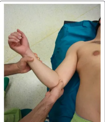
Reduction techniques Kocher method
Originally described by Kocher in 1870, Kocher’s method does not involve traction [5]. Kocher’s technique was per-formed and modified by Watson-Jones [6]. In this tech-nique, the patient was placed supine on the examining table with the physician standing at his/her side (Fig. 1).
The patient bends the affected arm at 90° at the elbow and adducts it against the body to allow the wrist and the point of the elbow to be grasped by the physician. The shoulder is slowly rotated externally between 70° and 85° until resistance is felt. The externally rotated upper arm is lifted in the sagittal plane as forward as possible, and the shoulder is internally rotated to bring the patient’s hand towards the opposite shoulder. The humeral head should now slip back into the glenoid fossa with pain eliminated during this process.
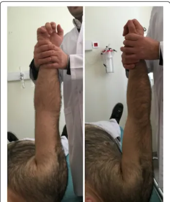
Spaso method
The patient is placed in the supine position; the affected arm is grasped around the wrist or the distal forearm and gently lifted vertically, applying gentle traction. While maintaining vertical traction, the shoulder is
slightly rotated externally. A clunk is heard and/or felt when the reduction is completed (Fig. 2).
Reduction will usually occur after a few minutes of gentle traction. If difficulty is experienced, it may be helpful to use one hand to palpate the head of the hu-merus and gently push it to assist the reduction, while maintaining traction with the other hand [7–9].
Chair method
The patient is asked to sit in a stable chair sideways using the backrest of the chair as a fulcrum in the axilla. If the backrest of the chair is not well-padded, it is sup-ported by a folded bed sheet or small, stiff pillow. Thus, the risks of an axillary nerve injury or iatrogenic fracture are minimized. The dislocated arm is allowed to hang over the backrest of the chair. The physician squats down behind the chair, holds the patient’s elbow with the left hand for a right shoulder dislocation, and in-duces the patient’s arm to gently flex at the elbow (Fig. 3).
The physician’s other hand holds the patient’s right hand without performing a maneuver. The patient is asked and encouraged to relax and be calm; traction is applied slowly by the left hand of the physician, and re-duction occurs at this stage. If the humeral head is stacked at the inferior margin of the glenoid, a slight amount of external rotation can be applied by the right hand of the physician [10, 11].
Matsen’s traction-countertraction method
The patient is placed on his/her back with a sheet around the chest and also around the assistant’s waist for countertraction. The physician stands on the side of the dislocated shoulder near the patient’s waist with the elbow of the dislocated shoulder bent to 90°. A second sheet, tied loosely around the physician’s waist and looped over the patient’s forearm, provides traction while the physician leans back against the sheet while grasping the forearm. Traction is applied to the arm with the shoulder in abduction, and the assistant applies firm countertraction to the body using a folded sheet (Fig. 4) [5].
Follow-up procedures
In patients who failed the first reduction maneuver, the reduction was achieved with one of the other reduction methods. Failed reduction maneuvers were recorded as failures, while successful second reductions were re-corded as successful. All patients were immobilized in internal rotation with a shoulder/arm sling for 3 weeks after the reduction. After 3 weeks, the patients under-went rehabilitation.
Demographic data, the number of dislocations, cause of injury, dislocation side, the dominant limb, pre- and post-reduction neurovascular examination findings, re-duction time, duration of emergency unit stay, the pres-ence of a tuberculum majus fracture, and complications during pre- and post-reduction period were recorded for all patients.
The visual analog scale (VAS) scoring system was used to assess intra- and post-reduction degree of pain, which was scored from 0 (no pain) to 10 (extremely severe). The same orthopedic surgeon who had performed the reduction asked the patients their level of pain and marked the answer on the VAS scoring system.
Fig. 2 Spaso method of reduction for shoulder dislocation
Fig. 3 Chair method of reduction for shoulder dislocation
Fig. 4 Matsen’s traction-countertraction method of reduction for shoulder dislocation
Statistical analyses
Study data were summarized using descriptive statistics (mean, standard deviation, frequency, percentage). Com-parison of categorical variables between reduction methods was performed using theχ2test or Fisher’s exact test. Ana-lysis of variance (ANOVA) was used to compare continu-ous data for the four reduction methods, followed by Tukey’s test for post hoc analyses. The level of statistical significance was set atp < 0.05. The statistical power to determine the probability of detecting 1.0 difference in the VAS score between four reduction techniques with 1.5 standard deviation and type I error of 0.05 changes be-tween 0.82 and 0.98 for a sample size of 27 to 57 per group, respectively. The power calculation for ANOVA was performed in www.statstodo.com (Statstodo Trading Pty Ltd). For statistical analyses, the MedCalc software (ver. 12.7.7; MedCalc Software bvba, Ostend, Belgium; http://www.medcalc.org; 2013) was used.
Results
In total, 153 patients (127 males, 26 females; mean age, 36.8 years; age range, 19–52 years) were included in the study. Of them, 39 patients (32 males, 7 females) were treated by the Spaso method, 47 patients (42 males, 5 females) by the Chair reduction method, 40 patients (31 males, 9 females) by Kocher’s method, and 27 patients (20 males, 7 females) by Matsen’s traction-countertraction method. Age and gender distributions of patients were similar between reduction methods (Table 1). Most pa-tients were right-handed in all groups, and dislocation was in the dominant arm in 60 % of patients without signifi-cant difference between reduction methods (Table 1). Body mass index of patients was also similar in all groups (Table 1).
No anesthetic or analgesic method was used during any reduction. Six patients who failed the initial reduc-tion technique were treated successfully by one of the
Table 1 Clinical data of shoulder dislocation patients in whom Chair, Matsen, Spaso, or Kocher reduction methods were applied
Chair method Kocher method Spaso method Matsen method p value n = 47 n = 40 n = 39 n = 27 Age (years) Female 41.4 ± 8.1 39.7 ± 11.4 42.1 ± 9.3 37.9 ± 10.4 0.98a Male 35.7 ± 12.4 32.4 ± 9.6 37.9 ± 13.3 34.6 ± 9.4 Gender Female 5 (10.6 %) 9 (22.5 %) 7 (17.9 %) 7 (25.9 %) 0.3379b Male 42 (89.4 %) 31 (77.5 %) 32 (82.1 %) 20 (74.1 %) Dominant arm Right 40 (85.1 %) 34 (85 %) 35 (89.7 %) 22 (81.4 %) 0.8165c Left 7 (14.9 %) 6 (15 %) 4 (10.3 %) 5 (18.6 %)
Dislocation in dominant arm 29 (61.7 %) 25 (62.5 %) 22 (56.4 %) 16 (59.2 %) 0.9509b
Body mass index (kg/m2) 29.3 ± 6.1 30.1 ± 5.4 28.1 ± 6.4 27.9 ± 7.1 0.825c
Number of dislocation First dislocation 28 (59.5 %) 23 (57.5 %) 24 (61.5 %) 18 (66.7 %) 0.8939b Recurrent 19 (40.5 %) 17 (42.5 %) 15 (38.5 %) 9 (33.3 %) Side Right 26 (55.3 %) 21 (52.5 %) 22 (56.4 %) 15 (55.5 %) 0.9871b Left 21 (44.7 %) 19 (47.5 %) 17 (43.6 %) 12 (44.5 %) Reason for dislocation
Sport trauma 24 (51 %) 20 (50 %) 20 (51.2 %) 15 (55.5 %) 0.9668c
Fall 21 (44.6 %) 17 (42.5 %) 18 (46.1 %) 11 (40.7 %) Traffic accidents 2 (4.4 %) 3 (7.5 %) 1 (2.7 %) 1 (3.8 %)
Tuberculus majus fracture 1 (2.1 %) 2 (5 %) 1 (2.5 %) 1 (3.7 %) 0.8848c
Pre-reduction neurologic deficit 5 (10.6 %) 3 (7.5 %) 4 (10.2 %) 3 (11.1 %) 0.9527c
Success rate 46 (97.8 %) 39 (97.5 %) 37 (94.8 %) 25 (92.5 %) 0.6509c a ANOVA test b Chi-square test c
other reduction techniques. A patient who failed the Kocher technique underwent the Chair method, another who failed the Chair underwent the Kocher method, two patients who failed the Matsen method underwent the Spaso method, and two patients who failed the Spaso underwent the Matsen method.
The number, side of dislocation, reason for dislocation, presence of tuberculum majus fracture, and pre-reduction neurological deficit ratio were similar among the four techniques (Table 1).
All four reduction techniques provided high success rates with no statistically significant difference among them (46/47, 39/40, 37/39, and 25/27, for Chair, Kocher, Spaso, and Matsen, respectively;p = 0.6509; Table 1).
Post-reduction radiographies revealed no displacement in any patients with tuberculum majus fractures, and frac-ture union was obtained with conservative treatment. Additionally, no new fracture was seen in post-reduction radiographies. Pre-reduction neurological deficits were im-proved after reduction in all patients. None of the patients had complications like proximal humeral fractures or neurological injuries.
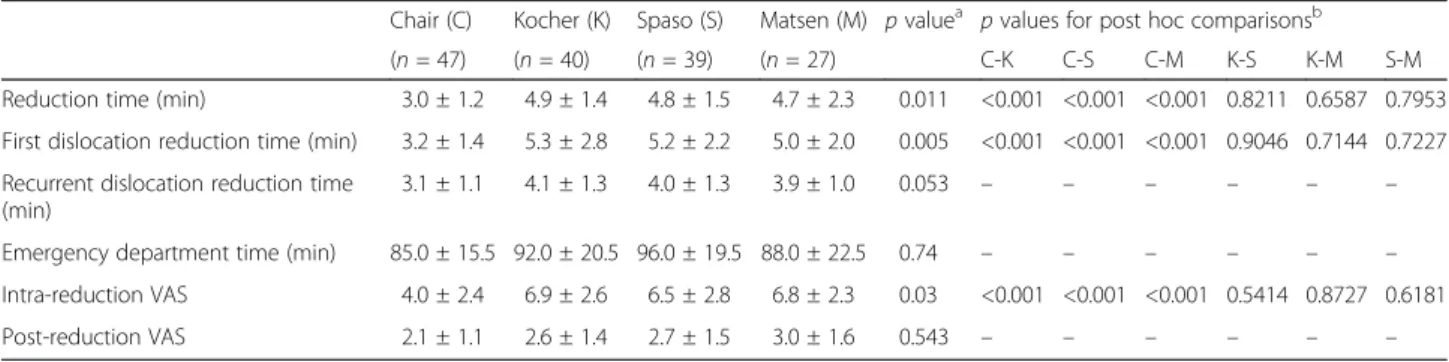
Reduction time, first dislocation reduction time, and intra-reduction VAS were significantly different among the groups, being lowest in patients who were treated with the Chair method (Table 2).
Discussion
Currently, no single shoulder reduction method has a 100 % success rate, and no technique has been found to be ideal in every shoulder dislocation situation. An ideal method should be simple, rapid, effective, painless, and free of complications and should facilitate rapid patient disposition [12, 13].
Today, many departments reduce shoulder disloca-tions either under general anesthetic or with the aid of parenteral analgesia or sedation [11, 14, 15]. However, this requires the use of further staff during the proced-ure and afterwards to observe the recovery period. Sed-ation/analgesia has usually been recommended when the
procedure has been unsuccessful [15]. Manipulation without sedatives or anesthetic allows rapid patient re-covery, reducing the time the patient spends in the de-partment and freeing medical and nursing staff for other tasks [16]. Any additional procedure such as sedation, local analgesics, or anesthesia during shoulder reduction can delay the reduction time and increase the length of stay in the emergency department. Furthermore, the benefits of reduction without sedation can save staff time in the emergency department and facilitate rapid patient disposition [17]. Tezel et al. reported that a lon-ger time was spent in the emergency department after shoulder reduction with suprascapular nerve block and sedation [18]. Other potential complications include re-sidual sedative effects, respiratory complications, cardio-vascular complications, permanent brain damage, and even death [19]. Unnecessary sedation should be avoided to reduce the chances of potential complications wher-ever possible. Chung et al. found a significantly shorter length of stay with the Oxford Chair method versus traditional methods without sedation [20]. In the present study, different reduction maneuvers were performed successfully without analgesics or anesthetics. None of our patients received sedation, anxiolytic treatment, or general anesthesia.
Many previous studies reported that muscle contrac-tion causes difficulty and pain during reduccontrac-tion [21–23]. The muscle contraction is usually caused by increase in pain during traction in patients with limited pain toler-ance, which then creates a common problem in reduc-tion [21–23]. The level of pain should be assessed and controlled for an effective shoulder reduction. The VAS is a widely used, validated scale for measuring pain and an effective instrument for surgical investigations. How-ever, studies that measure pain with VAS are limited. In a study comparing Stimson and Milch reduction tech-niques, Amar et al. recorded VAS pain score as 5.3 and 5.4, respectively [24]. Sayegh et al. reported VAS scores of 1.6, 4.9, and 5.4 with FARES, Hippocratic, and Kocher techniques [25]. In our study, VAS pain score was
Table 2 Outcome parameters for different reduction techniques against shoulder dislocation
Chair (C) Kocher (K) Spaso (S) Matsen (M) p valuea p values for post hoc comparisonsb
(n = 47) (n = 40) (n = 39) (n = 27) C-K C-S C-M K-S K-M S-M Reduction time (min) 3.0 ± 1.2 4.9 ± 1.4 4.8 ± 1.5 4.7 ± 2.3 0.011 <0.001 <0.001 <0.001 0.8211 0.6587 0.7953 First dislocation reduction time (min) 3.2 ± 1.4 5.3 ± 2.8 5.2 ± 2.2 5.0 ± 2.0 0.005 <0.001 <0.001 <0.001 0.9046 0.7144 0.7227 Recurrent dislocation reduction time
(min)
3.1 ± 1.1 4.1 ± 1.3 4.0 ± 1.3 3.9 ± 1.0 0.053 – – – – – – Emergency department time (min) 85.0 ± 15.5 92.0 ± 20.5 96.0 ± 19.5 88.0 ± 22.5 0.74 – – – – – – Intra-reduction VAS 4.0 ± 2.4 6.9 ± 2.6 6.5 ± 2.8 6.8 ± 2.3 0.03 <0.001 <0.001 <0.001 0.5414 0.8727 0.6181 Post-reduction VAS 2.1 ± 1.1 2.6 ± 1.4 2.7 ± 1.5 3.0 ± 1.6 0.543 – – – – – – a ANOVA b Tukey test
significantly lowest for the Chair procedure, followed in order by the Kocher, Matsen, and Spaso methods (4.0, 6.9, 6.8, and 6.5, respectively;p = 0.03).
Choosing the best technique for the reduction of a shoulder dislocation is often a multifactorial decision. The clinician must weigh the merits of each technique against factors such as sedation risks, availability of as-sistance, patient anxiety, and comfort level of the oper-ator. Sometimes, more than one technique will be attempted for the successful reduction of the shoulder [26]. Many methods for reduction of anterior shoulder dislocation have been reported, all of which including traction and minimal external rotation. Traction pro-vides sliding the humeral head from the anteroinferior glenoid rim; the humeral head is perched on the edge of the glenoid, and then humeral head rolls on the glenoid rim with external rotation [5]. In terms of reduction ma-neuvers, the Spaso method looks very simple. Although the Spaso method is associated with some disadvantages like having no mechanism to prevent muscle contraction and need for forward flexion of shoulder, high success rates (67.6–87.5 %) without complications have been re-ported [8, 9, 27]. In the Matsen traction-countertraction technique, a fully relaxed patient and an assistant for ap-plying strong countertraction are needed [28]. In the Kocher technique modified by Watson-Jones, in spite of need for a relaxed patient and increased risk of proximal humerus fractures, there is no need for countertraction and the success rate reaches to 68–93 % [21, 25, 29, 30]. The Chair technique has the disadvantage of need for a chair but reported a success rate over 90 % [11, 31]. The success rate in our study was 97.8, 97.5, 94.8, and 92.5 % for Chair, Matsen, Spaso, and Kocher techniques, re-spectively, without significant difference between tech-niques. However, initial reduction resulted in failure in six patients. Therefore, it should be noted that multiple reduction techniques need to be applied for anterior shoulder dislocations in emergency departments.
All the induction methods used in the present study include some traction and external rotation. All sur-geons involved in this study agreed that the Kocher and Matsen methods needed more force to reduce than the other methods. Patients may contract their muscle be-cause of pain with these two methods. The Chair method was found to be the easiest reduction method according to all physicians because the patients could not contract their muscles while sitting on a chair with the affected arm at their side. The duration of reduction varies for each technique, and in comparative studies, no superiority has been demonstrated for any technique [15, 16, 20, 32]. Sayegh et al. reported longer reduction duration in the Kocher technique (mean, 4.3 min) com-pared to the FARES technique [25]. Amar et al. recorded reduction duration as 8.82 and 4.68 min in Stimson and
Milch techniques, respectively [24]. On the other hand, in a comparative study by Chung et al., the mean reduc-tion durareduc-tion was 3 and 5 min in Chair and Kocher techniques, respectively [20]. In our opinion, each phys-ician has his/her own technique in which they have been trained, and typically uses it. We compared four differ-ent reduction techniques; the Chair method was found to be associated with significantly shorter reduction dur-ation. Compared to other methods, the Chair technique causes less pain during reduction, which allows the fast-est reduction and the shortfast-est reduction time (3.0 min for the Chair method; 4.9, 4.8, and 4.7 min for Kocher, Spaso, and Matsen, respectively, p = 0.011). The main disadvantage of the Chair method is the need for a chair. Finding an appropriate chair can be difficult in certain circumstances. Another drawback is the need for the pa-tient to be conscious and alert. This method cannot be used in non-cooperative patients and in the presence of other injuries preventing the patient from sitting com-fortably on a chair.
It is crucial to overcome the muscular resistance of the patient during these maneuvers. If a reduction man-euver is continued persistently against resistance, com-plications such as excessive pain or fracture (e.g., humoral neck fractures during reduction of anterior shoulder dislocation particularly in osteoporotic bones) and brachial plexus and axillary nerve damage may occur [4, 5, 7, 9, 10, 12–18, 21–23]. Hippocratic and Kocher techniques are commonly associated with axil-lary nerve injury, humeral shaft and neck fractures, and capsular damage [33]. Pectoralis major rupture has been reported as a rare complication of Kocher’s maneuver [34]. In our study, none of the patients had any such complications, probably due to long-term experience with the same technique of participating emergency clinics.
The main limitations of the study were its retrospect-ive design and low sample size to detect clinically im-portant but small differences between reduction methods, which both prevent reaching a definitive con-clusion. Furthermore, each of the four study groups was included from four different hospitals with different clin-ical practices; thus, the groups were heterogeneous. This study also did not evaluate any possible long-term soft tissue pathologies. Nevertheless, this study presents clin-ical comparison of multiple reduction techniques for shoulder dislocation.
In conclusion, multiple reduction techniques are avail-able for shoulder dislocation. Physicians working in the emergency departments should become familiar with many techniques for reducing anterior dislocations of the shoulder, because no single method has a 100 % suc-cess rate. On the basis of our findings, we suggest that the Chair method is an effective and fast reduction
maneuver that may be an alternative for the treatment of anterior shoulder dislocations. Further prospective studies with larger sample size are needed to compare safety of different reduction techniques.
Competing interests
The authors declare that they have no competing interests. Authors’ contributions
OG wrote the manuscript, analyzed the data, and gave final approval of the version to be published, drafting the manuscript or revising it critically for important intellectual content. SE wrote the manuscript and acquired the data. FA acquired the data. UT analyzed and interpreted the data. SC acquired and analyzed and interpreted the data. AU analyzed and interpreted the data and performed the statistical analysis. AP drafted the manuscript or revised it critically for important intellectual content. MM designed the study and checked the accuracy or integrity of all parts of the work and propriety. All authors read and approved the final manuscript.
Author details
1
Orthopedics and Traumatology Department, Medical Faculty, Medipol University, Atatürk Bulvarı. No:27 Unkapanı, 34083 Fatih, Istanbul, Turkey.
2
Orthopedics and Traumatology Department, Agrı Military Hospital, Agrı, Turkey.3Orthopedics and Traumatology Department, Malatya Military
Hospital, Malatya, Turkey.4Orthopedics and Traumatology Department, Etimesgut Military Hospital, Ankara, Turkey.5Orthopedics and Traumatology
Department, Gulhane Military Medical Academy Haydarpasa Training Hospital, Istanbul, Turkey.6Orthopedics and Traumatology Department,
Medical Faculty, Ondokuz Mayıs University, Samsun, Turkey.
Received: 4 March 2015 Accepted: 14 May 2015
References
1. Blake R, Hoffman J. Emergency department evaluation and treatment of the shoulder and humerus. Emerg Med Clin North Am. 1999;17:859–76. 2. Hill JA. Epidemiologic perspective on shoulder injuries. Clin Sports Med.
1993;2:241–7.
3. Kroner K, Lind T, Jensen J. The epidemiology of shoulder dislocations. Arch Orthop Trauma Surg. 1989;108:288–90.
4. Ideburg R. Fractures of the scapula involving the glenoid fossa. In: Bateman JE, Walsh RP, editors. Surgery of the shoulder. Philadelphia: BC Decker; 1984. p. 63–6. 5. Cunningham NJ. Techniques for reduction of anteroinferior shoulder
dislocation. Emerg Med Australas. 2005;17:463–71.
6. Watson-Jones R. Fractures and joint injuries. 6th ed. Edinburgh: Churchill Livingstone; 1982.
7. Miljesic S, Kelly AM. Reduction of anterior dislocation of the shoulder: the Spaso technique. Emerg Med. 1998;10:173–5.
8. Yuen MC, Yap PG, Chan YT, et al. An easy method to reduce anterior shoulder dislocation: the Spaso technique. Emerg Med J. 2001;18:370–2. 9. Ugras AA, Mahirogullari M, Kural C, et al. Reduction of anterior shoulder
dislocations by Spaso technique: clinical results. J Emerg Med. 2008;34:383–7.
10. Parisien VM. Shoulder dislocation: an easier method of reduction. J Maine Med Assoc. 1979;70:102.
11. Mahirogullari M, Akyildiz F, Koksal I, et al. Chair method: a simple and effective method for reduction of anterior shoulder dislocation. Acta Orthop Traumatol Turc. 2012;46:102–6.
12. Chung CH. Closed reduction techniques for acute anterior shoulder dislocation: from Egyptians to Australians. Hong Kong J Emerg Med. 2004;11:178–88.
13. Janecki CJ, Shahcheragh GH. The forward elevation maneuver for reduction of anterior dislocations of the shoulder. Clin Orthop Relat Res.
1982;164:177–80.
14. Jiang N, Hu YJ, Zhang KR, et al. Intra-articular lidocaine versus intravenous analgesia and sedation for manual closed reduction of acute anterior shoulder dislocation: an updated meta-analysis. J Clin Anesth. 2014;26:350–9. 15. Sahin N, Ozturk A, Ozkan Y, et al. A comparison of the scapular manipulation
and Kocher’s technique for acute anterior dislocation of the shoulder. Eklem Hastalik Cerrahisi. 2011;22:28–32.
16. Johnson G, Hulse W, McGowan A. The Milch technique for reduction of anterior shoulder dislocations in an accident and emergency department. Arch Emerg Med. 1992;9:40–3.
17. Descamps MJL, Gwilym S, Weldon D, et al. Prospective audit of emergency department transit times associated with entonox analgesia for reduction of the acute, traumatic dislocated shoulder. Accid Emerg Nurs. 2007;15:223–7. 18. Tezel O, Kaldirim U, Bilgic S, et al. A comparison of suprascapular nerve
block and procedural sedation analgesia in shoulder dislocation reduction. Am J Emerg Med. 2014;32:549–52.
19. Hesion PM, Joshi GP. Sedation: not quite that simple. Anesthesiol Clin. 2010;28:281–94.
20. Chung JYM, Cheng CH, Graham CA, et al. The effectiveness of a specially designed shoulder chair for closed reduction of acute shoulder dislocation in the emergency department: a randomised control. Emerg Med J. 2013;30:795–800. 21. Beattie TF, Steedman DJ, McGowan A, et al. A comparison of the Milch and
Kocher techniques for acute anterior dislocation of the shoulder. Injury. 1986;17:349–52.
22. Al Khayer A, Sedki I, Adra K.“Painless reduction of anterior shoulder dislocation by Kocher’s method” by Chitgopkar and Khan. Injury. 2006;37:672.
23. Ahmad R, Ahmed S, Bould M. latrogenic fracture of humerus—complication of a diagnostic error in a shoulder dislocation: a case report. J Med Case Rep. 2007;1.
24. Amar E, Maman E, Khashan M, et al. Milch versus Stimson technique for nonsedated reduction of anterior shoulder dislocation: a prospective randomized trial and analysis of factors affecting success. J Shoulder Elbow Surg. 2012;21:1443–9.
25. Sayegh FE, Kenanidis EI, Papavasiliou KA, et al. Reduction of acute anterior dislocations: a prospective randomized study comparing a new technique with the Hippocratic and Kocher methods. J Bone Joint Surg Am. 2009;91:2775–82.
26. Ufberg JW, Vilke GM, Chan TC, et al. Anterior shoulder dislocations: beyond traction-countertraction. J Emerg Med. 2004;27:301–6.
27. Fernández-Valencia JA, Cuñe J, Casulleres JM, et al. The Spaso technique: a prospective study of 34 dislocations. Am J Emerg Med. 2009;27:466–9. 28. Matsen FA, Thomas SC, Rockwood CA. The shoulder. In: Rockwood CA,
Matsen FA, editors. Glenohumeral instability. 2nd ed. Philadelphia, PA: WB Saunders; 1998. p. 611–54.
29. Uglow MG. Kocher’s painless reduction of anterior dislocation of the shoulder: a prospective randomised trial. Injury. 1998;29:135–7. 30. Ashton HR, Hassan Z. Best evidence topic report. Kocher’s or Milch’s
technique for reduction of anterior shoulder dislocations. Emerg Med J. 2006;23:570–1.
31. Gill EA, Noyes ME, Hubbard M. Anterior shoulder dislocation. A simple and rapid method for reduction. Am J Sports Med. 1995;23:369–71.
32. Gul M, Yavuz U, Sokucu S, et al. Flexion-adduction-external rotation method for shoulder dislocations. Acta Orthop Traumatol Turc. 2014;48:164–8. 33. Daya M. Shoulder. In: Marx JA, Hockberger RS, Walls RM, editors. Rosen’s
emergency medicine. Concepts and clinical practice. 5th ed. Mosby Inc: Missouri; 2002. p. 576–606.
34. Pimpalnerkar A, Datta A, Longhino D, et al. An unusual complication of Kocher’s manoeuvre. BMJ. 2004;329:1472–3.