IOF World Congress on Osteoporosis & 10th European Congress
on Clinical and Economic Aspects of Osteoporosis and Osteoarthritis
Poster Presentation Abstracts
statistical differences were observed, however these differences over the first 52 weeks of treatment were small and not deemed by the investigators as having major clinical importance. Both the 5 mg IR daily and the 35 mg OaW DR regimens were well toler-ated, and the overall frequency of adverse events was similar. Conclusions: Risedronate 35 mg OaW DR, whether taken before or after breakfast, provided similar efficacy and tolerability to risedronate 5 mg IR taken daily per the label.
Disclosure of Interest: M. McClung Consultant / Speaker’s bu-reau / Advisory activities with: Consultant, Advisory activities, J. Zanchetta Consultant / Speaker’s bureau / Advisory activities with: Consultant, C. Benhamou Consultant / Speaker’s bureau / Advisory activities with: Consultant, A. Balske Employee of: Company employee, J. Sarley Employee of: Company employee, R. Recker Consultant / Speaker’s bureau / Advisory activities with: Consultant, Advisory activities
P101 - GREATER CHILDHOOD VIGOROUS PHYSICAL ACTIVITY IS ASSOCIATED WITH IMPROVED STRUCTURAL PARAMETERS AND VOLUMETRIC DENSITY AT THE FEMORAL NECK
Z. A. Cole 1,*, N. Harvey 1, M. Kim 1, H. Inskip 1, K. Godfrey 1, E.
Dennison 1, U. Ekelund 2, N. Wareham 2, C. Cooper 1 and
South-ampton Women’s Survey Study Group
1MRC Epidemiology Resource Centre, Southampton University,
Southampton, 2MRC Epidemiology Unit, Cambridge University,
Cambridge, United Kingdom
Aims: To explore the relationships between objectively measured physical activity and hip geometry, strength and volumetric den-sity at 6 years old.
Methods: Children were recruited at 6 years old from the South-ampton Women’s Survey. They underwent measurement of bone mass by DXA (Hologic), including hip structure analysis (HSA), and by pQCT at the tibia (Stratec). Physical activity (PA) was as-sessed by accelerometry (Actiheart, Cambridge Neurotechnology Ltd, Cambridge, UK) for 7 continuous days.
Results: There were 215 children with PA data who underwent a DXA scan and of these 49 children also underwent pQCT assess-ment. Mean daily time spent in vigorous activity (VPA) was posi-P100 - EFFECT OF A DELAYED-RELEASE RISEDRONATE
35 MG ONCE-A-WEEK FORMULATION TAKEN WITH OR WITHOUT BREAKFAST ON BMD
M. R. McClung 1,*, J. R. Zanchetta 2, C. L. Benhamou 3, A. Balske 4, J. Sarley 5, R. R. Recker 6
1Oregon Osteoporosis Center, Portland, 4Procter & Gamble
Com-pany, 5Warner Chilcott, Mason, 6Creighton University,
Oma-ha, United States, 2IDIM, Buenos Aires, Argentina, 3INSERM,
Orleans, France
Aims: Oral bisphosphonates must be taken on an empty stomach at least 30 to 60 minutes before first food or drink. To address this restriction on food, a novel delayed release (DR) formulation of risedronate (RIS) 35 mg once-a-week (OaW) that can be taken with or without breakfast has been developed. One year efficacy and safety results of this new formulation are presented.
Methods: This phase III study was designed to test the non-infe-riority, based on the percent change in lumbar spine BMD from baseline at Endpoint (last observation carried forward at Week 52), of the RIS 35 mg OaW DR formulation taken before or after break-fast compared to the RIS 5 mg daily immediate release (IR) dose taken per label. Participants were postmenopausal women at least 50 years of age, ≥ 5 years since last menses, with a lumbar spine (LS) or total hip BMD corresponding to a 2.5 or a T-score<-2.0 and at least one prevalent vertebral fracture (T4 to L4). Patients were randomly assigned to RIS 35 mg OaW DR following breakfast (FB) (n=307), or RIS 5 mg IR daily (n=307) or RIS 35 mg OaW DR at least 30 minutes before breakfast (BB) (n=308).
Results: At 52 week endpoint, the mean percent change in lum-bar spine BMD was 3.1% (95% CI, 2.71% to 3.53%) in the 5 mg IR daily group, 3.4% (95% CI, 2.94% to 3.77%) in the 35 mg DRFB group and 3.4% (95% CI, 3.01% to 3.82%) in the 35 mg DRBB group. The mean difference (95% CI) between IR - DRBB was -0.296% (-0.873, 0.281) and between IR - DRFB was -0.233% (-0.816, 0.349). Because the upper limit of the 95% 2-sided CI of the treatment differences did not exceed the pre-defined non-infe-riority margin of 1.5% (chosen based on data from previous stud-ies), the RIS 35 mg OaW DR formulation, whether taken before or after breakfast, was shown to be non-inferior to the 5 mg IR daily. The mean percent changes in BMD in the hip regions were similar across groups. The magnitude of bone turnover marker response (NTX, CTX, and BAP) was similar across groups; some
tively associated with femoral neck and intertrochanteric section modulus (r=0.23, p=0.001; r=0.23, p=0.001 respectively), cross sectional area (CSA) (r=0.26, p=0.0002; r=0.24, p=0.0009) and cortical thickness (r=0.17, p=0.02; r=0.19, p=0.009). These rela-tionships were independent of maternal and childhood dietary, lifestyle and anthropometric factors. Similar associations for VPA with section modulus (r=0.17, p=0.02) and CSA (r=0.16, p=0.02) were observed at the femoral shaft. In the subset that underwent pQCT, VPA was positively related to cortical volumetric bone mineral density (r=0.29, p=0.05).
Conclusions: Higher levels of vigorous physical activity in child-hood are associated with increased femoral neck strength, both in terms of geometric shape and volumetric mineral density. This work supports the notion that increasing physical activity in childhood is likely to be a potential public health strategy to improve childhood skeletal development.
Disclosure of Interest: None Declared
P102 - HEALTH-RELATED QUALITY OF LIFE AFTER TOTAL KNEE OR HIP REPLACEMENT: A 7-YEAR PROSPECTIVE STUDY
O. Bruyere 1,*, M. Vanoverberghe 1, A. Neuprez 1, O. Ethgen 1, B.
Zegels 1, P. Gillet 1, J. Huskin 1, J. Reginster 1 1University of Liege, Liege, Belgium
Aims: Arthritic conditions are extremely painful for the patient and are associated with a significant reduction in health related quality of life (HRQOL), mainly in term of physical and func-tional impairment. Therefore, the demand for total joint replace-ment (TJR) is increasing as patients gain substantial pain relief and increased mobility and HRQOL, at least over a short-term period. However, few long-term studies are available. The objec-tive of the present study was to assess the long-term effect of TJR on HRQOL.
Methods: We conducted a prospective study with 7 years of fol-low-up. Patients experiencing a TJR at the level of the hip or the knee because of arthritic condition were eligible for this study. Generic HRQOL was assessed with the Short-Form 36 (SF36) and specific HRQOL with the Western Ontario and McMaster Universities Osteoarthritis Index (WOMAC). These question-naires were administered preoperatively and after 3 months, 6 months and 7 years of follow-up.
Results: Out of the 65 subjects included in this study, 45 (69.2%) completed all follow-up visits. Preoperative characteristics, in-cluding age, sex, body mass index, number of co-morbidities and reason for TJR, were not significantly different between compl-eter and non-complcompl-eter groups. Patients who completed all visits were aged (mean±SD) 64.2±12.6 years, have a body mass index of 27.6±4.1 and were predominantly women (75.6%). Out of the 45 subjects, 26 (57.8%) experienced a hip replacement surgery. Six months after surgery, there was a significant improvement, com-pared to preoperative score, in 3 of the 8 dimensions of the SF-36 (i.e. physical function, role-physical and pain). Surprisingly, there was a significant worsening in the general health dimensions of the SF-36. After 6 months of follow-up, pain and physical function dimensions of the WOMAC were significantly improved but there
was no significant change in the stiffness score. Changes in SF-36 scores from month 6 to 7 years showed a significant improvement in physical function (p<0.001), physical (p<0.001), role-emotional (p<0.01) and pain (p<0.05). From month 6 to year 7, all scores of the WOMAC improved (p<0.001 for pain, p<0.001 for stiffness and p<0.01 for physical function).
Conclusions: The improvement observed in HRQOL over a short-term period after surgery is at least maintained over a 7-year follow-up period.
Disclosure of Interest: None Declared
P103 - EPIGENETIC MODULATION OF THE ENOS PROMOTER AT BIRTH PREDICTS BONE MINERAL CONTENT AT AGE 9 YEARS
J. Rodford 1, A. Sheppard 2, 3, S. Earl 4,*, K. Lillycrop 1, J.
Slater-Jef-feries 1, M. Hanson 1, K. Godfrey 1, 4, N. Harvey 4, P. Gluckman 2, 5,
C. Cooper 4 and EpiGen Working Group
1Institute of Developmental Sciences, 4MRC Epidemiology
Re-source Centre, University of Southampton, Southampton, United Kingdom, 2Liggins Institute, University of Auckland, Auckland, 3AgResearch, Ruakura Research Centre, Hamilton, New Zealand, 5Singapore Institute for Clinical Sciences, Singapore
Aims: Endothelial nitric oxide synthase (eNOS) has been impli-cated in the regulation of osteoblast and osteoclast function (1). We investigated whether epigenetic changes within the promoter of the eNOS gene in human umbilical cords were associated with altered bone development, assessed by the DXA of the child at age 9 years.
Methods: Participants were drawn from a Southampton birth cohort, with appropriate institutional ethics committee approval and participants’ informed consent. Methyl-DNA Immunopre-cipitation followed by a commercial tiled oligomer microarray in 15 human umbilical cords was used to identify potentially in-formative genomic regions in a panel of gene promoters, includ-ing eNOS, with strong correlations between methylation status and bone mineral content at age 9-years, assessed by DXA (Lunar DPXL). We used Sequenom MassARRAY to carry out in-depth analysis of the methylation status of part of the eNOS promoter in the umbilical cords of 66 children from the same cohort. Results: Methylation at particular CpG sites varied greatly. After taking account of the child’s age and sex, there was a positive as-sociation between the methylation status of a particular CpG site within the eNOS promoter and whole body bone mineral content at 9 years of age (rp=0.36, p=0.003).
Conclusions: We have demonstrated that perinatal epigenetic variations within the eNOS promoter are associated with altered bone mineral content in childhood. These findings provide fur-ther support for a role of eNOS in bone metabolism and suggest that its contribution may at least in part have its origins in altered bone development.
EpiGen Working Group: Cameron McLean, Bright Starling Em-erald, Catharine Gale, Sarah Crozier
References: 1. Zaman G et al, J Bone Miner Res 1999;14:1123. Disclosure of Interest: None Declared
P104 - ODANACATIB TREATMENT OF POSTMENOPAU-SAL WOMEN WITH LOW BONE MINERAL DENSITY: 3-YEAR CONTINUED THERAPY AND RESOLUTION OF EFFECT
R. Rizzoli 1,*, J. A. Eisman 2, H. Bone 3, D. Hosking 4, M. McClung 5, I. Reid 6, H. Resch 7, N. Verbruggen 8, A. Santora 9, C. Hustad 9,
A. Lombardi 9
1University Hospital Cantonal, Geneva, Switzerland, 2Garvan
In-stitute of Medical Research, St Vincent’s Hospital, Sydney, Aus-tralia, 3Michigan Bone and Mineral Center, Detroit, 5Oregon
Osteoporosis Center, Portland, 9Merck Research Laboratories,
Rahway, United States, 4Nottingham City Hospital, Nottingham,
UK, Nottingham, United Kingdom, 6University of Auckland,
Auckland, New Zealand, 7St. Vincent Hospital, Vienna, Austria, 8Merck Research Laboratories, Brussels, Belgium
Aims: Odanacatib (ODN), a selective cathepsin K inhibitor, pro-gressively increased bone mineral density (BMD) and decreased bone resorption markers during 2 years of treatment in a dose-ranging study of postmenopausal women with low BMD. A 1-year extension study assessed the efficacy and safety of ODN and the effects of discontinuing therapy.
Methods: In the base study, postmenopausal women with BMD T-scores between -2.0 and -3.5 at the lumbar spine, femoral neck, trochanter or total hip (n=399) received placebo or ODN at 3, 10, 25 or 50 mg weekly. After 2 years, patients (n=189) remaining blinded were re-randomized to ODN 50 mg weekly or placebo for an additional year, and 169 completed 3 years. Endpoints included BMD at the lumbar spine (primary), total hip and hip subregions, and 1/3 radius; levels of bone turnover markers; and assessments of safety and tolerability.
Results: Continued treatment with 50 mg ODN for 3 years pro-duced significant increases from baseline and during the third year in spine (8% and 2%), total hip (6% and 2%), femoral neck (5% and 2%), and trochanter (7% and 3%) BMD and maintained BMD at the 1/3 radius. Urine NTx remained suppressed at Month 36 (-50%), but BSAP rose to slightly above baseline (18%) after the initial decrease. Treatment discontinuation resulted in bone loss at all sites, with higher rates in the initial 6 months after switching to placebo. At the end of Year 3, mean BMD among patients who took 50 mg ODN for 2 years and placebo in the third year was still above baseline at the femoral neck, was near baseline at the spine, and did not differ from placebo for total hip, trochanter, and 1/3 radius. Following ODN discontinua-tion, biochemical markers of bone remodeling increased rapidly above baseline values. This rebound in bone turnover occurred promptly after treatment discontinuation and largely resolved with time. For example, mean urine NTx increased to 50% above baseline by Month 30, but was only approximately 28% above baseline by Month 36. No differences in the overall incidence of adverse events were observed between the pooled placebo and ODN treatment groups.
Conclusions: Three years of ODN treatment resulted in progres-sive increases in lumbar spine and femur BMD and was generally well-tolerated. Bone resorption markers remained suppressed, whereas formation markers were relatively unaffected. ODN ef-fects were reversible: bone resorption increased and BMD de-creased following discontinuation of treatment.
Disclosure of Interest: R. Rizzoli Grant / Research Support from: Merck Research Laboratories, Consultant / Speaker’s bureau / Advisory activities with: Merck Research Laboratories, J. Eisman Grant / Research Support from: Merck Research Laboratories, Consultant / Speaker’s bureau / Advisory activities with: Merck Research Laboratories, H. Bone Grant / Research Support from: Merck Research Laboratories, Consultant / Speaker’s bureau / Advisory activities with: Merck Research Laboratories, D. Hosk-ing Grant / Research Support from: Merck Research Laboratories, Consultant / Speaker’s bureau / Advisory activities with: Merck Research Laboratories, M. McClung Grant / Research Support from: Merck Research Laboratories, Consultant / Speaker’s bu-reau / Advisory activities with: Merck Research Laboratories, I. Reid Grant / Research Support from: Merck Research Labora-tories, Consultant / Speaker’s bureau / Advisory activities with: Merck Research Laboratories, H. Resch Consultant / Speaker’s bureau / Advisory activities with: Merck Research Laboratories, N. Verbruggen Employee of: Merck Research Laboratories, A. Santora Employee of: Merck Research Laboratories, C. Hustad Employee of: Merck Research Laboratories, A. Lombardi Em-ployee of: Merck Research Laboratories
P105 - THE INCIDENCE OF VERTEBRAL FRACTURES IN ITALY: RESULTS FROM A 3-YEARS MULTICENTRIC STUDY
P. Piscitelli 1,*, M. L. Brandi 1, U. Tarantino 2
1University of Florence, Florence, 2University of Rome
Torver-gata, Rome, Italy
Aims: vertebral fractures or deformities are the most common osteoporotic fractures. However, two-thirds of vertebral frac-tures do not come to clinical attention because most of them are asymptomatic (although they are also with increased risk of mor-bidity and mortality). Therefore, it is very difficult to assess their incidence among general population. Our aim was to assess the incidence of vertebral fractures in Italy.
Methods: We have carried out a survey on about 28,000 fractured patients referring to Emergency departments of ten major Ital-ian hospitals in different northern, central and southern regions of the country. Physicians involved were asked to systematically record specific data concerning the fractures observed between 2004 and 2006: the skeletal site of the fracture, the gender and age of the patient, the type of trauma suffered from the patient (low energy trauma). Fractures occurred because of low energy trau-ma were considered as osteoporotic fragility fractures. Orthopae-dic surgeons involved in the study had to record if the patient was discharged from emergency department after having been treated or if the patient was hospitalized because of the fracture. The hospitalization rate was computed on the whole sample and applied to the overall hospitalization occurred in Italy because of vertebral fractures, thus enabling us to estimate the number of clinical vertebral fractures all over the country. We assumed these clinical fractures to represent 30% of the overall deformities oc-curring each year in Italy in order to provide a final estimation of the annual incidence.
Results: We have estimated about 170,000 vertebral fragility frac-tures to occur each year in Italy, with 48,000 of them requiring
medical attention (clinical vertebral fractures) and 13,000 result-ing in hospital admissions.
Conclusions: On the basis of our multicentric study, vertebral deformities currently represent the most incident type of fragility fracture in Italy.
Disclosure of Interest: None Declared
P106 - TYPE 2 DIABETES AND SELF-REPORTED HIP AND FOREARM FRACTURES IN WOMEN AND THE ROLE OF BONE MINERAL DENSITY: THE HUNT STUDY
L. Forsén 1,*, K. Midthjell 2, H. Meyer 3, S. Forsmo 2, A.
Langham-mer 2, B. Schei 2
1Division of epidemiology, 3Norwegian Institute of Public Health,
Oslo, 2Norwegian University of Science and Technology,
Trond-heim, Norway
Aims: Even if several studies have shown increased risk of hip fracture1,2, studies also have shown elevated bone mineral density
(BMD) in type 2 diabetes. The aim of the present study was to examine the relation between self-reported hip and forearm frac-ture and type 2 diabetes in women, and the role of BMD. Methods: In the Nord-Trøndelag Health Study 1995-97 (HUNT 2) 12903 women had BMD of the non-dominant forearm meas-ured with single-energy X-ray absorptiometry. Among these 11889 answered questions on ever having had hip and/or fore-arm fractures or not, had their height and weight measured and had blood samples analysed for serum C-peptide and anti-GAD. The latter analyses were performed in order to avoid misclassifi-cation of type 1 and type 2 diabetes which had been difficult in earlier studies1,2. Correct classification was important since type
2 diabetes results in hyperinsulinemia - an anabolic agent in bone while type 1 diabetes results in insulinopenia which has a nega-tive effect on bone. Logistic regression was used to study the age adjusted relation between type 2 diabetes and self-reported frac-tures (hip and forearm) and linear regression was used to study the age adjusted relation between type 2 diabetes and BMD. Due to power consideration, the combined endpoint of hip fracture and forearm fracture was used.
Results: The mean age was 58 years (SD=17). Those with type 2 diabetes had an age adjusted OR of fracture of 0.61(0.56-0.66) compared to those without type 2 diabetes, and an OR of 0.65 (0.47-0.90) after additional adjustment for BMI and BMD. The age adjusted difference in BMD between those with and without type 2 diabetes was 23 (17-30) mg/cm2, p<0.001 which decreased
to 15 (9-22) mg/cm2, p<0.001 after additional adjustment for
BMI.
Conclusions: Our results may indicate that the increased BMD in type 2 diabetics prevent fractures at the early stage of the disease. A follow-up with respect to fractures is needed to further study the role of time since diagnosis.
References: 1) Forsén et al. Diabetologia 1999;42:920; 2) de Liefde et al. Osteoporosis Int 2005;16:1713.
Disclosure of Interest: None Declared
P107 - BENEFICIAL EFFECTS OF STRONTIUM RANELATE COMPARED TO ALENDRONATE ON BONE MICRO-STRUCTURE – A 2-YEAR STUDY
R. Rizzoli 1,*, D. Felsenberg 2, M. Laroche 3, M. Krieg 4, I. Frieling 5, T. Thomas 6, R. Chapurlat 7
1University Hospital, Geneva, 4Department of Muskuloskeletal
Medicine, CHUV, Lausanne, Switzerland, 2Charite-Campus
Ben-jamin Franklin, Berlin, 5Osteoporosezentrum, Hamburg,
Ger-many, 3Rangueil Hospital, Toulouse, 6INSERM U890, University
Hospital, Saint Etienne, 7Edouard Herriot Hospital, Lyon, France Aims: In a head-to-head study, we compared the effects of stron-tium ranelate (SrRan) and alendronate (ALN), anti-osteoporotic agents with antifracture efficacy, on bone microstructure, a com-ponent of bone quality, hence of bone strength.
Methods: In a randomised, double-dummy, double-blind con-trolled trial, 88 postmenopausal osteoporotic women were ran-domised to SrRan 2g/day or ALN 70mg/week for 2 years. Micro-structure of the distal radius and distal tibia were assessed by HR-pQCT after 3,6,12,18 and 24 months of treatment. Primary endpoint was HR-pQCT variables relative changes from baseline. An ITT analysis was applied.
Results: Baseline characteristics were similar in both groups (mean ±SD): age: 63.6±7.5 vs. 63.7±7.6 yrs; L1-L4T-Score: -2.7±0.8
vs. -2.8±0.8g/cm², Cortical Thickness (CTh), trabecular bone fraction (BV/TV) and cortical density=721±242 vs. 753±263µm, 9.5±2.5 vs. 9.3±2.7%, and 750±87 vs. 745±78mg/cm3 respectively.
Over 2 yrs, distal radius values changes were within 1 to 2% with-out significant differences except cortical density. In contrast dis-tal tibia CTh, BV/TV, trabecular and cortical densities increased significantly more in the SrRan group than in the ALN group (Table). No significant between-group differences were observed for the remaining measured parameter (trabecular number, trabecular spacing, and trabecular thickness). After 2 years, L1-L4 and hip aBMD increases were similar to results from pivotal trials (L1-L4:+6.5% and +5.6%;total hip:+4.1% and +2.9%, in Sr-Ran and ALN groups, respectively). In the SrSr-Ran group, bALP increased by a median of 18% (p<0.001) and sCTX decreased by a median of –16% (p=0.005) while in the ALN group, bALP and CTX decreased by median of –31% (p<0.001) and –59% (p<0.001) respectively.
Relative changes from baseline to last observation (%)
SrRan ALN Estimated
between group difference p value CTh (µm) 6.29±9.53 0.93±6.23 5.411±1.836 0.004 BV/TV (%) 2.48±5.13 0.84±3.81 1.783±0.852 0.040 Trabecular density (mgHA/cm3) 2.47±5.07 0.88±4.00 1.729±0.859 0.048 Cortical density (mgHA/cm3) 1.43±2.77 0.36±2.14 1.137±0.530 0.045
The two treatments were well tolerated.
Conclusions: Within the constraints related to HRpQCT tech-nology, it appears that strontium ranelate has greater effects than alendronate on distal tibia cortical thickness, trabecular and cor-tical bone densities in women with postmenopausal osteoporosis
after two years of treatment. A concomitant significant increase in bone formation marker is observed in the SrRan group. Disclosure of Interest: R. Rizzoli Grant / Research Support from: Investigator of the study, D. Felsenberg Grant / Research Support from: Investigator of the study, M. Laroche Grant / Research Sup-port from: Investigator of the study, M. Krieg Grant / Research Support from: Investigator of the study, I. Frieling Grant / Re-search Support from: Investigator of the study, T. Thomas Grant / Research Support from: Investigator of the study, R. Chapurlat Grant / Research Support from: Investigator of the study P108 - INHIBITION OF SCLEROSTIN BY SYSTEMIC TREATMENT WITH A SCLEROSTIN MONOCLONAL ANTIBODY ENHANCES FRACTURE HEALING IN RODENT AND PRIMATE MODELS
M. S. Ominsky 1,*, X. Li 1, W. S. Simonet 1, C. Paszty 1, H. Z. Ke 1 1Metabolic Disorders, Amgen Inc., Thousand Oaks, United
States
Aims: Inhibition of sclerostin by a sclerostin antibody (Scl-Ab) stimulates bone formation and increases bone mineral density in animal models of osteopenia and in both men and women. Here, the effects of systemically administrated Scl-Ab on bone healing in a mouse osteotomy and a non-human primate fibular osteoto-my model were investigated.
Methods: Two groups of 9-week-old male CD1 mice underwent osteotomy on their right femurs and were injected subcutane-ously (sc) with vehicle (Veh) or a murine Scl-Ab at 25 mg/kg, 2x/ week for 4 weeks (n=9-10/group). Additionally, male cynomolgus monkeys (aged 4-5 years) underwent bilateral fibular osteotomies with intramedullary fixation, and were injected sc with Veh or Scl-Ab, 25 mg/kg, biweekly for 10 weeks (n=18-21/group). Results: In the mouse model, maximum load and stiffness were increased by 117% and 195% (p<0.05) respectively, in the frac-ture site of Scl-Ab-treated mice, compared with Veh controls. In the monkey model, Scl-Ab significantly increased serum bone formation markers, BMD at the lumbar spine, hip and distal ra-dius, and bone strength of the lumbar vertebral body. At the frac-ture site, Scl-Ab significantly increased total callus bone mineral content (BMC), hard callus area, and BMC (all p<0.05 vs. Veh). The Scl-Ab-mediated improvements in callus maturity were as-sociated with 48% greater mean torsional stiffness compared to Veh (p<0.05), while maximum torque was increased by a non-significant 32% (n=12/group). Histology analysis of fracture sites (n=10/group) showed that Scl-Ab-treated fibulae had a higher rate of complete union compared to Veh (90% vs. 45%). The cal-luses in the Scl-Ab group contained less cartilage as measured by both semi-quantitative scoring (-52%) and histomorphometry (-80%) compared to Veh. The osteotomy gaps in the Scl-Ab group had less fibrous/granulation tissue area (-94%) and were more filled with bone by both semi-quantitative scoring (+59%) and histomorphometry (+37%). Total gap area was significantly lower in the Scl-Ab group (-59%) compared with Veh.
Conclusions: Inhibition of sclerostin by Scl-Ab improved bone healing in two different animal models. In addition, Scl-Ab sig-nificantly increased bone formation and bone mass in
non-frac-tured skeletal sites. These results suggest that Scl-Ab may have therapeutic benefits in enhancing fracture healing and increasing bone formation and bone mass in non-fractured skeletal sites, thereby preventing fractures at other skeletal sites.
Disclosure of Interest: M. Ominsky Employee of: Amgen Inc., Stock ownership or royalties of: Amgen Inc., X. Li Employee of: Amgen Inc., Stock ownership or royalties of: Amgen Inc., W. Si-monet Employee of: Amgen Inc., Stock ownership or royalties of: Amgen Inc., C. Paszty Employee of: Amgen Inc., Stock ownership or royalties of: Amgen Inc., H. Ke Employee of: Amgen Inc., Stock ownership or royalties of: Amgen Inc.
P109 - ADHERENCE TO TREATMENT OF OSTEOPOROSIS AND FRACTURE RISK: THE SWEDISH ADHERENCE REGISTER ANALYSIS (SARA)
O. Ström 1, 2,*, E. Landfeldt 2, S. Robbins 3, F. Borgström 1, 2 1Medical Management Center, Karolinska Institute, 2I3 Innovus,
Stockholm, Sweden, 3Amgen GmbH, Zug, Switzerland
Aims: To estimate the association between persistence and com-pliance to osteoporosis treatment and fracture risk in a Swedish population
Methods: The sample was an open cohort from the Swedish pre-scription, inpatient care, and cause-of-death registers. It com-prised all patients >50 years of age who filled prescriptions for one or more osteoporosis treatments (alendronate, risedronate, strontium, and raloxifene) between 2005 and 2007, and including those who switched therapy. Patients with secondary osteoporo-sis or cancer were excluded. Patients were considered perosteoporo-sistent if they remained on treatment without prescriptions gaps greater than 8 weeks. Compliance was quantified as medication posses-sion ratio (MPR), only measuring prescription gaps while patients were still persistent. The association between persistence/compli-ance and fracture risk was estimated using parametric survival analysis allowing multiple fractures and controlling for available covariates.
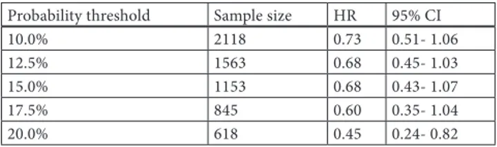
Results: The sample consisted of 37,394 patients (86% females) with a mean age of 71 years, sustaining 1,151 fractures. 95% of persistent patients had a MPR>80%. Mean MPR and follow-up were 94.6% (SD±7) and 400 days (max 760), respectively. Non-persistence was associated with increased 2-year risk of fracture compared with the risk associated with less than 1 month’s treat-ment (p<0.001). In persistent patients, however no association between MPR and fracture risk was found. The hazard ratios in each interval indicate that at least 6 months of treatment is neces-sary to achieve an anti-fracture effect (Figure 1). Patients persist-ent for 24 months had similar relative risk reductions to those found in clinical trials. Increased fracture risk was also found in patients who were institutionalized (HR2.43, p<0.001), switched treatment (HR1.31, p=0.002), had a prior fracture (HR1.63, p<0.001) or co-morbidity (HR1.57, p<0.001).
Conclusions: At least 6 months of treatment was necessary to reduce fracture incidence over 24-months. Th ere was no clear association with compliance, as assessed by MPR, and fracture risk. Lack of persistence was associated with increased cumula-tive fracture risk.
Disclosure of Interest: O. Ström Grant / Research Support from: sponsored by Amgen, E. Landfeldt Grant / Research Support from: sponsored by Amgen, S. Robbins Employee of: Amgen, F. Borgström Grant / Research Support from: sponsored by Amgen P110 - ANNUAL HIGH-DOSE ORAL VITAMIN D FOR FALLS AND FRACTURES IN ELDERLY WOMEN: A RANDOMISED, DOUBLE-BLIND, PLACEBO-CONTROL-LED TRIAL (VITAL D STUDY)
K. M. Sanders 1,*, A. L. Stuart 1, E. J. Williamson 2, J. A. Simpson 2,
M. A. Kotowicz 1, G. C. Nicholson 1
1Clinical and Biomedical Sciences; Barwon Health, University
of Melbourne, Geelong, 2Centre for Molecular, Environmental,
Genetic and Analytic Epidemiology, University of Melbourne, Parkville, Australia
Aims: We hypothesised that an annual dose of 500,000 IU chole-calciferol administered orally to older women would reduce their risk of falls and fracture.
Methods: In this double-blind, placebo-controlled trial, 2,256 com-munity-dwelling women (median age 76 years, IQR 73, 80.0 years) were randomly assigned to receive a single oral dose of cholecal-ciferol 500,000IU or placebo each autumn/winter for 3 to 5 years. Falls and fractures were ascertained using monthly calendars and details confi rmed by telephone interview. Fractures were radiologi-cally confi rmed. In a sub-study, 137 randomly-selected participants underwent serial blood sampling at baseline as well as 12-months post-dose (coinciding with immediate pre-dose for the current year). In 2006 and 2007 blood sampling was also done at one- and three-month post-dose. Serum 25-hydroxyvitamin D (25D) levels were measured in batches using DiaSorin immunoassay.
Results: Th e vitamin D group had a 15% higher rate of falling (Incidence Rate Ratio (IRR): 1.15; 95%CI 1.02, 1.30; p=0.03) and 26% higher rate of fracture (IRR: 1.26; 1.00, 1.59; p=0.05) compared to the placebo group. Th e increased rate of falling in the vitamin D group was higher in the fi rst three months follow-ing dosfollow-ing (p=0.02). Th is temporal pattern was also apparent in
fracture rates although statistical signifi cance was not reached (interaction p=0.36). Th e cumulative incidence of fi rst fall and fi rst fracture were both increased in the vitamin D group (haz-ard ratios; 95% CI: Falls 1.16; 1.05, 1.28 p=0.003; Fractures 1.26; 0.99, 1.59 p=0.057). In the sub-study, the median baseline serum 25D was 49nmol/L. Less than 3% of the sub-study participants had baseline levels <25nmol/L. In the vitamin D group, one- and 12-month post dose levels were 3-fold and 1.4-fold higher than baseline, respectively.
Conclusions: Th e results indicate that high-dose vitamin D ad-ministered orally once yearly to elderly community-dwelling women causes harm by increasing falls and fractures.
Disclosure of Interest: None Declared
P111 - COST-EFFECTIVENESS OF DENOSUMAB COMPARED WITH ORAL BISPHOSPHONATES IN THE TREATMENT OF POSTMENOPAUSAL OSTEOPOROTIC WOMEN
M. Hiligsmann 1,*, J. Y. Reginster 1 1University of Liège, Liège, Belgium
Aims: In addition to the therapeutic value of a new treatment, it becomes important to evaluate whether it represents good value for money compared with the most relevant alternative treat-ments. Health economic evaluations play an increasing role to assist health policy decision-makers. Th e present study aims to estimate the cost-eff ectiveness of denosumab compared with oral bisphosphonates (branded and generic drugs) in the treatment of postmenopausal osteoporotic women.
Methods: Th e cost-eff ectiveness of 3-year denosumab was com-pared with branded risedronate, branded and generic alendro-nate using an updated version of a previously validated Markov microsimulation model [1]. Th e model was populated with rel-evant cost, adherence and epidemiological data for Belgium from a health-care perspective and the results were presented in cost (€2009) per quality-adjusted life-year (QALY) gained. Analyses were performed in populations (over 60 years) where osteoporo-sis medications are currently reimbursed in many European coun-tries, i.e. bone mineral density T-score is below 2.5 or prevalent vertebral fracture. Patients on denosumab were assumed to have a 50% lower risk of discontinuation than those on oral bisphos-phonates and the eff ect of denosumab aft er treatment cessation was assumed to decline linearly for a maximum of 1 year. Results: Denosumab was cost-eff ective compared with all other therapies, assuming a willingness to pay of €40,000 per QALY gained. In particular, denosumab was found to be cost-eff ective compared with branded alendronate and risedronate at a thresh-old value of €30,000 for a QALY and denosumab was cost-saving (i.e. lower cost and greater eff ectiveness) compared with rise-dronate from the age of 70 years in women with densitometric osteoporosis. Th e cost-eff ectiveness of denosumab compared with generic alendronate was estimated at €38,875 €20,690 and €26,153 per QALY for women with T-score ≤-2.5 aged 60, 70 and 80 years, respectively. Th e equivalent values were €37,856 €18,764 and €17,309 per QALY for women with prevalent vertebral frac-tures.
Conclusions: This study suggests that denosumab is a cost-effec-tive strategy compared with oral bisphosphonates (including ge-neric alendronate) for the treatment of postmenopausal Belgian osteoporotic women, aged 60 years and above.
References: [1] Hiligsmann et al, Value Health 2009;12:687. Acknowledgement: This study was supported by an unrestricted educational grant from Amgen Inc.
Disclosure of Interest: M. Hiligsmann Grant / Research Support from: Amgen, J. Y. Reginster Grant / Research Support from: Amgen, Consultant / Speaker’s bureau / Advisory activities with: Amgen P112 - PREGNANE X RECEPTOR KNOCKOUT MICE DIS-PLAY OSTEOPENIA WITH REDUCED BONE FORMATION AND ENHANCED BONE ABSORPTION
K. Azuma 1,*, S. Casey 2, S. Kirchner 2, M. Ito 3, T. Urano 1, 4,
K. Horie 5, Y. Ouchi 1, B. Blumberg 2, S. Inoue 1, 4, 5
1Department of Geriatric Medicine, 4Department of Anti-aging
Medicine, University of Tokyo, Tokyo, 3Division of Radiology,
Nagasaki University Hospital, Nagasaki, 5Division of Gene
Regu-lation and Signal Transduction, Research Center for Genomic Medicine, Saitama Medical University, Saitama, Japan, 2
Depart-ment of DevelopDepart-mental and Cell Biology, University of Califor-nia, Irvine, CaliforCalifor-nia, United States
Aims: SXR (Steroid and Xenobiotic Receptor) and its murine or-tholog PXR (Pregnane X Receptor) are nuclear receptors which are expressed mainly in the liver and the intestine where they function primarily as xenobiotic sensors. Recently, we found SXR/PXR func-tions as a receptor for vitamin K2 (J Biol Chem 2003;278:43919, J Biol Chem 2006;281:16927), which is clinically used for treatment of osteoporosis in Japan and other Asian countries. In this study, we investigated the functions of SXR/PXR in the bone tissue by analyzing phenotypes of PXR knockout (PXRKO) mice.
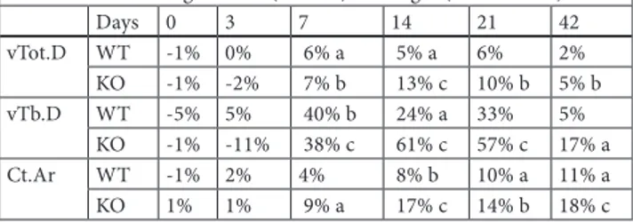
Methods: Femoral and tibial bones of 4 month-old PXRKO fe-male mice (129/Sv strain) and age-matched control wild type (WT) female mice were used for this study. Mice were injected intraperitoneally with tetracycline hydrochloride 5 days and cal-cein 2 days tissue collection. 30 hours after calcal-cein injection, mice were euthanized and both legs were dissected. The bone mineral densities (BMD) of the right femoral bones were measured by dual-energy X-ray absorptiometry. Micro-computed tomography scanning was performed on the right femoral bones. Bone histo-morphometry was performed on undecalcified sections with the Villanueva Bone Stain.
Results: BMD of PXRKO mice was significantly decreased com-pared with BMD of WT mice in micro-CT analysis of femoral trabecular bones, the 3-dimensional bone volume fractions of PKRKO mice were markedly reduced compared with those of WT mice. The histomorphometrical analysis of trabecular bones from the proximal tibia revealed a remarkable reduction of bone mass in PXRKO mice. As for bone turnover, BFR/BS was decreased in PXRKO mice, whereas ES/BS was enhanced in PXRKO mice. This indicates that bone formation is reduced and bone resorp-tion enhanced in PXRKO mice. Histomorphometrical analysis of femoral cortical bones also revealed decreased cortical area and width in PXRKO mice.
Conclusions: These results indicate that SXR/PXR is required for proper bone homeostasis. Signaling through SXR/PXR protects bone structure by promoting bone formation and suppressing bone resorption.
Disclosure of Interest: None Declared
P113 - ASSOCIATION BETWEEN 25(OH)-VITAMIND3 LEVELS AND DISEASE ACTIVITY IN PATIENTS WITH ESTABLISHED RHEUMATOID ARTHRITIS
M. Bernardes 1,*, M. J. Martins 2, G. Terroso 1, A. Bernardo 1,
L. Sampaio 1, L. Silva 1, S. Pimenta 1, C. Gonçalves 3, F.
Simões-Ventura 4
1Rheumatology, Sao Joao Hospital, 2Biochemistry, 3Laboratório
Nobre, 4Rheumatology, Oporto Medical School, Porto,
Portu-gal
Aims: 1,25(OH)2-vitaminD3 may have a role in modulating rheumatoid arthritis (RA) activity in experimental settings. Vita-min D Receptor (VDR) has been demonstrated in macrophages, chondrocytes and synoviocytes in RA synovium and sites of carti-lage erosion in RA patients, but not in controls. There may also be a relationship between VDR gene polymorphisms and RA onset and activity. Although two previous studies have failed to show a link between 25(OH)-vitaminD3 (25(OH)vit.D3) and CRP or ESR in established RA, a recent one reported an inverse associa-tion between RA activity and vitamin D in early polyarthritis.We intended to determine if vitamin D adequacy state contributes to differences in RA activity and bone turnover markers in estab-lished RA.
Methods: Clinical data and blood samples were collected in the last monitoring visit. The Portuguese version of the Standford Health Assessment Questionnaire (HAQ), Disease Activity Score (DAS28), three and four variables, 68 tender and swollen joint count were obtained. We measured ESR and CRP, 25(OH)vit.D3, serum β-C-telopeptide of collagen1 cross-links (β-CTX1), os-teocalcin (OC), Dkk-1 (ELISA, Biomedica) and osteoprotogerin (OPG)(ELISA, Biomedica). SPSS14.0 was used for statistical data analysis.
Results: We evaluated 185 RA patients, 125 (68%) women, 91 (49%) under biologics, 81 (44%) under TNFα blockers, with mean ages of 53±12 years, 14±10 years of disease duration, mean DAS28(4v) of 4,25±1,32 and a mean HAQ of 1,252±0,683. 13% of the patients had vitamin D deficiency (<15ng/ml) and 53% had inadequate (between 15 and 30 ng/ml) serum levels. In a multi-variate modelling (adjusted for age, age at diagnosis, disease du-ration, daily dose of prednisone, years of corticosteroid use and anti-resorptive therapy) 25(OH)vit.D3 deficiency or inadequacy were associated with higher DAS28(4V) (p<0,01), DAS28(3v) (p<0,01), 68 swollen joint count (p<0,01), patient global activ-ity (p<0,01), OC (p<0,05) and β-CTX1 levels (p=0,05). We did not find any association with ESR, CRP, Dkk-1, OPG, rheumatoid factor or anti-CCP antibodies levels.
Conclusions: Vitamin D deficiency and inadequacy were highly prevalent in our population. There was also evidence that vitamin D levels seem to play an immunomodulatory role in established rheumatoid arthritis as was previously reported for early
polyar-thritis, reinforcing the need to monitor and correct its serum lev-els in clinical daily practice and in an individual base.
Disclosure of Interest: None Declared
P114 - SERUM RECEPTOR ACTIVATOR OF NUCLEAR FACTOR ΚB LIGAND (RANKL) AS BIOMARKER OF CHRONIC HEART FAILURE IN ELDERLY MALES
B. Bozic 1,*, G. Loncar 2, N. Prodanovic 3, V. Cvorovic 4,
Z. Radojicic 5, S. Dimkovic 2, V. Popovic-Brkic 6
1University of Belgrade, 2Cardiology Department Clinical
Medi-cal Center Zvezdara, 3Clinic for Rheumatology and Clinical
Im-munology, Military Medical Academy, 4Clinical Hospital Center
Zemun, 5Institute for Statistics, Faculty of Organizational
Scienc-es, University of Belgrade, 6Institute of Endocrinology, University
Clinical Center of Serbia, Belgrade, Serbia
Aims: Chronic heart failure (CHF) and osteoporosis are common conditions in frail individuals. The osteoprotegerin (OPG)/ re-ceptor activator of nuclear factor κB ligand (RANKL) system is thought to play an important role in bone remodeling and cardio-vascular disease as well. This study aimed to investigate whether OPG/RANKL is associated with neuroendocrine activation in CHF.
Methods: 75 elderly males with mild to moderate CHF and 20 age-, sex- and BMI-matched healthy subjects. Serum RANKL, OPG, NT-pro-BNP, adiponectin, leptin, clinical and echocardi-ography parameters were evaluated.
Results: In comparison to the control group, the CHF patients showed significantly increased RANKL levels [126.8 (122.6) vs. 47.8 (44.4) pg/ml, p<0.0001]. In the CHF patients, the trans-formed values of RANKL levels correlated positively with the log-transformed values of the serum NT-pro-BNP and adiponectin levels (p=0.004, r=0.326 and p=0.037, r=0. 241, respectively). In multivariate regression model, RANKL was a significant deter-minant of NT-pro-BNP independent of age, BMI and creatinine clearance (p=0.002, R2=0.546).
Conclusions: In conclusion, our study suggests that in elderly males with CHF serum RANKL is associated with the established biomarker of CHF, the NT pro-BNP, as well as with the new bi-omarker of CHF, adiponectin. It would be tempting to target OPG/RANKL system with human monoclonal antibody against RANKL, denosumab, with the aim to improve cardiac perform-ance.
Disclosure of Interest: None Declared
P115 - MORPHOGENESIS OF THE BONE SYSTEM AFTER IMPLANTATION BIOLOGICAL HYDROXYAPATITE (ОC-015) IN TO THE TIBIA DEFECT
V. Luzin 1,*, A. Lubenets 1, V. Stry 1, B. Rudoy 1 1State Medical University, Luhansk, Ukraine
Aims: Researched of the bone morphogenesis, after tibia defects filling by Ukrainian material “Ceramic osteoapatite OC-015”, based on the biological hydroxyapatite.
Methods: Researched 126 white rats with initial mass of 130-135 grams, divided into 3 groups: 1st group intact animals, 2nd group
rats with 2.2 mm defect on the border between proximal meta-physic of the tibia and diaphysis. In the 3rd group, into the bone
defect was implanted blocks of the biological hydroxyapatite 2.2 mm diameters (OC-015).The observations terms from 7 to 180 days. After the end of the experiment, were separated the hu-merus, pelvic bones and also the 3rd lumbar vertebrae, and their
osteometry was taken by caliper.
Results: In the 1st group, was observed the continuous growth
of all sizes of the researched bones. During the observed time the lengths of the humerus increased from 22,89±0,23 mm to 27,24±0,26 mm, pelvis bone from 33,43±0,25 mm to 41,01±0,33 mm, height of the vertebral body was also increased from 5,10±0,05 mm to 6,56±0,10 mm. In 2nd group of rats was observed
deceleration of the bones growth speed. Length of the humerus was less than in 1st group, from 30 till 90 days by 3,21%, 2,44%
and 5,43% respectively. Height of the vertebral body after 30 days observations was also less than in 1st group-by 3,05. Largest
thick-ness of the humerus, was less than in group 1st by 5,64%, 5,42%,
8,83%, 6,87% and 6,14%, respectively. In the 3rd group growth
speed was also decelerated, however less than in 2nd group.
Lon-gitudinal growth speed was decelerated only for humerus. The length of the humerus was less than in 1st group, by the 7, 30, 60
90 days by 2,69%, 3,16%, 3,06% and 4,20% respectively. Largest thickness of the humerus, was less than in group 1st on the 7,22%,
3,31% and 6,45% respectively from 15 to 60 days.
Conclusions: The tibia defect, are accompanied with decreasing skeletal bones growth speed. Decreasing skeletal bones growth speed depended on reparative regeneration activity degree in bone defect zone. Probably we can have influence on the repara-tive regeneration process in the defect zone, filling the biological hydroxyapatite (OС-015) and systemic skeletal reactions, when we change composition in the implanted material.
Disclosure of Interest: None Declared
P116 - OXYTOCIN MEDIATES SKELETAL ACTIONS OF ESTROGENS
G. Colaianni 1,*, C. Cuscito 1, G. Greco 1, A. Di Benedetto 1, R.
Tamma 1, S. Dell’Endice 1, L. Mancini 1, I. Bab 2, L. Sun 3, M. Zaidi 3, A. Zallone 1
1Human Anatomy and Histology, University of Bari, Bari, Italy, 2Bone Laboratory, The Hebrew University of Jerusalem,
Jerusa-lem, Israel, 3The Mount Sinai Bone Program, Mount Sinai School
of Medicine, New York, United States
Aims: We have already demonstrated the presence of functional oxytocin receptors (OTR) on osteoblasts (OBs) and the role of the hormone as an enhancer of bone cell activity(1). Here we show
that the anabolic effects of estrogen on the skeleton and on OBs activities are oxytocin mediated. Our findings indicate a local Oxytocin source and indeed we found by northern blot, RT-PCR and immunofluorescence that OT mRNA as well as the peptide synthesis (demonstrated by western blot by the level of its intrac-ellular precursor neurophysin I) were induced by 17-b- estradiol after 6-12 hours treatment in human and mice OBs.
Methods: To better understand which genes are regulated by E2
or by E2-induced Oxytocin, we utilized an OTR silenced MC3T3
OBs line or primary cells obtained from OTR-KO mice in which the treatment with E2 should activate only oxytocin-independent
genes. The ex-vivo and in-vivo experiments with OTR null mice were performed as 21-days culture in mineralizing conditions and one month injections with anabolic doses of 17- b -Estradiol, respectively.
Results: Estrogen-induced Oxytocin expression requires an intact MAPK kinase signal transduction pathway. Interfering with the MAPK signaling cascade ablates the ability of estrogen to induce OT mRNA. Furthermore the treatment with BSA conjugated-E2,
unable to permeate plasma membrane, still increases OT-mRNA indicating a non-canonical signaling pathway. After estrogen treatment, the up-regulation of many osteoblast relevant genes as Osteopontin, BSP, Osteocalcin, the transcription factors ATF4, Runx2 and Osterix and the members of the Shnurri family: Shn2 and Shn3 was obtained only if intact OTRs were present. BMP2 expression at the contrary was not affected. The result on OBs differentiation ex vivo revealed a striking mineralization reduc-tion in OTR-/- calvaria osteoblasts. Exposure to 17- b-Estradiol increased mineralized bone nodules only in wild type cells, but failed to rescue the defect in OBs from OTR-/- mice. The admin-istration of 17- b -Estradiol to wild type mice increased the bone mineral density, as expected, but didn’t fully restore the BMD of OTR null mice, further confirming that many estradiol actions on bone are oxytocin-mediated.
Conclusions: This study suggest that OT could represent a novel tissue specific pharmacologic therapy for postmenopausal oste-oporosis when, in the absence of estrogen, is not directly pro-duced by bone cells.
References: (1) Tamma R et al, PNAS, 2009 Disclosure of Interest: None Declared
P117 - VITAMIN D LEVELS AND ENDOTHELIAL FUNCTION IN HEALTCARE PROFFESIONALS O. Asmaz 1,*, D. Gogas Yavuz 2
1Internal Medicine, 2Endocrinology and Metabolism, Marmara
University School of Medicine, Istanbul, Turkey
Aims: Vitamin D deficiency is a worldwide problem. Hospital staff is one of the risk groups of vitamin D deficiency. The aim of this study was to evaluate vitamin D status and its association with an early atherosclerosis marker endothelial function in Mar-mara university Hospital staff which is located in a sunny city Istanbul.
Methods: Eighty heath care professionals (doctors and nurses) of Marmara University hospital (F/M:50/30,35±4yrs) also 70 (F/M 45/35) volunteers from nonhealtcare professionals were included as controls. Vitamin D levels measured with HPLC, serum cal-cium and Phosphor were measured. Endothelial function evalu-ated with flow medievalu-ated dilatation (FMD)by ultrasonography. All parameters were studied during October 2009.
Results: Serum calcium and phosphor levels were not differ-ent between hospital staff and controls. Vitamin D levels were 59.7±25 nmol/l and 67±28 nmol/L(p> 0.05) for study and
con-trols respectively. In the hospital staff group±5 have vitamin D levels<20 nmol/L and±52.5 have vitamin D levels<40 nmol/L. Flow mediated dilation were not different between the study group (% 12.2±9.4) and controls (±11.7± 9.8).Vitamin D levels were not different between men (55±22 nmol/L) and women (62± 27 nmol/L). FMD measurements were±8.9± 7.5 and±14.4±10 for men and women respectively (P=0.07).
Conclusions: We observed that more than±50 of the hospital staff have vitamin D insufficiency at the end of the summer in a sunny geographic area. Although effects of vitamin D deficiency on endothelial function needs to be studied in larger groups Hos-pital staff needs vitamin D replacement also in summer time Disclosure of Interest: None Declared
P118 - RECOMBINANT LEPTIN THERAPY INCREASES SKELETAL MUSCLE MASS IN AGED MICE: IMPLICA-TIONS FOR THE PREVENTION OF SARCOPENIA, FALLS AND FRACTURES
M. Hamrick 1,*, P. Arounleut 1, A. Shiver 1, Q. Mi 2, C. M. Isales 3 1Department of Cellular Biology & Anatomy, 3Orthopaedic
Sur-gery, Medical College of Georgia, Augusta, 2Henry Ford Hospital,
Detroit, United States
Aims: Age-associated loss of muscle mass, or sarcopenia, con-tributes directly to frailty and an increased risk of falls and frac-tures among the elderly. Aged mice and elderly adults both show decreased muscle mass as well as relatively low levels of the fat-derived hormone leptin. Here we test the hypothesis that leptin treatment can improve muscle mass and fiber size in aged mice. Methods: C57BL/6 mice 12 and 24 months of age received daily subcutaneous injections of either vehicle (saline) or recombinant leptin (10 µg/day) for 10 days. Mice were euthanized after the 10 day treatment period and body weight and quadriceps mass record-ed. The extensor digitorum longus (EDL; predominantly type II, or fast-twitch fibers) and soleus (primarily type I, or slow-twitch fib-ers) muscles were dissected free, embedded in OCT medium, and snap frozen. Cryostat sections of the EDL and soleus were stained with H&E and muscle fiber cross-sectional areas measured. Results: Body weight data demonstrate that the aged mice weighed significantly less than the younger mice (P<.05), and that leptin treatment did not significantly alter body weight in mice of either age group. Quadriceps muscle weights were significantly lower (P<.001) in the aged mice. Leptin treatment did, however, significantly increase quadriceps muscle mass both absolutely (P<.05) and relative to body mass (P<.01) in the aged mice but not in the younger mice. Muscle fiber cross-sectional areas of the extensor digitorum longus muscle (EDL) were slightly lower in aged mice, and leptin treatment significantly (P<.05) increased EDL fiber area in the aged mice but not the young mice. Mus-cle fiber cross-sectional areas of the soleus musMus-cle were similar between young and aged mice, and leptin treatment produced a slight but non-significant increase in soleus fiber area in mice from each age group.
Conclusions: We hypothesize that a decline in musculoskeletal function with age is due in part to a decline in nutrient-activated anabolic signals, and that leptin is a key factor linking nutrient
intake with normal musculoskeletal function. Results from our study support this hypothesis by revealing that a nutrient-related peptide (leptin) can have anabolic effects in aging skeletal muscle. Recombinant leptin treatment may therefore have potential as a novel therapeutic approach for the prevention of sarcopenia, falls and fractures.
Disclosure of Interest: None Declared
P119 - EARLY CHANGES IN BONE SPECIFIC TURNOVER MARKERS DURING THE HEALING PROCESS AFTER VER-TEBRAL FRACTURE
H. Hashidate 1,*, M. Kamimura 2, H. Nakagawa 3, K. Takahara 4, S.
Uchiyama 1, H. Kato 1
1Orthopedic Surgery, Shinshu University, 2Center of Spinal
Dis-order and Osteoporosis, Kamimura Clinic, Matsumoto, 3
Ortho-pedic Surgery, Suwa Red Cross Hospital, Suwa, 4Orthopedic
Sur-gery, Ina Central Hospital, Ina, Japan
Aims: The present study measured longitudinal changes in bone turnover markers in elderly patients with vertebral fracture and investigated the relationship among bone turnover markers, du-ration of bed rest and bone mineral density (BMD) during heal-ing process.
Methods: Criteria for patient selection were aged 50 years and older, and presence of vertebral fracture. Serum BAP was meas-ured as a marker of bone formation. Urinary NTX was measmeas-ured as a marker of bone resorption. In principle, samples of venous blood and spot urine were collected six times as closely as pos-sible according to the following schedule: just after injury, within 24 h, and 1, 2, 3, 5 and 8 weeks after. We also measured duration of bed rest and bone mineral density.
Results: The study population consisted of 42 cases (mean 77.7 years). The average BMD of the lumbar vertebrae was 0.670±0.174 g/cm2. Bed rest period was 17.9±8.8 days. BAP was significantly
higher value at 2 and 3 weeks compared with the baseline val-ue. Thereafter, BAP decreased at 8 weeks. Urinary NTX was in-creased soon after onset with the same patterns in BAP. Urinary NTX reached a peak at 3 weeks then after and kept significantly high value compared with baseline within 8 weeks. The peak val-ue of bone formation marker was affected by differences in the duration of bed rest, although the peak value of bone resorption marker was not affected. And the peak value of serum BAP and urinary NTX showed a negative correlation to the BMD value at onset.
Conclusions: Bone turnover markers remained higher even at 8 weeks after vertebral fracture, and had an affect on physical activ-ity and BMD. In osteoporosis patients with high turnover, verte-bral fracture might affect the level of bone turnover markers. Disclosure of Interest: None Declared
P120 - HISTOMORPHOMETRIC EVALUATION OF TWO POTENTIAL OSTEOGENIC PROTEINS IN CRITICAL SIZED DEFECTS
J. P. M. Issa 1,*, J. B. Volpon 1, H. L. A. Defino 1, J. C. Netto 1,
M. M. Iyomasa 2
1Biomechanics and Medicine, 2Morphology, Stomatology and
Physiology, University of São Paulo, Ribeirão Preto, Brazil Aims: The aim of the present study was to evaluate the quantity and quality of the new bone formation in a critical sized defect (6mm of diameter) in the calvaria region by surgical technique, in 70 Wistar rats (250g), by histomorphometrical technique. Methods: The animals were randomly distributed in five groups with seven rats in each, and sacrificed by intracardiac perfusion according to the period of time, 2 and 4 weeks. The treatments employed are the following: group I- 5µg of the rhBMP-2 pure; group II- 5µg of the Hevea brasiliensis pure; group III- 5µg of the rhBMP-2/monoolein gel; group IV- 5µg of the Hevea
brasilien-sis/monoolein gel; group V- monoolein gel. The obtained samples
were submitted to histological processing and the histomorpho-metrical results evaluated by statistical methods.
Results: Results showed significant differences (p<0.05) in the following comparisons: 1-) group V (2 weeks) compared to the group III (4 weeks), IV (2 weeks) and I (2 and 4 weeks); 2-) group V (4 weeks) compared to the group I (2 and 4 weeks); group II (2 weeks) compared to the group I (2 and 4 weeks); 3-) group III (4 weeks) compared to the group V(2 weeks); 4-) group IV (2 weeks) compared to the group V (2 weeks); 5-) group I (4 weeks) compared to the groups V (2 and 4 weeks) and II (2 weeks); group I (2 weeks) compared to the groups V (2 and 4 weeks) and II (2 weeks).
Conclusions: It can be concluded based on this methodology and using this experimental animal model that occurred a significant difference for the group factor (p<0.001), but it was not observed significance for the time factor (p=0.139), and it was not observed interaction between these two factors (p=0.707).
Acknowledgement: The authors are grateful to Fapesp for the fi-nancial support
Disclosure of Interest: None Declared
P121 - EVALUATION OF RHBMP-2 AND NATURAL LATEX AS POTENTIAL OSTEOGENIC PROTEINS IN CRITICAL SIZE DEFECTS BY HISTOMORPHOMETRIC METHODS J. P. M. Issa 1,*, H. L. A. Defino 1, J. B. Volpon 1, S. C. H. Regalo 2,
M. M. Iyomasa 2, S. Siéssere 2, D. L. Pitol 2, F. Cerni 1
1Biomechanics and Medicine, 2Morphology, Stomatology and
Physiology, University of São Paulo, Ribeirão Preto, Brazil Aims: This in vivo study evaluated the osteogenic potential of two proteins, recombinant human bone morphogenetic protein-2 (rhBMP-2) and a protein extracted from natural latex (Hevea
bra-siliensis, P-1), and compared their effects on bone defects when
combined with a carrier or a collagen gelatin.
Methods: Eighty-four (84) Wistar rats were divided into two groups, with and without the use of collagen gelatin, and each of these were divided into six treatment groups of seven animals
each. The treatment groups were: (1) 5 µg of pure rhBMP-2; (2) 5 µg of rhBMP-2/monoolein gel; (3) pure monoolein gel; (4) 5 µg of pure P-1; (5) 5 µg of P-1/monoolein gel; (6) critical bone de-fect control. The animals were anesthetized and a 6 mm diameter critical bone defect was made in the left posterior region of the parietal bone. Animals were submitted to intracardiac perfusion after 4 weeks and the calvaria tissue was removed for histomor-phometric analysis.
Results: Group 1 (rhBMP-2) associated with collagen gelatin pre-sented higher levels of newly formed bone (P<0.05) than all other groups. Also, with collagen gelatin, Groups 1 and 2 presented sig-nificant higher levels of new bone formation (P<0.05). When the collagen gelatin was not used, Group 1 presented higher levels of newly formed bone (P<0.05).
Conclusions: In this experimental study, it was concluded that rhBMP-2 allowed greater new bone formation than P-1 protein and this process was more effective when the bone defect was covered with collagen gelatin (P <0.05).
Acknowledgement: FAPESP (Fundação de Amparo à Pesquisa do Estado de São Paulo)
Disclosure of Interest: None Declared
P122 - ANALYSIS OF BIOAVAILABLE PHOSPHORUS CONTENT IN DIFFERENTLY PROCESSED CEREALS S. T. Itkonen 1,*, V. E. Kemi 1, P. Ekholm 1, C. J. Lamberg-Allardt 1 1Department of Food and Environmental Sciences, University of
Helsinki, Helsinki, Finland
Aims: The amount of bioavailable phosphorus in different food-stuffs is unknown and is expected to vary. In Western countries the intake of phosphorus is higher than recommended which can be detrimental to bone health. To get information on bioavailable phosphorus (BP), a new in vitro method for analysis was devel-oped. The amounts of total phosphorus (TP) were also analysed and compared to the values of Finnish food database Fineli®. We
focused on the analysis of cereals.
Methods: Total and bioavailable phosphorus contents of wheat flour, rye flour, oat flakes, barley grits, barley porridge, self-made wheat sourbread, wheat bread and rye sourbread, and one bakery wheat bread and two bakery rye breads were analysed (N=5). For the BP analysis the samples were first processed by simulating the processing of chyme with alimentary enzymes and incubating at room temperature. The samples were dialyzed against water in room temperature and phosphorus analysis was made from the dialyzate by inductively coupled plasma mass spectrometry de-vice (ICP-MS). TP analysis was also made by ICP-MS.
Results: In each sample, the amount of BP was significantly lower (P<0.05) than the amount of TP. In wheat breads the amounts of BP were about 50± of TP and did not significantly differ from wheat flour (P=0.086 and P=0.154). In rye breads the BP part of TP was 69-78± and in rye flour it was 45±, and they differed sig-nificantly from each other (P<0.05). Measurement uncertainty was 6-7±.
Conclusions: BP contents in cereals differed from TP which sug-gests that the estimations based on food databases do not give an accurate estimation of the phosphorus intake. This method seems
to be useful for BP analysis. TP values of Fineli® seem to require
evaluating. BP contents in foodstuffs need further investigation and the method should be validated against an in vivo method. Disclosure of Interest: None Declared
P123 - COMBINED AND SEPARATE EFFECTS OF WRIST FLEXOR AND EXTENSOR MUSCLES ON DISTAL RADIUS BONE MINERAL DENSITY
M. A. Sariyildiz 1, A. Ozen 2, I. Karacan 1,*, O. Ergin 1
1Physical Medicine and Rehabilitation Dept, 2Nuclear Medicine
Dept, Vakif Gureba Training and Research Hospital, Istanbul, Turkey
Aims: To examine the combined and separate effects of wrist flexor and extensor muscles on distal radius bone mineral density (BMD).
Methods: Twenty-six young-adult healthy males, aged 22-42, were included in this study. Dominant hand was right in all cases. Distal radius BMD was bilaterally measured by dual energy X-ray absorptiometry. Isokinetic torque was measured in the right and left forearm with the Cybex extremity-testing system. The mus-cles tested included the right and left wrist flexors and extensors. A flexor/extensor strength ratio (flexor muscle strength/extensor muscle strength) was calculated.
Results: There were no correlations between distal radius BMDs and the wrist flexor or extensor muscles strength in both sides. A significant correlation between ultradistal radius BMD and the flexor/extensor ratio was found in the right and left forearm (re-spectively, R=0.518, p=0.007 and R=0.392, p=0.048). There was also a significant correlation between total radius BMD and the flexor/extensor strength ratio in the right and left forearm (re-spectively, R=0.449, p=0.022 and R=0.466, p=0.016).
Conclusions: The wrist flexor and extensor muscles do not contract independently of each other in activity of daily living. Present study suggest that combined effects of wrist flexor and extensor muscles may be more important for distal radius BMD. Disclosure of Interest: None Declared
P124 - EFFECTS OF BONE EXPOSED TO CYCLIC
MECHANICAL LOADING ON ELECTRICAL ACTIVITY OF MUSCLES
I. Karacan 1,*, M. A. Sariyildiz 1, C. Bahadir 2, A. Ozen 3
1Physical Medicine and Rehabilitation Dept, Vakif Gureba
Train-ing and Research Hospital, 2PMR Dept, The Ministry of Health
Erenkoy Physical Medicine and Rehabilitation Hospital, 3Nuclear
Medicine Dept, Vakif Gureba Training and Research Hospital, Istanbul, Turkey
Aims: Based on the bone myoregulation reflex, bone sensing me-chanical stimuli can send the signals to central nervous system and may neuronally regulate muscle activity1. Aim of this study
was to determine whether radius bone exposed to cyclic mechan-ical loading affects background muscle electrmechan-ical activity of m. flexor carpi radialis (FCR) in healthy adult volunteers.