The utility of first trimester uterine artery Doppler, placental volume and
PAPP-A levels alone and in combination to predict preeclampsia
Burak Yücel
a,⇑, Ali Gedikbasi
a, Oznur Dündar
b, Yusuf Olgac
c, Dogukan Yıldırım
a, Gokhan Yıldırım
a,
Ibrahim Polat
aa
Department of Obstetrics and Gynecology, Kanuni Sultan Suleyman Training and Research Hospital, Istanbul, Turkey
bDepartment of Obstetrics and Gynecology, Medipol University Medicine Faculty, Istanbul, Turkey c
Department of Obstetrics and Gynecology, Umraniye Training and Research Hospital, Istanbul, Turkey
a r t i c l e i n f o
Article history:Received 22 February 2016
Received in revised form 16 April 2016 Accepted 25 April 2016
Available online 3 May 2016
a b s t r a c t
In this study, we aimed to evaluate the detection of pre-eclampsia (PE) by integrating uterine artery Doppler, placental volume, and pregnancy-associated plasma protein A (PAPP-A) levels in the first trime-ster. We prospectively recruited 602 women that underwent 11–13 weeks’ aneuploidy screening. The mean pulsatility index (PI) of the uterine arteries and the placental volume were measured by ultra-sonography. Measurement of PAPP-A levels has been performed at the same day of ultrasonographic examinations. The 90th percentile of uterine artery PI and the 10th percentile of placental volume and PAPP-A levels were used as cut-offs. Uterine artery PI, placental volume, and PAPP-A levels had similar sensitivities in predicting PE (53.66%, 63.41%, and 70.73%, respectively). Use of the parameters in combi-nation had better sensitivity. If one parameter was positive, the sensitivity was 92.68% with 85.20% speci-ficity. If at least two parameters were positive, the sensitivity was 85.37% with 98.89% specispeci-ficity. In conclusion, the combination of increased PI of uterine artery with low placental volume and low PAPP-A levels in the first trimester achieved better results than either test alone in the prediction of PE. Ó 2016 Published by Elsevier B.V. on behalf of International Society for the Study of Hypertension in Pregnancy.
1. Introduction
Preeclampsia (PE) is a pregnancy-specific disorder that compli-cates between 2% and 8% of pregnancies[1]. Hypertensive disor-ders were the second most common cause of maternal death after hemorrhages in the developing countries. Even in countries with low maternal mortality, PE and eclampsia is still one of the leading cause of women dying during pregnancy. In addition, PE is also associated with worse fetal outcomes. It is the most com-mon cause for iatrogenic prematurity, and through the association with abruptio placentae and intrauterine fetal growth restriction is an important contributor to perinatal death[2,3].
There are increasing evidences of a relationship between impaired placentation and the subsequent development of PE. The underlying pathology is present at first trimester, however, the symptoms of the disorder are generally present in the late second to third trimester[4].
Early risk stratification may allow for more appropriate alloca-tion of resources, thus; this may allow the diversion of follow-up to patients most likely to benefit from them. Multiple tests have been proposed as screening tests for PE, including analysis of maternal serum biochemical markers and feto-maternal sono-graphic parameters. Doppler study of the uterine arteries at 22–24 gestational weeks proved to be a reliable method to predict PE in the second trimester[5]. Doppler studies to predict PE have been also performed during the first trimester, however, such stud-ies have reported a poorer performance when compared to second trimester recordings[6]. Metzenbauer et al.[7]firstly, concluded that small placental volumes in first trimester were more common in cases of adverse pregnancy outcome. Furthermore, previous studies have suggested that low maternal serum levels of pregnancy-associated plasma protein A (PAPP-A) levels at 11–14 weeks of pregnancies may be an early sign of impaired placental development leading as pregnancy complications such as PE[8–10].
No single test has been demonstrated a sufficient predictive value for PE in the clinical use. These tests appear to be most useful in combination with other parameters instead. PE has a nature of
http://dx.doi.org/10.1016/j.preghy.2016.04.007
2210-7789/Ó 2016 Published by Elsevier B.V. on behalf of International Society for the Study of Hypertension in Pregnancy.
⇑ Corresponding author at: Kanuni Sultan Suleyman Egitim ve Arastirma Hastanesi Atakent Mh, Turgut Ozal Cd, No. 1, 34303 Altınsehir, Kücükcekmece, Istanbul.
E-mail address:[email protected](B. Yücel).
Contents lists available atScienceDirect
Pregnancy Hypertension: An International Journal of
Women’s Cardiovascular Health
PE, thus; a combination of two or more independent biomarkers, each reflecting a different pathologic process, should potentially increase the possibility of suitable predictive algorithms. Previ-ously, uterine artery Doppler, placental volume and PAPP-A levels, separately, studied in predicting PE in the first trimester. To best of our knowledge a combination of these potential markers has been not studied, yet.
The aim of this study was to determine the combination of uterine artery Doppler, placental volume and PAPP-A levels in the first trimester is able to predict PE.
2. Materials and methods
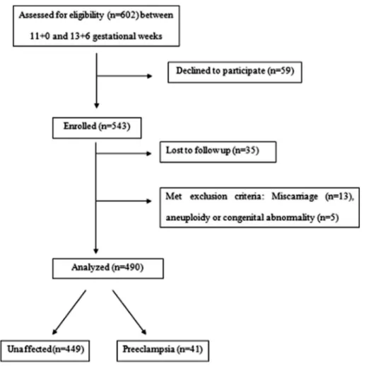
This is a prospective cohort study of pregnant women followed by the Gynecologic and Prenatal Diagnosis Unit of a third level reference hospital with around eighteen thousand deliveries each year. Approval for the study was obtained from the institutional review board. Women with singleton pregnancies between 11 and 14 weeks’ gestation attending for first-trimester aneuploidy screening of the routine antenatal care were invited to participate in the study. A diagram outlining the selection and follow-up of patients is shown inFig. 1.
A written informed consent was collected and enrolled women were interviewed during the first visit. Maternal history was recorded (women were asked to provide information on age, weight and height, previous pregnancies, history of chronic hyper-tension, diabetes and previous pregnancy with PE, cigarette smok-ing dursmok-ing pregnancy). The last menstrual period was used to calculate gestational age and it was confirmed by crown-rump
length measurement. Doppler ultrasound scan of uterine arteries, volumetry of placenta were performed and blood samplings were collected. A single experienced observer, who had obtained The Fetal Medicine Foundation Certificate of Competence in Placental and Fetal Doppler, performed all two-dimensional (2D), three-dimensional (3D) ultrasound and Doppler scans using a Voluson 730 Expert (GE Healthcare, Milwaukee, WI, USA) with a transab-dominal 2–5 MHz transducer. The uterine arteries were identified by Color flow mapping at the level of the isthmus and pulsed wave Doppler was used to obtain the waveforms. The pulsatility index (PI) was calculated after manual tracing of the waveforms. The average PI of the left and the right uterine arteries were recorded. The placenta was examined. The borders of placenta were drawn at six levels manually by using roller ball and the size of the volume box was adapted in such a way that the placenta fitted into it completely. Placental volume was then automatically calculated by the equipment’s software like described in a previous study[11]. Each patient provided a blood sample for first trimester screening. The blood samples were allowed to clot and centrifuged at 1500g for 15 min. The serum was then removed and aliquots were stored at 80°C until the use. Concentrations of PAPP-A were then transformed as multiples of median (MoM) and adjusting for gestational age, ethnicity, maternal weight, insulin-dependent diabetes, history of anticonvulsant use and history of a previous fetal neural tube defect by the commercial software used for first trimester screening at our institution. Samples were analyzed by an examiner blinded to the clinical outcomes.
The exclusion criteria were late miscarriages (miscarriages between 14 and 24 weeks of pregnancy), major fetal abnormalities
(such as aneuploidy and multiple congenital abnormality syndromes).
2.1. Diagnosis of preeclampsia
Participants were recruited consecutively and followed from first visit to delivery. We considered PE as a main outcome and used the definition of the American College of Obstetricians and Gynecologists for the diagnosis [12]: Hypertension (>140/90 mmHg) measured on two separate occasions, >6 h apart developing after 20 weeks of gestation in a pregnancy with previously normal blood pressure and co-existing significant proteinuria (>0.3 g in a 24-h urine specimen) is defined as PE. 2.2. Statistical analysis
Demographic characteristics, ultrasound findings and the results of biochemical testing were entered into a computerized database. Patients’ individual medical records at delivery were reviewed to obtained the data on pregnancy outcomes. The 10th and 90th percentiles of uterine artery mean PI, placental volume and PAPP-A levels were calculated. These cut of were used for prediction of PE. Specify, sensitivity, negative predictive value (NPV) and positive predictive value (PPV) results for each measure-ment were examined to the diagnostic test performances. Statistical analyses were performed using SPSS (Statistical Package for the Social Sciences) software (SPSS 19.0 for Windows; SPSS Inc. Chicago, IL). Kolmogorov–Smirnov tests of normality were used to evaluate the distributions. Non-normally distributed metric variables were analyzed by Mann–Whitney U-test. Qualitative data
were compared using Fisher’s exact test. Statistical significance was considered as p < 0.05.
3. Results
A total of 602 pregnant were enrolled consecutively in our study. Fifty-nine women declined to participate. We excluded 18 women; include all cases of late miscarriages, aneuploidy and mul-tiple congenital abnormality syndromes. Thirty-five women were lost at follow-up. In the remaining 490 pregnancies, 41 women developed PE (8.37%).
Maternal characteristics are summarized inTable 1. There was no difference in age, body mass index, number of parity, and num-ber of smokers between groups. Nulliparous women and pregnant with a history of PE in previous pregnancies were higher in PE group. Median of MoM values of PAPP-A levels and placental volume measurements were significantly lower and median of uterine artery PI values were significantly higher in PE group compared to control group. Delivery week and fetal weight were also significantly lower in PE group.
PE screening characteristics of uterine artery mean PI >90th centile, placental volume <10th centile and MoM of PAPP-A levels <10th centile used alone or in combination are showed inTable 2. An abnormal PI in uterine arteries or a PAPP-A levels had similar sensitivities and specificities in predicting PE but the sensitivities and specificities for placental volume below the 10th centile was lower for predicting PE. The higher sensitivity values were reached when uterine arteries PI, placental volume and PAPP-A levels were used in combination considering the result of the test positive such as at least one or two of the parameters was abnormal.
Table 1
Demographic characteristics of the preeclampsia and control (not-affected) groups.
Preeclampsia group Control group (Non-affected) P value Number of participants 41 (8.37%) 449 (91.63%)
Age (years) 28 (18–42) 28 (18–45) 0.819
Body mass index (kg/m2
) 23.24 (17.78–38.2) 23.83 (17.07–42.15) 0.989 Number of parity 1 (0–5) 1 (1–7) 0.472 Nulliparous 15 (36.59%) 97 (21.6%) 0.034 Smokers 9 (21.95%) 62 (13.81%) 0.16 History of preeclampsia 5 (12.2%) 13 (2.9%) 0.012 PAPP-A (MoM) 0.26 (0.06–1.47) 0.75 (0.22–3.42) <000.1 Placental volume (ml) 34 (16.40–74.63) 62 (15–131) <000.1 Uterine artery PI 2.74 (0.8–5.12) 1.24 (0.02–6.39) <000.1 Delivery week 36 (28–38) 38 (28–41) <000.1 Fetal weight (g) 2400 (740–3700) 3110 (1080–4720) <000.1
Continuous variables were given as medians (ranges: minimum and maximum) and categorical variables were given as number (percentages). Bold represents the significant P-values.
Table 2
Preeclampsia screening characteristics of uterine artery mean PI >90th centile, placental volume <10th centile and MoM of PAPP-A measurement <10th centile used alone or in combination.
Sensitivity Specificity PPV NPV
Uterine PI >90th centile 70.73% 95.32% 58.00% 97.27 %
(54.46–83.87%) (92.94–97.08%) (43.21–71.81%) (95.28–98.58%) Placental volume <10th centile 53.66% 93.99% 44.90% 95.69%
(37.42–69.34%) (91.37–96.00%) (30.67–59.77%) (93.35–97.39%) PAPP-A measurement <10th centile 63.41% 94.88% 53.06% 96.60 %
(46.94–77.88%) (92.41–96.73%) (38.27–67.47%) (94.45–98.08%) At least one parameter is abnormal 92.68% 85.20% 36.54% 99.22 %
(80.08–98.46%) (81.56–88.37%) (27.31–46.55%) (97.73–99.84%) At least two parameters are abnormal 85.37% 98.89 % 87.50% 98.67%
(70.83–94.43%) (97.42–99.64%) (73.20–95.81%) (97.12–99.51%) All values are given as % (95% confidence interval), PPV: positive predictive value, NPV: negative predictive value.
4. Discussion
This study analyzes the role of uterine artery Doppler, placental volume, measurement of PAPP-A levels in maternal serum and their combined use in the first trimester for the prediction of PE in an unselected population. These parameters were separately studied in previous studies [11,13,14]; however, to the best of our knowledge, our study is the first that investigated the effective-ness of combination of these three parameters in predicting PE.
In previous studies[15,16], it has been showed that the higher uterine artery PI, the lower placental volume and the lower PAPP-A levels in the first trimester were correlated with PE. In light of these studies, we determined 10th centile lower cut-off for placen-tal volume and PAPP-A results and 90th centile higher cut-off val-ues for uterine artery PI. This method was used in several studies
[11,17,18]. Our study revealed that these parameters had low sen-sitivity and specificity values when they were used separately in prediction of PE. On the other hand, when we used the combina-tion of parameters, such as at least one or two of three parameters were positive, tests reached adequate sensitivity and specificity values. We found a prevalence of PE of 8.37% a value significantly higher than those reported in other studies[6,19]. Possible expla-nations for this finding may be the demographic characteristics of the included patients who are attended to a tertiary hospital or limitations in the sample size. Higher ratio of nulliparous women and women with history of PE within study group might also cause higher prevalence.
Although there are still many unanswered questions, the patho-physiology of pre-eclampsia involves combination of maternal, fetal and placental factors. The most commonly suggested hypoth-esis strongly relies on impaired invasion of fetal trophoblast cells into the maternal decidua. Decreased invasion leads to an impeded transformation of the spiral arteries into the low resistance vessels, causes to reduced placental blood flow, leads to hypoxic damage of the endothelial vessels, at last, initiates a range of disorders culmi-nating in placental infarction. The physiological response of to this phenomenon is increased maternal blood pressure and fetal growth retardation to continue sufficient maternal–fetal circula-tion. Consequently, placental developmental and functional impairment during the first trimester appears to affect the development of hypertension in second or third trimester of pregnancy[20].
At present, Doppler examination might give us relatively early information about impaired trophoblast invasion of the uterine arteries. High impedance of the uterine arterial blood flow reveals an insufficient physiological conversion of the spiral arteries. The best time in pregnancy to perform uterine artery Doppler scans to predict PE is between 22 and 24 weeks, although this is rela-tively late for the start of an efficient prophylaxis and treatment
[21]. Since an earlier identification of pregnancies at risk should be beneficial to promptly start closer follow-up and prophylactic treatment. In recent studies, Doppler examinations have been also performed during the first trimester. Such studies [11,17] have reported a poorer performance when compared to late second tri-mester recordings with an overall sensitivity of 25% to 79%. In our study, we found 70.73% sensitivity when we determined cut-off in 90th centile.
With the introduction of three-dimensional ultrasound to ante-natal care, there has been interest in measurement of placental volume as a screening test for PE. Hafner et al. [18] reported a screening study based on placental quotient (a ratio of placental volume to crown-pump length) measurements at around 12 weeks, at a false positive rate of 11%, the sensitivity in predict-ing PE was 20% for placental quotient below the 10th percentile. Our study revealed that placental volume measurement had a 55.66% sensitivity for values below <10th percentile.
PAPP-A may affect placental function through its action on insulin-like growth factor (IGF) and IGF binding protein 4. IGFs are thought to play a key role in regulating fetal growth and tro-phoblast invasion of the decidua. The IGF binding protein 4 inhibit the action of IGFs. PAPP-A is a protease for IGF binding protein 4
[22]. This mechanism is considered as the most plausible explana-tion for the reduced PAPP-A levels in predicting PE[17]. In the literature, sensitivity of first trimester PAPP-A levels in predicting PE was reported between 23% and 51.7% [14,23,24], we found 63.41% sensitivity with 53.06% PPV for PAPP-A levels in predicting PE. This relatively high detection rate is correlated with relatively high incidence of PE in our study population.
As it is now generally accepted that finding a single effective test with sufficient accuracy to be clinically useful is highly unli-kely interest in combining several tests into multi-parametric models has been growing in recent years[4,25,26]. We observed that high sensitivity and specificity values were reached when we used uterine artery PI, placental volume and PAPP-A levels in combination compared to when they were used alone.
In conclusion, this study revealed that pregnancies complicated by PE are associated with increased uterine artery PI and decreased placental volume and PAPP-A levels in first trimester. Certainly, use of parameters in the combination improves prediction over the use of parameters alone. Further studies are required in larger populations to establish the real potential of first trimester PAPP-A levels, placental volume and uterine artery PI in predicting PE.
References
[1]Geographic variation in the incidence of hypertension in pregnancy. World Health Organization International Collaborative Study of Hypertensive Disorders of Pregnancy, Am. J. Obstet. Gynecol. 158 (1988) 80–83.
[2]K.S. Khan, D. Wojdyla, L. Say, et al., WHO analysis of causes of maternal death: a systematic review, Lancet 367 (2006) 1066–1074.
[3]C. Briceno-Perez, L. Briceno-Sanabria, Gracia P. Vigil-De, Prediction and prevention of preeclampsia, Hypertens. Pregnancy 28 (2009) 138–155. [4]A.T. Papageorghiou, S. Campbell, First trimester screening for preeclampsia,
Curr. Opin. Obstet. Gynecol. 18 (2006) 594–600.
[5]K. Harrington, D. Cooper, C. Lees, et al., Doppler ultrasound of the uterine arteries: the importance of bilateral notching in the prediction of pre-eclampsia, placental abruption or delivery of a small-for-gestational-age baby, Ultrasound Obstet. Gynecol. 7 (1996) 182–188.
[6]G. Rizzo, A. Capponi, O. Cavicchioni, et al., First trimester uterine Doppler and three-dimensional ultrasound placental volume calculation in predicting pre-eclampsia, Eur. J. Obstet. Gynecol. Reprod. Biol. 138 (2008) 147–151. [7]M. Metzenbauer, E. Hafner, D. Hoefinger, et al., Three-dimensional ultrasound
measurement of the placental volume in early pregnancy: method and correlation with biochemical placenta parameters, Placenta 22 (2001) 602– 605.
[8]C.Y. Ong, A.W. Liao, K. Spencer, et al., First trimester maternal serum free beta human chorionic gonadotrophin and pregnancy associated plasma protein A as predictors of pregnancy complications, BJOG 107 (2000) 1265–1270. [9]L. Dugoff, J.C. Hobbins, F.D. Malone, et al., First-trimester maternal serum
PAPP-A and free-beta subunit human chorionic gonadotropin concentrations and nuchal translucency are associated with obstetric complications: a population-based screening study (the FASTER trial), Am. J. Obstet. Gynecol. 191 (2004) 1446–1451.
[10]P. Meloni, I. D’Angeli, J. Piazze, et al., First trimester PAPP-A levels associated with early prediction of pregnancy induced hypertension, Hypertens. Pregnancy 28 (2009) 361–368.
[11]K. Schuchter, M. Metzenbauer, E. Hafner, K. Philipp, Uterine artery Doppler and placental volume in the first trimester in the prediction of pregnancy complications, Ultrasound Obstet. Gynecol. 18 (2001) 590–592.
[12] Bulletins-Obstetrics ACoP, ACOG practice bulletin. Diagnosis and management of preeclampsia and eclampsia. Number 33, January 2002, Obstet. Gynecol. 99 (2002) 159–167.
[13]G. Di Lorenzo, M. Ceccarello, V. Cecotti, et al., First trimester maternal serum PIGF, free beta-hCG, PAPP-A, PP-13, uterine artery Doppler and maternal history for the prediction of preeclampsia, Placenta 33 (2012) 495–501. [14]K. Spencer, N.J. Cowans, I. Chefetz, et al., First-trimester maternal serum PP-13,
PAPP-A and second-trimester uterine artery Doppler pulsatility index as markers of pre-eclampsia, Ultrasound Obstet. Gynecol. 29 (2007) 128–134. [15]A. Youssef, F. Righetti, D. Morano, et al., Uterine artery Doppler and
biochemical markers (PAPP-A, PIGF, sFlt-1, P-selectin, NGAL) at 11 + 0 to 13 + 6 weeks in the prediction of late (>34 weeks) pre-eclampsia, Prenat. Diagn. 31 (2011) 1141–1146.
[16]A.O. Odibo, K.R. Goetzinger, K.M. Huster, et al., Placental volume and vascular flow assessed by 3D power Doppler and adverse pregnancy outcomes, Placenta 32 (2011) 230–234.
[17]K. Spencer, C.K. Yu, N.J. Cowans, et al., Prediction of pregnancy complications by first-trimester maternal serum PAPP-A and free beta-hCG and with second-trimester uterine artery Doppler, Prenat. Diagn. 25 (2005) 949–953. [18]E. Hafner, M. Metzenbauer, D. Hofinger, et al., Comparison between
three-dimensional placental volume at 12 weeks and uterine artery impedance/ notching at 22 weeks in screening for pregnancy-induced hypertension, pre-eclampsia and fetal growth restriction in a low-risk population, Ultrasound Obstet. Gynecol. 27 (2006) 652–657.
[19]A.M. Martin, R. Bindra, P. Curcio, et al., Screening for pre-eclampsia and fetal growth restriction by uterine artery Doppler at 11–14 weeks of gestation, Ultrasound Obstet. Gynecol. 18 (2001) 583–586.
[20]G. Urban, P. Vergani, A. Ghidini, et al., State of the art: non-invasive ultrasound assessment of the uteroplacental circulation, Semin. Perinatol. 31 (2007) 232– 239.
[21]A.T. Papageorghiou, C.K. Yu, R. Bindra, et al., Multicenter screening for pre-eclampsia and fetal growth restriction by transvaginal uterine artery Doppler at 23 weeks of gestation, Ultrasound Obstet. Gynecol. 18 (2001) 441–449.
[22]J.B. Lawrence, C. Oxvig, M.T. Overgaard, et al., The insulin-like growth factor (IGF)-dependent IGF binding protein-4 protease secreted by human fibroblasts is pregnancy-associated plasma protein-A, Proc. Natl. Acad. Sci. U.S.A. 96 (1999) 3149–3153.
[23]L.C. Kenny, M.A. Black, L. Poston, et al., Early pregnancy prediction of preeclampsia in nulliparous women, combining clinical risk and biomarkers: the Screening for Pregnancy Endpoints (SCOPE) international cohort study, Hypertension 64 (2014) 644–652.
[24]P. Wu, C. van den Berg, Z. Alfirevic, et al., Early pregnancy biomarkers in pre-eclampsia: a systematic review and meta-analysis, Int. J. Mol. Sci. 16 (2015) 23035–23056.
[25]E.A. Steegers, P. von Dadelszen, J.J. Duvekot, R. Pijnenborg, Pre-eclampsia, Lancet 376 (2010) 631–644.
[26]S. Sep, L. Smits, M. Prins, L. Peeters, Prediction tests for recurrent hypertensive disease in pregnancy, a systematic review, Hypertens. Pregnancy 29 (2010) 206–230.