DESIGN AND SYNTHESIS OF SELF-ASSEMBLING PEPTIDES FOR
FABRICATION OF FUNCTIONAL NANOMATERIALS
A DISSERTATION SUBMITTED TO
THE GRADUATE SCHOOL OF ENGINEERING AND SCIENCE OF BILKENT UNIVERSITY
IN PARTIAL FULFILLMENT OF THE REQUIREMENTS FOR THE DEGREE OF
DOCTOR OF PHILOSOPHY IN
MATERIALS SCIENCE AND NANOTECHNOLOGY
By
Mohammad Aref Khalily December 2016
ii
DESIGN AND SYNTHESIS OF SELF-ASSEMBLING PEPTIDES FOR FABRICATION OF FUNCTIONAL NANOMATERIALS
By Mohammad Aref Khalily December 2016
We certify that we have read this dissertation and that in our opinion it is fully adequate, in scope and in quality, as a thesis for the degree of Doctor of Philosophy.
Engin Umut Akkaya (Advisor)
Mustafa Özgür Güler (Co-Advisor)
Emrah Özensoy
Hasan Tarık Baytekin
Özdemir Doğan
Canan Ünaleroğlu
Approved for the Graduate School of Engineering and Science:
Ezhan Karaşan
iii ABSTRACT
DESIGN AND SYNTHESIS OF SELF-ASSEMBLING PEPTIDES FOR FABRICATION OF FUNCTIONAL
NANOMATERIALS
Mohammad Aref Khalily
PhD in Materials Science and Nanotechnology Supervisor: Engin Umut Akkaya
December 2016
Self-assembling peptides are a class of supramolecular polymers, which exploit noncovalent interactions such as hydrogen bonding, hydrophobic, electrostatic, charge-transfer complex, -, and van der Waals interactions to generate well-defined supramolecular nanostructures including nanospheres, nanosheets, nanotubes, and nanofibers. These versatile peptide-based supramolecular nanomaterials have been utilized in variety of applications including catalysis, sensing, light harvesting, optoelectronic, bioelectronic and tissue engineering.
In this thesis, use of supramolecular peptide nanofibers formed by specially designed short peptide sequences that can form sheet-like hydrogen bonded structures for controlled synthesis of nanometer scale functional materials were explored. Specifically, n-type and p-type β-sheet
forming short peptide sequences were synthesized, which assemble separately into well-ordered nanofibers in aqueous media. These p-type and n-type nanofibers coassemble via hydrogen bonding and electrostatic interactions to generate highly uniform supramolecular n/p-coassembled 1D nanowires. This smart molecular design ensures alternating arrangement of D and A chromophores within coassembled supramolecular nanowires. Supramolecular
n/p-iv
coassembled nanowires were found to be formed by alternating A-D-A unit cells having an association constant of (KA) of 5 x 10⁵ M-1.
Moreover, I designed and synthesized β-sheet forming peptide nanofibers to fabricate different metal and metal oxide nanostructures in highly controlled manner using wet chemistry and atomic layer deposition techniques. These hybrid organic-inorganic nanostructures were employed in model Suzuki coupling, alkyne-azide cycloaddition and hydrolysis of ammonia borane reactions.
Keywords: Supramolecular chemistry, self-assembling peptides, nanostructured materials, optoelectronics, atomic layer deposition, catalysis
v ÖZET
FONKSİYONEL NANOMALZEMELERİN ÜRETİMİ İÇİN KENDİLİĞİNDEN DÜZENLENEN PEPTİTLERİN
TASARIM VE SENTEZİ
Mohammad Aref Khalily
Malzeme Bilimi ve Nanoteknoloji, Doktora Tez Danışmanı: Engin Umut Akkaya
Aralık 2016
Kendiliğinden düzenlenen peptitler, nanoküreler, nanotabakalar, nanotüpler ve nanofiberleri içeren iyi tanımlanmış supramoleküler nanoyapıları oluşturmak için, hidrojen bağı, hidrofobik, elektrostatik, yük-transfer kompleksi, -, ve Van der Waals etkileşimleri gibi non-kovalent etkileşimleri kullanan supramoleküler polimerlerin bir sınıfıdır. Bu çok yönlü peptit temelli supramoleküler nanomalzemeler, kataliz, algılama, ışık-hasatı, optoelektronik, biyoelektronik ve doku mühendisliğini içeren çeşitli uygulamalarda kullanılmaktadır.
Bu tezde, nanometre boyutunda fonksiyonel malzemelerin kontrollü sentezi için, özel olarak tasarlanmış, tabakaya benzer hidrojen bağlı yapılar oluşturabilen kısa peptit dizilimlerinin meydana getirdiği supramoleküler peptit nanofiberlerin kullanımını araştırılmıştır. Özellikle, sulu ortamda düzenli nanofiberleri ayrı ayrı biraya getiren, n tipi ve p tipi β-yaprağı oluşturan kısa peptit sekansleri sentezlendi. Bu n tipi ve p tipi nanofiberler, hidrojen bağı ve elektrostatik etkileşimler aracılığıyla, oldukça düzenli supramoleküler n/p-düzenli, 1D nanoteller oluşturmak
vi
için bir araya gelmektedirler. Bu akıllı moleküler tasarım, n/p-düzenli supramoleküler nanoteller içinde D ve A kromoforlarının alternatif şekilde düzenlenmesini sağlar. Supramoleküler n/p-düzenli nanotellerin, birleşme sabiti (KA) of 5 x 10⁵ M-1 olan, değişimli A-D-A birim
hücrelerinden oluştuğu bulunmuştur.
Ayrıca, farklı metal ve metal oksit nanoyapıları, sıvı kimyası ve atomik katman kaplama teknikleri kullanarak, oldukça kontrollü biçimde üretmek için, β-sheet yapan peptit nanofiberleri tasarlandı ve sentezlendi. Bu hibrit organik-inorganik nanoyapılar, Suzuki kenetlenme, azid-alkin siklo katılma ve amonyum boran’ın hidroliz reaksiyonlarında kullanılmıştır.
.
Anahtar kelimeler: Supramolekular kimya, kendiliğinden düzenlenen peptitler nanomalzemeler optoelektronik, atomik katman kaplama, kataliz
vii
Acknowledgements
I would like to express my deep appreciation to my advisors, Assoc. Prof .Dr. Mustafa O. Güler and Prof. Dr. Engin U. Akkaya for their valuable guidance and endless support during my PhD research. I was very lucky to be a part of their productive research groups. I have acquired precious experiences which will help me a lot in my new life as an independent researcher.
I would also express my appreciation to Assoc. Prof. Dr. Ayse B. Tekinay, Assoc. Prof. Dr. Aykutlu Dana and Assoc. Prof. Dr. Emrah Özensoy for their guidance, support and valuable scientific discussions.
I would like to acknowledge TÜBİTAK (The Scientific and Research Council of Turkey) for grant number 114Z753 and BIDEB 2215 scholarship. I would also like to thank TÜBİTAK for generous financial support for attending many national and international conferences.
I would like to express my deepest appreciation and gratitude to my lovely mom and dad for their endless patience and support throughout my life. They have always trusted and supported me for my decisions. I hope I will deserve their love and satisfy their expectations.
I would like to thank my friends at Bilkent University and in Turkey to make my life happier and never felt alone. Specially, the members of BML and NBT (Ruslan Garifullin, Hepi Hari, , Meryem Hatip, Elif Arslan, Ilke Oya Senturk, Berna Senturk, Begum Dikecioglu, Aygul Zengin, Ahmet Emin Topal, Alper Devrim Özkan, Yasin Tümtaş, Nuray Gündüz, Büşra Mammadov, Rashad Mammadov, Seher Üstün Yaylacı, Melike Sever, Gökhan Günay, Egemen Deniz Eren, Seren Hamsici, Zeynep Orhan, İslam Oğuz Tuncay, Merve Şen, Çağla Eren, İdil Uyan, İbrahim
viii
Çelik, Canelif Yılmaz, Melis Şardan, and Göksu Çınar) provided a very nice and warm friendship environment which made my research joyful and essay going.
I would like to thank my close and special friends Melek, Nisar, Ismail, Ziya, Ali, Uzeyir, Serdar, Emrah, Seyithan and Hamit for their friendship and I am very lucky to have them in my life.
Lastly, I would like to thank Zeynep Erdoğan and Mustafa Güler for their immense support and help during my PhD research. They were more than a technician for me.
ix
Contents
ABSTRACT ... III ÖZET ... V Acknowledgements ... VII Contents ... IX List of Figures ... XV List of Tables ... XXII Abbreviations ... XXIIIChapter 1 ... 1
Introduction: Designing Peptide Based Nanomaterials ... 1
1.1 Amyloid-inspired peptide nanostructures ... 4
1.2 Peptide bolaamphiphile based nanostructures ... 6
1.3 Cyclic peptide based nanostructures ... 8
1.4 Peptide amphiphile based nanostructures ... 9
Applications of self-assembled peptide nanostructures ... 12
1.5 Supramolecular Self-Assembled Peptide Catalysts ... 12
1.6 Supramolecular Semiconductor Peptide Nanostructures ... 17
1.7 Self-Assembled Peptide Templated Synthesis of Inorganic Nanomaterials ... 23
x
Fabrication of Supramolecular n/p- Nanowires via Coassembly of Oppositely Charged
Peptide-Chromophore Systems in Aqueous Media ... 28
2.1 Introduction ... 28
2.2 Experimental Section ... 32
2.2.1 Synthesis of 7-butyl-1H-isochromeno[6,5,4-def]isoquinoline-1,3,6,8(7H)-tetraone (n-Bu-NTA) ... 32
2.2.2 Synthesis of n-Bu-NTA- ß alanine ... 32
2.2.3 Synthesis of Peptide Molecules ... 36
2.2.4 Synthesis of pPC molecule ... 36
2.2.5 Synthesis of nPC molecule ... 36
2.2.6 Preparative High Performance Liquid Chromatography ... 37
2.2.7 Liquid Chromatography-Mass Spectrometry (LC-MS) ... 37
2.2.8 UV-Vis Spectroscopy ... 37
2.2.9 Fluorescence Spectroscopy... 38
2.2.10 Circular Dichroism (CD) Spectroscopy ... 38
2.2.11 X-ray Photoelectron Spectroscopy (XPS) ... 39
2.2.12 Ultrafast Pump Probe Spectroscopy ... 39
2.2.13 Fourier Transform Infrared (FT-IR) Spectroscopy ... 39
2.2.14 Transmission Electron Microscopy (TEM) ... 40
xi
2.2.16 Isothermal Titration Calorimetry (ITC) ... 40
2.2.16 Atomic Force Microscopy (AFM) ... 40
2.3 Results and Discussion ... 41
2.4 Conclusion ... 59
Chapter 3 ... 60
Supramolecular Peptide Nanofiber Templated Pd Nanocatalyst for Efficient Suzuki Coupling Reactions in Aqueous Conditions ... 60
3.1 Introduction ... 60
3.2 Experimental Section ... 62
3.2.1 Materials ... 62
3.2.2 Peptide Synthesis ... 63
3.2.3 Liquid Chromatography ... 63
3.2.4 Circular Dichroism (CD) Spectroscopy ... 63
3.2.5 Rheology ... 64
3.2.6 Transmission Electron Microscopy ... 64
3.2.7 Scanning Electron Microscopy/Critical Point Dryer ... 65
3.2.8 Synthesis of Palladium Nanostructures ... 65
3.2.9 Characterization of Pd Nanostructures ... 65
3.2.10 X-Ray Diffraction Analysis ... 66
xii
3.2.12 Suzuki Coupling Reactions ... 66
3.3 Results and Discussion ... 68
3.4 Conclusion ... 81
Chapter 4 ... 82
Biocompatible Supramolecular Catalytic One-Dimensional Nanofibers for Efficient Labeling of Live Cells ... 82
4.1 Introduction ... 82
4.2 Experimental Section ... 84
4.2.1 Synthesis of Peptide Amphiphile Molecule ... 84
4.2.2 Preparation of PA-CuII Complex ... 85
4.2.3 Liquid Chromatography-Mass Spectrometry ... 85
4.2.4 Determination of Critical Aggregation Concentration (CAC) ... 85
4.2.5 Circular Dichroism (CD) Spectroscopy ... 86
4.2.6 Rheological Analysis ... 86
4.2.7 Isothermal Titration Calorimetry (ITC) ... 86
4.2.8 Elemental Analysis ... 87
4.2.9 Inductively Coupled Plasma-Mass Spectrometry (ICP-MS) ... 87
4.2.10 X-Ray Photoelectron Spectroscopy (XPS) Analysis ... 87
4.2.11 Transmission Electron Microscopy (TEM) ... 88
xiii
4.2.13 Synthesis of benzyl azide ... 88
4.2.14 Synthesis of 3-aminopropyl-1azide ... 90
4.2.15 Synthesis of Biotin-Azide ... 92
4.2.16 Synthesis of N-succinimidyl-4-pentynoate ... 94
4.2.17 Synthesis of N-4-Pentynoylmannosamine (ManNAl) ... 96
4.2.18 Synthesis of 1.3.4.6-Tetra-O-Acetyl-N-4-Pentynoylmannosamine (Ac4ManNAl) ... 96
4.2.19 Catalytic Reactions ... 99
4.2.20 Cell Culture and Maintenance ... 99
4.2.21 Cell Viability Assay... 99
4.2.22 Microscopic Analysis of Fluorescent Labeling in Fixed Cells ... 100
4.2.23 Microscopic Analysis of Fluorescent Labeling of Cells ... 100
4.2.25 Flow Cytometry Analysis of Fluorescent Labeling ... 101
4.3 Results and Discussion ... 102
4.4 Conclusion ... 123
Chapter 5 ... 124
Facile Synthesis of Three-Dimensional Pt-TiO2 Nanonetworks: Highly Active Catalyst for Hydrolytic Dehydrogenation of Ammonia Borane ... 124
5.1 Introduction ... 124
5.2 Experimental Section ... 127
xiv
5.2.2 Circular Dichroism (CD) Spectroscopy ... 127
5.2.3 Rheological Analysis ... 127
5.2.4 Scanning Electron Microscopy/Critical Point Dryer ... 128
5.2.5 Transmission Electron Microscopy (ALD) ... 128
5.2.6 Atomic Layer Deposition (ALD) ... 128
5.2.7 Inductively Coupled Plasma-Mass Spectrometry (ICP-MS) ... 129
5.2.8 X-Ray Photoelectron Analysis (XPS) ... 129
5.2.9 X-Ray Diffraction Analysis (XRD) ... 129
5.2.10 Catalytic hydrolysis of AB using Pt@TiO2 nanocatalysts ... 130
5.2.11 Determination of the most active Pt ALD cycle for Pt@TiO2 nanocatalysts used in hydrolysis of AB ... 130
5.2.11 Catalyst durability study of hydrolysis of AB catalyzed by Pt@TiO2 nanocatalysts130 5.3 Results and Discussion ... 131
5.4 Conclusion ... 145
Chapter 6 ... 146
Conclusion and Future Prospects ... 146
xv
List of Figures
Figure 1.1 Supramolecular structures formed via short peptide self-assembly. ... 2
Figure 1.2 Chemical structures of natural amino acids. Different peptide-based nanostructures with some potential applications.. ... 3
Figure 1.3 Proposed mechanism for the self-assembly of Ac-KLVFFAE-Am into nanotubes (left). High resolution cryo-TEM images of nanotubes (right). ... 5
Figure 1.4 Self-assembly of FF into different morphologies. ... 6
Figure 1.5 Self-assembly of bolaamphiphile monomers into nanotube morphology. ... 7
Figure 1.6 Self-assembly of cyclic D, L- peptide into nanotubes morphology. ... 8
Figure 1.7 Typical chemical structure of peptide amphiphile and its self-assembly into various supramolecular nanostructures... 9
Figure 1.8 Standard solid-phase peptide synthesis procedure based on Fmoc chemistry ... 11
Figure 1.9 Chemical structures of peptide molecules (A and B), schematic representation of self-assembled nanofibers (C) and spherical aggregates (D) ... 13
Figure 1.10 Schematic representation of L-proline catalyzed Henry nitroaldol reaction. ... 15
Figure 1.11 Semiconducting oligomers conjugated to peptide molecules ... 20
Figure 1.12 Biocatalytic self-assembly of acceptor-appended peptides to form charge-transfer nanostructures. ... 22
Figure 1.13 Scheme of Au nanowire fabrications. (a) Modification of preassembled nanofibers by histidine-rich peptide (b) Formation of gold nanoparticles at the histidine sites of the nanofibers. ... 24
xvi
Figure 2.1 STEM images of pPC (a and b) and nPC (c and d) nanofibers with diameters of 11±1
nm. ... 31
Figure 2.2 Schematic presentation of synthesis of n-Bu-NTA- ß alanine ... 33
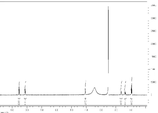
Figure 2.3 1H NMR of n-Bu-NTA ... 33
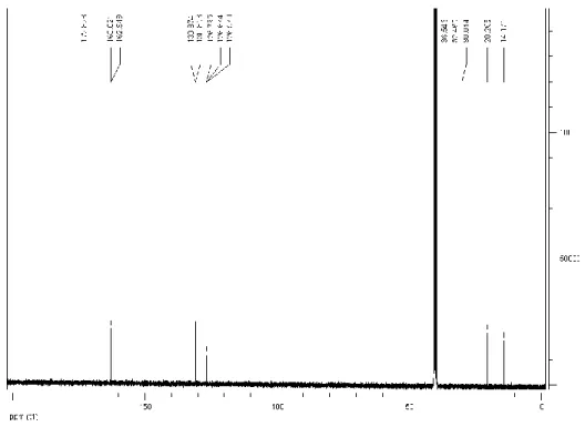
Figure 2.4 13C NMR of n-Bu-NTA ... 34
Figure 2.5 1H NMR of n-Bu-NTA- ß alanine ... 34
Figure 2.6 13C NMR of n-Bu-NTA- ß alanine ... 35
Figure 2.7 Mass spectrum of n-Bu-NTA- ß alanine ... 35
Figure 2.8 Chemical structure of pPC molecule ... 41
Figure 2.10 Mass spectrum of pPC molecule ... 42
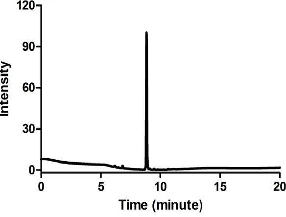
Figure 2.9 Liquid chromatogram of pPC molecule ... 42
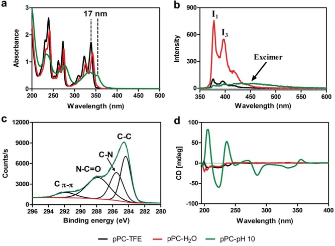
Figure 2.11 Spectroscopic characterization of pPC assembly in aqueous media. (a) UV-vis absorption and (b) fluorescence emission spectra (excitation wavelength = 340 nm) of pPC. (c) XPS analysis of assembled pPC powder and (d) CD spectrum of pPC molecules in different conditions. ... 43
Figure 2.12 Chemical structure of nPC molecule ... 45
Figure 2.14 Mass spectrum of nPC molecule ... 46
Figure 2.13 Liquid chromatogram of nPC molecule ... 46
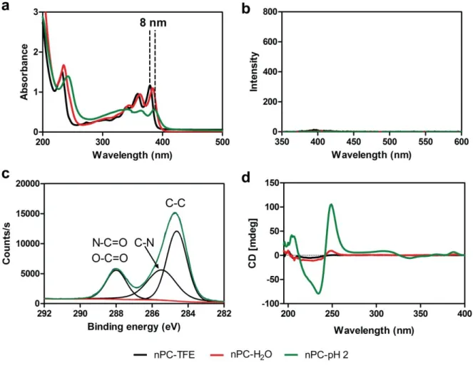
Figure 2.15 Spectroscopic characterization of nPC assembly in aqueous media. (a) UV-vis absorption spectrum and (b) fluorescence emission spectra (excitation wavelength = 340 nm) of nPC. (c) XPS analysis of assembled nPC powder and (d) CD spectrum of nPC molecule in different conditions. ... 47
xvii
Figure 2.16 Spectroscopic characterization of pPC and nPC coassembly in aqueous media. (a) UV-vis absorption spectra and inset shows appearance of CT band. (b) Fluorescence emission spectra (excitation wavelength = 340 nm) of pPC-nPC CT complex. (c) Decay curves of pPC-pH 2, pPC-pH 10 samples and pPC-nPC CT complex pair at 477 nm probe wavelength. (d) CD spectra of pPC and nPC coassembly in aqueous media. ... 49 Figure 2.17 Uv-vis absorption of Charge-Transfer Complex formation (a), fluorescence quenching measurements of pPC by nPC (b) and quenching of pPC by nPC under 254 nm illumination (c)... 50 Figure 2.18 Imaging of pPC-nPC CT complex nanowires. STEM images (a and b) and AFM images (c and d) of n/p-coassembled supramolecular nanowires ... 52 Figure 2.19 XPS analysis of pPC (a), nPC (b), and pPC-nPC (c) powders. FT-IR analysis (d) .. 53 Figure 2.20 NOESY spectrum of pPC-nPC CT complex. Close contact is shown by red circle . 55 Figure 2.21 (a) UV-vis absorbance spectra of pPC and nPC molecules mixed at different mole ratios. (b) Plot of absorbance at 520 nm (CT band) versus mole ratio of nPC (XnPC) ... 56 Figure 2.22 (a) Gaussian deconvolution of the N1s signal acquired by XPS. (b) Isothermal calorimetry titration of nPC (0.1 mM) by pPC (1 mM) ... 57 Figure 2.23 Mass spectrum at positive mode showing formation of nPC-pPC-nPC (A-D-A) CT complex ... 58
Figure 3.1 (a) TEM and (b. SEM images of PA at pH 7.0 ... 68 Figure 3.3 (a) Time sweep graph of peptide amphiphile gel. (b) Strain sweep graph of peptide amphiphile gel ... 69 Figure 3.2 CD spectra of peptide amphiphile ... 69 Figure 3.4 TEM of Pd nanostructures after first reduction cycle ... 70
xviii
Figure 3.5 TEM images of Pd nanostructures after third reduction cycle ... 71
Figure 3.6 (a) HRTEM image of Pd nanostructures. (b) XRD pattern of Pd nanostructures ... 72
Figure 3.7 Gas Chromatogram of 4-methoxybiphenyl (first run)... 78
Figure 3. 8 Gas Chromatogram of 4-methoxybiphenyl (second run) ... 78
Figure 3.9 Gas Chromatogram of 4-methoxybiphenyl (Third run) ... 79
Figure 3.10 Gas Chromatogram of 4-methoxybiphenyl (Fourth run) ... 79
Figure 3.11 Gas Chromatogram of 4-methoxybiphenyl (Fifth run) ... 80
Figure 3.12 Mass spectrum of 4-methoxybiphenyl ... 80
Figure 4.1 1H NMR of benyl azide ... 89
Figure 4.2 13C NMR of benyl azide ... 89
Figure 4.3 13C NMR of benyl azide ... 90
Figure 4.4 1H NMR of 3-aminopropyl-1azide ... 91
Figure 4. 5 13C NMR of 3-aminopropyl-1azide ... 91
Figure 4.6 HRMS spectrum of Biotin-NHS ... 93
Figure 4.7 1H NMR of Biotin-azide ... 93 Figure 4.8 13C NMR of Biotin-azide... 94 Figure 4.9 1H NMR of N-succinimidyl-4-pentynoate ... 95 Figure 4.10 13C NMR of N-succinimidyl-4-pentynoate ... 95 Figure 4.11 HRMS of N-4-Pentynoylmannosamine ... 96 Figure 4.12 HRMS of Ac4ManNAl ... 97
Figure 4.13 1H spectrum of Ac4ManNAl ... 98
Figure 4.14 13C spectrum of Ac4ManNAl ... 98
xix
Figure 4.16 Liquid chromatogram of peptide amphiphile ... 103
Figure 4.17 Mass spectrum of peptide amphiphile ... 103
Figure 4.18 Chemical structure of Ac-HH-Am molecule ... 104
Figure 4.19 Liquid chromatogram of Ac-HH-Am molecule ... 104
Figure 4.20 Mass spectrum of Ac-HH-Am molecule ... 104
Figure 4.21 CAC plots for C12VVAGHH-Am peptide amphiphile ... 105
Figure 4.22 (a) CD analysis of PA and PA-CuII,( b) STEM image of the PA nanofibers, (c) XPS and (d) STEM image of PA-CuII nanofibers ... 106
Figure 4.23 Preparation of of PA-CuII ... 107
Figure 4.24 Rheological characterization of the PA-CuII hydrogel. (a) Time sweep, (b) ... 107
Figure 4.25 STEM (a, b), and SEM images of PA-CuII nanofibers (c) and EDS of PA-CuII nanofibers (d) ... 108
Figure 4.26 High resolution XPS analysis of copper (PA-CuII) ... 109
Figure 4.27 Isothermal titration calorimetry of Ac-HH-Am by CuSO4 solution ... 110
Figure 4.28 Gas chromatogram of crude triazole (CuSO4 as catalyst, 37 oC) ... 113
Figure 4.29 Mass spectrum of crude triazole (CuSO4 as catalyst, 37 oC) ... 113
Figure 4.31 Mass spectrum of crude triazole (His-CuII as catalyst, 37 oC) ... 114
Figure 4.30 Gas chromatogram of crude triazole (His-CuII as catalyst, 37 oC) ... 114
Figure 4.33 Mass spectrum of crude triazole (HH-CuII as catalyst, 37 oC) ... 115
Figure 4.32 Gas chromatogram of crude triazole (HH-CuII as catalyst, 37 oC) ... 115
Figure 4.35 Mass spectrum of crude triazole (PA-CuII as catalyst, 37 oC) ... 116
Figure 4. 34 Gas chromatogram of crude triazole (PA-CuII as catalyst, 37 oC) ... 116
xx
Figure 4.37 Mass spectrum of crude triazole (PA-CuII as catalyst, 25 oC) ... 117
Figure 4.38 Viability of MCF-7 cells in the presence of PA-CuI and CuI (a), microscopic analysis of fixed cells labeled with PA-CuI nanofibers after 6 h (b), and after 24 h reaction (c) and non-treatment group after 24 h reaction (d). (Scale bars = 20 µm) ... 119
Figure 4.39 Fluorescent imaging of fixed cells labeled with (a) PA-CuI, (b) CuI and (c) non-treatment sample after 6 h and 24 h reaction ... 120
Figure 4.40 Confocal microscopy images of labeled cells with PA-CuI nanofibers (a), CuI (b) and non-treated (c) groups after 6 h reaction. Flow cytometry analysis of MCF-7 cells (green histogram, cells treated with PA-CuI; blue histogram, cells treated with CuI; black histogram, non-treated cells) (d). Comparison of cell labeling efficiency by flow cytometry (e). To quantify labeling signals, the mean FITC intensities were calculated for >10000 cells for each condition, averaged and compared. (Mean ± SEM) ... 121
Figure 5.1 Chemical Structure of Peptide Molecule (K-PA) ... 131
Figure 5.3 Mass spectrum of peptide molecule (K-PA) ... 131
Figure 5.2 Liquid Chromatogram of Peptide Molecule (K-PA) ... 131
Figure 5.4 CD spectra of K-PA in acidic (pH 2) and basic medium (pH 10) (a), rheological analysis of K- PA gel (b), SEM image of peptide 3D aerogel (c) and TEM image of peptide nanofibers ... 132
Figure 5.5 SEM (a) and STEM (b) images of 3D TiO2 nanonetwork. Powder XRD pattern (c), and BET analysis (d) of 3D TiO2 nanonetwork ... 134
Figure 5.6 SEM images (a-c), STEM images (d-e) and EDS (f) of 3D TiO2 nanonetwork ... 134 Figure 5.7 STEM images and size distribution of Pt5@TiO2 (a, g), Pt10@TiO2 (b, h) Pt15@TiO2 (c, i), Pt20@TiO2 (d, j), Pt25@TiO2 (e, k) and Pt30@TiO2 (f, l). Effect of Pt cycle on Pt
xxi
nanoparticle size (m) and Pt loading (n). Histograms of particle size distribution were constructed by counting 100 nanoparticles for 5 cycle and 200 nanoparticles for the rest of cycles ... 135 Figure 5.9 XPS analysis of (a) Pt5@TiO2, (b) Pt10@TiO2, (c) Pt15@TiO2, (d) Pt20@TiO2, (e) Pt25@TiO2 and Pt30@TiO2 ... 136 Figure 5.8 STEM images and EDS of Pt5@TiO2 (a, g and m), Pt10@TiO2 (b, j and n), Pt15@TiO2 (c, i and o), Pt20@TiO2 (d, j and p), Pt25@TiO2 (e, k and q) and Pt30@TiO2 (f, l and r) ... 136 Figure 5.10 High resolution XPS and deconvolution analysis of (a) Pt5@TiO2, (b) Pt10@TiO2, (c) Pt15@TiO2, (d) Pt20@TiO2, (e) Pt25@TiO2 and (f) Pt30@TiO2. Table shows summary of deconvolution analysis of Pt ... 137 Figure 5.11 XRD of 3D Pt@TiO2 nanonetworks... 138 Figure 5. 12 HRTEM of 3D Pt@TiO2 nanonetwork ... 139 Figure 5.13 Turnover frequencies of (a) Pt5@TiO2, (b) Pt10@TiO2, (c) Pt15@TiO2, (d) Pt20@TiO2, (e) Pt25@TiO2 and (f) Pt30@TiO2. Effect of Pt cycle (g) and Pt nanoparticle size (h) on TOF ... 140 Figure 5.14 STEM images of (a, g) Pt5@TiO2, (b, h) Pt10@TiO2 (c, i), Pt15@TiO2 (d, j), Pt20@TiO2 (e, k) Pt25@TiO2 and (f, l) Pt30@TiO2 after recycling ... 144 Figure 5.15 High resolution XPS and deconvolution analysis of fresh and deactivated Pt25@TiO2 ... 145
xxii
List of Tables
Table 1 Suzuki-Miyaura coupling of aryl halides with Pd@Peptide ... 72 Table 2 Suzuki-Miyaura coupling of aryl iodides with Pd@Peptide Nanocatalyst ... 73 Table 3 Miyaura coupling of aryl bromide with Pd@Peptide ... 74 Table 4 Suzuki-Miyaura coupling of aryl bromides with Pd@Peptide Nanocatalyst ... 75 Table 5 Recyclability test of bromobenzene with 4-methoxyphenylboronic at 80 0C. ... 77 Table 6 Comparison of catalyst efficiency in CuAACa ... 111 Table 7 Characteristics of 3D Pt@TiO2 nanonetworks and their turnover frequency (TOF, min-1) in hydrolysis of AB at 25.0 ± 0.1 °C. ... 141 Table 8 Turnover frequency value (TOF) of the platinum catalysts reported for the hydrolysis of AB at 25.0 ± 0.1°C. ... 143
xxiii
Abbreviations
CD : Circular dichroism
DCM : Dichloromethane
DMF : N, N-Dimethylformamide
FBS : Fetal bovine serum
dd : Double distilled
ACN : Acetonitrile
EtOH : Ethanol
D : π-electron donor
A : π-electron acceptor
HPLC : High performance liquid chromatography
LC-MS : Liquid chromatography-mass spectrometry
GC-MS : Gas chromatography-mass spectrometry
PA : Peptide amphiphile
PBS : Phosphate-buffered saline
Q-TOF : Quadrupole time of flight
xxiv SEM : Scanning electron microscopy
XRD : X-ray diffraction
STEM : Scanning transmission electron microscopy
TIS : Triisopropylsilane
TCP : Tissue culture plate
TEM : Transmission electron microscopy
TFA : Trifluoroacetic acid
XPS : X-ray photoelectron spectroscopy
NMR : Nuclear magnetic resonance spectroscopy
FT-IR : Fourier transform-infrared spectroscopy
ITC : Isothermal titration calorimetry
TOF : Turnover frequency
HBTU : N,N,N′,N′-Tetramethyl-O-(1H-benzotriazol-1-yl)uranium hexafluorophosphate
FMOC : Fluorenylmethyloxycarbonyl DIEA : N,N-Diisopropylethylamine
1
Chapter 1
Introduction: Designing Peptide Based Nanomaterials
Bottom-up and top-down processing are the two strategies to fabricate nanomaterials with well-defined size, shape, composition and property [1, 2]. In latter approach, sophisticated techniques such as e-beam and atomic force microscopy based lithography are required to construct nanomaterials from bulk. While former approach, exploits noncovalent interactions such as hydrogen bonding, hydrophobic, electrostatic, charge-transfer complex, -, and van der Waals interactions to organize molecular elements into desired supramolecular nanostructures. Particularly, biomolecules such as peptides, nucleic acids, and lipids interact and self-organize through noncovalent interactions to produce complex structures like collagen, keratin, pearl, shell, coral,calcite microlenses, DNA and RNA which perform important functions in biological systems[3]. Although these noncovalent interactions are weak in nature, when combined together as a whole, they shape biomolecules with stable structures and crucial functions. Self-organization of biomolecules through molecular self-assembly has been of a source of inspiration for many research groups to utilize peptides, proteins, lipids, DNA and RNA as building blocks to synthesize novel nanomaterials with a broad range of applications including tissue engineering, catalysis, sensing, light harvesting, optoelectronic, and bioelectronic [4-7]. Owing to complex structure and complicated synthetic protocols of these giant biomolecules, researchers have developed bioinspired minimalistic designs based on short sequence peptides which self-assemble through noncovalent interactions into hierarchical structures. Self-assembling short peptides are a class of supramolecular polymers, which exploit supramolecular interactions to generate well-defined nanostructures including nanospheres, nanosheets,
2 nanotubes, and nanofibers (Figure 1) [8]. These versatile peptide-based supramolecular nanomaterials have been utilized in variety of applications which will be discussed in next section .
Figure 1.1 Supramolecular structures formed via short peptide self-assembly. (Figure adapted from ref. 8)
There are 20 natural amino acids (Figure 1.2) which are the building blocks for synthesis of peptides and proteins in biological systems. All amino acids have a stereogenic center with L configuration accept glycine. The basic chemical structure of amino acids is similar with different side chains in central carbon (Cα). Based on diverse side chains, amino acids can be classified as hydrophobic (A, V, L, l, and M), aromatic (F, W and Y), negatively charged (D and E), positively charged (H, K and R) and hydrophilic (S, N, Q and T) (Figure 1.2) [9].
3 Figure 1.2Chemical structures of natural amino acids. Different peptide-based nanostructures with some potential applications. (Figure adapted from ref. 9).
The chemical nature of side chains of amino acids is highly important in governing the structure and function of peptides and proteins in biological environment. Due to large number of amino acids with rich functional groups, the number of peptide sequences and structures is endless. Therefore, rational arrangement of amino acid sequence is extremely vital to get the desired nanostructures. Several research groups have put effort to design self-assembling short sequences
4 based on dipeptides, tripeptides, and longer peptide sequences to acquire different supramolecular nanostructures. Self-assembling peptide designs to produce nanostructures will be explained in coming section.
1.1 Amyloid-inspired peptide nanostructures
Formation of amyloid nanostructures which is associated with neurodegenerative diseases including Alzheimer’s disease and Parkinson’s disease has long been under study. Amyloids are produced from different peptide and protein aggregates. Structural studies and mechanistic understanding of amyloid structures have come out with various peptide sequences which can self-assemble into β-fibrils. Heptapeptide Aβ (16–22), Ac-KLVFFAE-Am, cationic core section of the Alzheimer’s disease can assemble into β-sheets and forming nanotubes [10]. These well-defined nanotubes have a 52 nm cross-sectional diameter and lengths in microns (Figure 1.3). Many groups have been inspired by these short self-assembling peptide sequences and made trivial modifications to get different nanostructures. For example, Ac-E-FFAA-E-Am and Ac-K-FFAA-K-Am sequences can form nanofibers having negative and positive charges respectively[11]. FF in AAKLVFF-OH can be replaced by aromatic moieties like thiophene (2-Thi)(2-Thi)VLKAA) to generate well-ordered nanofibers in methanol [12].
5 Figure 1.3 Proposed mechanism for the self-assembly of Ac-KLVFFAE-Am into nanotubes (left). High resolution cryo-TEM images of nanotubes (right). (Figure adapted from ref. 10)
Another interesting bioinspired self-assembling peptide molecule is diphenylalanine (FF) dipeptide, the core recognition motif of the β-amyloid polypeptide, self-organizes via π-π interactions between phenyl groups to form highly-ordered nanostructures [13]. Diphenylalanine dipeptide is one of the simplest self-assembling sequences which can aggregate into different structures from nanoscale to microscale (Figure 1.4). Owing to its structural simplicity, facile synthesis, and widespread applications, it is a widely studied self-assembling peptide. FF supramolecular nanotubes can easily be formed by diluting the concentrated solution of FF in fluorinated alcohol by water. The morphology of the FF nanostructures can be readily manipulated and controlled by experimental conditions such as pH, temperature, peptide concentration and solvent to get vesicles, tubes, rods and strips at nano to microscale [14].
6 1.2 Peptide bolaamphiphile based nanostructures
Bolaamphiphilic peptides are another well studied design composed of peptidic and non-peptidic parts. In this design, a hydrophobic molecule (aliphatic or aromatic molecules) is integrated polar or charged amino acids from two arms (Figure 1.5). When dissolved in aqueous media, the bolaamphiphilic monomers arrange themselves in such way that charged motifs expose to water while hydrophobic segments escapes from water and forming nanotubes. The driving force for the formation of nanotubes in this peptide design is either hydrogen bonding and/or π-π interactions [10, 15]. Depending on the design of monomer molecules, the nanotubes can adopt diameters ranging from nanometers to micrometers. Bolaamphiphilic peptide design is especially important in construction of supramolecular organic nanowires in aqueous media by providing Figure 1.4 Self-assembly of FF into different morphologies. (Figure adapted from ref. 13 Copyright 2015 American Chemical Society)
7 efficient cofacial π-π interactions among hydrophobic semiconducting chromophores. Efficiency of charge transport, energy migration and mobility of charge carriers in these supramolecular one-dimensional (1D) nanowires is directly dependent on supramolecular order of π-conjugated chromophores. Interestingly, this molecular design ensures H-type π-π interactions among hydrophobic semiconducting chromophores in aqueous medium. More examples and explanations will be provided in section 1.6.
Figure 1.5 Self-assembly of bolaamphiphile monomers into nanotube morphology. (Figure adapted from ref. 15. Copyright © 2010 WILEY‐VCH Verlag GmbH & Co.)
8 1.3 Cyclic peptide based nanostructures
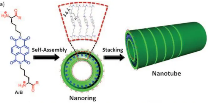
In 1993 Ghadari [16] demonstrated that stacking of cyclic D, L-peptide rings can produce high respect ratio hollow nanostructures (Figure 1.6). The sequence of octapeptide cyclo [-(l-Gln-d-Ala-l-Glu-d-Ala)2-] dissolved in basic solution could not aggregate due to columbic charge repulsion among carboxylate groups (COO-). Well-ordered nanotubes were formed upon charge neutralization by controlled addition of acid into solution of cyclo [-(l-Gln-d-Ala-l-Glu-d-Ala)2-]. The divining force behind self-assembly of cyclic D, L-peptide rings is anti-parallel β-sheet hydrogen bonding to form nanotubes with a van der Waals internal diameter of approximately 7 Å. Several derivatives of cyclic D, L-peptide were synthesized to be utilized in wide range of applications like sensing, targeted drug delivery and optoelectronics [16, 17].
Figure 1.6Self-assembly of cyclic D, L- peptide into nanotubes morphology. (Figure adapted from ref. 16. Copyright © 2001 WILEY‐VCH Verlag GmbH & Co.)
9 1.4 Peptide amphiphile based nanostructures
Another interesting class of short self-assembling peptides is amphilphilic peptides [18]. Typical peptide amphiphile molecule is consisted of hydrophobic and hydrophilic segments. Hydrophobic fragment is composed of an aliphatic tail or an aromatic moiety plus β-sheet hydrogen bonding amino acids such a valine and alanine. While charged amino acids like glutamic acid or lysine constitutes the rest of peptide amphiphile fraction (Figure 1.7).
Figure 1.7Typical chemical structure of peptide amphiphile and it’s self-assembly into various supramolecular nanostructures.
10 The peptide sequence in figure 1.7 with an alkyl tail and β-sheet sequence prefers to form cylindrical nanofibers. Replacing β-sheet forming residues (valine and alanine) with hydrogen breaking moieties such as proline give rise to spherical nanostructures. Slight changes in chemical structure of peptide amphiphile or experimental conditions such as pH, temperature, sonication, ionic gradient, peptide concentration and solvent can produce well-defined nanostructures with diverse morphologies (Figure 1.7) [19].
Besides above mentioned self-assembling peptide designs, many research groups have designed and synthesized novel self-assembling peptide sequences based on pure amino acids, peptide-small molecule and peptide-polymer conjugates to obtain supramolecular nanostructures having various morphologies for variety of applications [18, 20] .
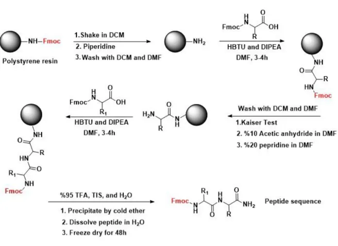
One of the main reasons for the widespread studying and utilization of short self-assembling peptides is the development of well established synthetic procedures to get peptides with high purity and efficiency without rigorous purification and work-up processes. Solid-phase peptide synthesis (SPPS), pioneered by Robert Bruce Merrifield, is now the classic synthetic method to manufacture peptides and proteins [21]. There are mainly two forms of SPPS- Fmoc and Boc. Chemically stable and insoluble polystyrene microbeads (resin) are used as solid support to grow peptide chains step by step. These polymer based solid supports are functionalized by linkers having amine or hydroxyl groups. Peptides can be obtained with amide or carboxylic acid C-terminal depending on choice of functionalized polystyrene solid supports. Unlike natural synthesis of proteins in ribosome, peptides are synthesized from C to N-terminal. Fmoc protected amino acids are now commercially available in large scales for suitable prices. To start peptide synthesis (Figure 1.8), polystyrene bead is swelled in DCM for 30 minutes then carboxylic acid group of N-terminal Fmoc protected amino acid is activated by HBTU and then reacted with free
11 amine groups on solid resin to form amide bonds. A Kaiser test is performed after thoroughly washing of resin with DMF and DCM to test if the amino acid is successfully conjugated on resin. For the conjugation of second amino acid, Fmoc of first amino acid is removed by solution of piperidine to get free amine groups to react with carboxylic acid groups of second amino acid. Using this simple procedure a peptide sequence up to 70 amino acids can readily be grown on solid resins (Figure 1.8). The peptide sequence can be cleaved from solid resin in the presence of concentrated TFA solution. After cold ether precipitation, pure peptide powder can be obtained in high yield.
12 Applications of self-assembled peptide nanostructures
This part is partially described in the following publication:
Melis Sardan Ekiz, Goksu Cinar, Mohammad Aref Khalily and Mustafa O Guler “Self-assembled peptide nanostructures for functional materials” Nanotechnology 27 (2016) 402002
In this section, I will review technological applications of short self-assembled peptide nanostructures.
1.5 Supramolecular Self-Assembled Peptide Catalysts
Extraordinary catalytic performance and superior selectivity of enzymes in organic transformations under physiological conditions have always been a source of inspiration to scientists. Complex structure and function of biocatalysts has enforced scientists to develop minimalistic approaches and mimic a fragment of multifaceted biocatalysts such as active site, hydrophobic pocket, or structure [22]. Of those minimalistic approaches, supramolecular self-assembled peptide nanostructures have been utilized to assemble essential functional groups within a nanoenviroment to catalyze desired organic transformations in an efficient manner.
Stupp showed self-assembling peptide amphiphiles which aggregated into well-defined catalytic nanostructures in aqueous media [23]. In this study, four different peptide sequences containing histidine residue as reactive site were designed and synthesized (Figure 1.9A and B). The peptide 1 having alanine and valine residues promoted formation of β-sheet and palmitoyl tail assisted hydrophobic collapse of the peptide molecules into high respect ratio 1D nanofibers as shown in Figure 1.9C.
13 The same peptide sequence without palmitoyl tail (3) formed nanospheres (Figure 1.9D), meanwhile, the peptide sequences having proline residues (2 and 4) also formed polydisperse nanospheres. Hydrolysis of 2, 4-dinitrophenyl acetate (DNPA) by these nanostructures have shown that 1D nanofibers promoted highest hydrolysis rate when compared to nanospheres or soluble histidine residue. Interestingly, 1D nanofibers showed a Michaelis-Menten enzyme-like behavior whereas the other peptide sequences (2-4) demonstrated a linear rate increase with substrate concentration increase. This study showed that assembling reactive sites within a nanoenviroment could afford a synergistic effect which enhanced the rate of hydrolysis of DNPA.
Figure 1.9 Chemical structures of peptide molecules (A and B), schematic representation of self-assembled nanofibers (C) and spherical aggregates (D).
14 This study was one of the first examples of supramolecular assembled peptide catalyst which inspired many research groups to develop new catalytic supramolecular peptide nanostructures. Escuder and Miravet research groups have extensively studied the structure and catalytic activity of proline based supramolecular gelators [24]. L-proline is a well-known stereoselective organocatalyst to catalyze C-C bond formation reactions, such as aldol and Michael addition reactions [25]. A series of bolaamphiphilic molecules consisting of two L-prolines were designed and used in aldol reactions [26]. Due to presence of hydrogen-bond promoting valine residues in the structure of bolaamphiphilic peptide molecules, they could encapsulate large amount of organic solvents (acetonitrile and ethyl acetate) to form organogels. These peptide molecules catalyzed the aldol reaction in solution state while racemisation of the products was observed in gel state when the reactions were left for several days. The reason for racemisation was explained by increase of basicity of the solution due to the close assembly of prolines in the gel state. The authors exploited the emergence of new property (increased basicity) caused by cooperativity of neighboring prolines in gel state [27]. They chose Henry (nitro-aldol) reaction which requires a basic catalyst for deprotonation of nitroalkane and then addition to carbonyl functional group to form new C-C bond. A bolaamphiphile peptide molecule was used to form gel in nitromethne and nitroethane. This basic catalytic gel promoted the addition of nitroalkane to 4-nitrobenzylaldehyde as shown in Figure 1.10. The assembled bolaamphiphile peptide molecules into fibrous networks showed excellent catalytic activity while dispersed peptide molecules showed poor catalytic activity.
15 Performing organic reactions in aqueous media motivated the researchers to synthesize a new proline conjugated amphilphilic peptide molecules which formed hydrogels [28]. This catalytic hydrogel assisted the direct aldol reaction between cyclohexanone and 4-nitrobenzaldehyde. The reactants dissolved in toluene were gently dropped on pre-formed peptide hydrogel showed high stereoselectivity (anti: syn 92:8, 88% ee). The catalytic hydrogel was recycled three times without loss of efficiency and stereoselectivity.
Beside L-proline based supramolecular catalysts, the researchers have also developed different novel approaches to mimic active sites of enzymes. Junqiu Liu and his co-workers demonstrated that co-assembly of two different amino acid residues could enhance the rate of hydrolysis [29]. They synthesized two short amphilphilic peptide sequences one bearing histidine (Fmoc-FFH-CONH2) and other bearing arginine residues (Fmoc-FFR-CONH2). The Fmoc-FFH-CONH2 molecules aggregated into nanotube in aqueous medium could catalyze p-nitrophenyl acetate in efficient manner. Upon co-assembly of Fmoc-FFH-CONH2 and Fmoc-FFR-CONH2 peptides into nanotubes, the catalytic activity was further enhanced. A similar study was conducted by Liang
16 research group [30]. Co-assembly of histidine and arginine bearing self-assembled peptide nanofibers could enhance the hydrolysis of catalyze p-nitrophenyl acetate. These two studies underlined the importance of arginine residues which stabilized the transition state of the hydrolytic reaction.
Metal ions play an important role as cofactors in metalloenzymes. The metal ion based cofactors are present in the active site of enzymes which aid the chemical reactions to proceed [31]. Mimicking a part of metalloenzyme by short sequenced self-assembled supramolecular peptide nanostructures decorated metal ions is another interesting research field. Liu and coworkers have designed and synthesized glutamic acid bearing bolaamphiphile nanotubes which copper (II) ions could bind via electrostatic interactions. The produced nanotubes induced chirality to copper (II) ions could catalyze the Diels-Alder reaction between cyclopentadiene and an aza-chalcone [32]. Korendovych research group conducted a more detailed and systematic study on short sequenced metallopeptides to mimic metalloenzyme. A series of short amyloid-forming peptides which have binding affinity towards Zn (II) ions were designed and synthesized [33]. A β-sheet forming heptapeptide sequence (LKLKLKL) was modified at position 2, 4 and 6 by different amino acids. Hydrophobic leucine (L) required for self-assembly kept unchanged while Lysine (K) residues at positions 2 and 4 were replaced by Zn+2-binding histidines. K at position 6 was replaced by either acidic (Asp, Glu), neutral (Gln, Tyr) or basic residues with various pKa values (His, Lys, Arg). The peptide sequence Ac-LHLHLRL-CONH2 in the presence of ZnCl showed Michaelis–Menten behavior with catalytic efficiency of Kcat/KM= 18±4 M-1 s-1. When the R was replaced by Q residue (Ac-LHLHLQL-CONH2) the catalytic efficiency was further increased to kcat/KM = 30±3 M-1 s-1. Peptides having Asp, Glu or His residues at position 6 had little catalytic activity. When the hydrophobic L was replaced by isoleucine (I) in the heptapeptide sequence
17 keeping Gln at 6th position (Ac-IHIHIQI-CONH2) further enhancement of catalytic efficiency (kcat/KM = 62±2 M-1 s-1) was observed.
We recently also developed a β-sheet forming peptide amphiphile which had high binding affinity towards copper ions. This short peptide sequence assembled into nanofibers having histidine residues over the surface. The copper ions bound on pre-assembled peptide nanofibers could catalyze click alkyne- azide cycloaddition in aqueous media [34]. The conversion of the reactants into product was almost quantitive (95%) by our supramolecular nanocatalyst while soluble histidine-Cu complexes showed a moderate efficiency of 65%. Active sites demonstrated positive cooperativity when assembled within a nanoenviroment on peptide nanofibers. Supramolecular nanocatalyst did not only show superior catalytic activity but also lowered the cytotoxicity of the copper ions. Therefore, we utilized the metallopeptide to label alkyne decorated live cells by click alkyne- azide cycloaddition.
1.6 Supramolecular Semiconductor Peptide Nanostructures
Organic electronics can be divided into three major field’s namingly supramolecular, plastic, and molecular electronics [35]. The latter two fields are studied in detail and Nobel Prize in chemistry was awarded to discovery of electrically conductive polymers (plastic electronics) in year 2000. While supramolecular electronics is much lesser studied research field but promising research progress has been achieved in last decade. Supramolecular chemistry is a powerful tool to design and construct well-defined nanowires for nanosized optoelectronic devices using noncovalent interactions. The rich chemistry of 20 natural amino acids makes them an eye-catching tool to build novel supramolecular soft semiconducting materials which could be
18 employed for bioelectronics [36], light harvesting systems [37], OFETS, nanosized devices and nanosensors [38].
The main strategy to fabricate a nanosized semiconducting wire is to conjugate hydrophobic, pi conjugated p-n type semiconductor molecules to hydrophilic or charged self assembling peptide sequences. Upon hydrophobic collapse in aqueous media, the semiconducting conjugated peptide molecules can form variety of nanostructures. This strategy also allows working with highly hydrophobic molecules in aqueous media. Parquette and co-workers conjugated a n-type semiconducting molecule (1,4,5,8-naphthalenetetracarboxylic acid diimide, NDI) to s series of dipeptides (Ac-KK-NH2) and studied their self-assembly in aqueous medium and trifluoroethanol (TFE) [39]. The NDI semiconductor peptide conjugate assembly was studied by UV and circular dichroism spectroscopies. The NDI-peptide molecule dissolved in TFE showed absorption peaks at 240 nm (band II) and in the range of 300-400 nm (band I) which are characteristic absorption peaks of molecular NDI molecules. When TFE changed with water there was a decrease in absorption intensities followed by large red shift in the absorption bands of NDI. This change showed the formation of j-aggregates in water. The IR bands at 1612 and 1608 cm-1, and XRD analysis proved the presence of β-sheet in Ac-KK (NDI)-NH2 andin Ac-K(NDI)-KNH2 peptides. TEM and AFM images of Ac-KK(NDI)-NH2 peptide molecules showed helical nanofibers while Ac-K(NDI)-KNH2 peptides showed flattened, twisted nanoribbons morphology. The same research group conducted a similar study by conjugating two lysine moieties to the two arms of NDI forming n-type bolaamphiphile [15]. Bolaamphiphile molecules formed hydrogel when in dissolved 1% (w/w). The designed molecule formed nanotubes as imaged by TEM and AFM. Uv-vis absorption spectroscopy demonstrated a red shift in the absorption bands showing the formation of NDI j-aggregates in assembled state. CD
19 spectra revealed peaks at the absorption bands of NDI molecule demonstrating induced chirality rooted from supramolecular organization of NDI molecules within the assembled nanotubes. Fluorescence decay of 350 nm excitated nanotubes was followed by time-correlated single photon counting at 410 and 505 nm. The life time (64 ps) of excited NDI nanotubes were slightly longer than the molecular NDI. The excimer emission at 505 nm showed much longer decay life time (197 ps (86%) and 950 ps (14%)) which is due to closely stacked and packed NDI molecules in nanotube.
Tovar [40-45] and others [46-48] have developed semiconducting oligomers conjugated to peptides (Figure 1.11) assembled into well defined nanostructures in aqueous media. In this design, oligomers are located at middle and peptide sequences are conjugated at two sides. This strategy facilitates face to face aggregation of the semiconducting molecules which can enhance the hole or electron transporting properties as a consequence amplified conductivities could be achieved. Tovar research group also developed a straightforward method to conjugate oligomers molecules on solid phase resin. On resin synthesis avoids multiple synthetic routs and complex purification techniques. Charge transport properties of assembled soft fibrous nanonetwork were measured by constructing a field-effect transistor. The peptide conjugated quaterthiophene was used as semiconductor layer and mobility of 10-3 -10-5 cm2 /(V s) was measured. When the nanowires were aligned via shear-assembly the mobility were increased to 0.03 cm2 /(V s).
20 Ashkenasy and her workers conducted a detailed study of conductivities of self-assembled semiconducting peptide nanostructures [12, 49]. A well-known amyloid β-peptide AAKLVFF was studied by incorporating 2-thienylalanine and 3-thienylalanine moieties instead of FF. They investigated the self-assembly of these molecules in different solvents and different morphologies were formed. They observed that peptide molecule forming long range and straight nanofibers show highest conductivity. In another research work, Ashkenasy demonstrated that the both protons and electrons are responsible for charge transport process in self-assembled nanostructures. In highly humid environment proton conductivity dominated electron conductivity. Hodgkiss et.al. has recently constructed a water-processed bio-organic field-effect transistor (biOFET) based on bioinspired assembled peptide-pyrelene conjugates [50]. The peptide sequence IKHLSVN which is responsible for homo dimmerization of peroxiredoxin protein was modified as IRHLSVN. Three glumatic acid residues were conjugated to IRHLSVN to enhance the solubility of IRHLSVN-perylene molecules in water. The
21 assembly of these bioinspired molecules were studied by CD and Uv-vis spectroscopy to see whether the β-sheet forming characteristic of native IKHLSVN sequence was affected by the modifications and perylene conjugation. Long nanofibers of assembled IRHLSVN-perylene were imaged by TEM and AFM. Interestingly, the semiconducting nanofibers showed p-type behavior upon increasing positive gate bias a decreasing current trend was measured.
One component semiconductor-peptide self-assembly was further extended to two components including co-assembly of p-n type semiconductors to achieve charge-transfer or photo (conductive) supramolecular nanomaterials. Ulijn group [51-53] have conducted a series of research on biocatalytic self of n-type semiconductor conjugated peptides that could form charge-transfer supramolecular nanostructures with variety of p-type semiconductors (Figure 1.12). NDI-functionalized tyrosine (NDI-Y) was conjugated to phenylalanine-amide (F-NH2) by addition of thermolysin to trigger the self-assembly and forming thermodynamic nanostructures. At the presence of p-type dihydroxy/alkoxy naphthalene donors, NDI-Y forms polydisperse spherical nanostructures when thermolysin was absent as imaged by TEM and AFM. Upon addition of biocatalyst to solution of NDI-Y and naphthalene donors, a yellowish gel was formed and spherical nanostructures were converted to highly-ordered long nanofibers. The change in color of solution followed by donor emission quenching and appearance of new absorption at 550 nm was the evidence for formation of charge-transfer complex. The same group conjugated a well-known p-type tetrathiafulvalene (TTF) to a self-assembling peptide sequence (-FF-NH2). TTF-FF-NH2 molecules formed self standing gel in chloroform and dried gels showed conductivity of 1.9 × 10⁻¹⁰ S cm−1. When the organogel was doped by an acceptor molecule (TCNQ) the conductively was increased to 3.6 × 10⁻⁴ S cm−1. However, iodine vapor doping did not enhance the conductivity to a great extent as in case of acceptor doping [54]. There are also
22 some studies showing the energy transfer within self-assembled peptide nanostructures through donor-acceptor interactions [55, 56].
Recently, Martín research group has exploited electrostatic interactions to assemble donor- acceptor molecules in highly organized manner within a peptide nanofiber [57]. Electron donor tetrathiafulvalene was conjugated to anionic peptide sequence bearing glutamic acid while electron-acceptor perylene-bisimide was conjugated to cationic peptide sequence bearing residue. These two complimentary p-n nanofibers could co-assemble into highly alternating p-n stacks in the single nanofiber. The co-assembled p-n nanofibers showed photoconductivity of 0.8 cm2 V−1 s−1. These kinds of novel and straightforward methodologies are promising for design and fabrication of next generation nanodevices.
Figure 1.12Biocatalytic self-assembly of acceptor-appended peptides to form charge-transfer nanostructures.
23 1.7 Self-Assembled Peptide Templated Synthesis of Inorganic Nanomaterials
Synthesis of inorganic nanomaterials with precise shape, size and composition has potential industrial applications. Biomolecules having well-defined shapes and sizes are remarkable platforms for synthesis of inorganic functional nanomaterials [58]. Cutting-edge developments have been achieved by utilizing biotemplates such as viruses [59-61], bacteria [62, 63], nucleic acids [64] (DNA or RNA) and proteins [65, 66]to mineralize clear-cut functional nanomaterials for catalysis, sensing, imaging, therapy, energy storage/ conversion, electronic and piezoelectric devices. Self-assembling peptide templated synthesis of functional nanostructures is another emerging area which has been extensively studied.
Matsui et. al designed a bolaamphiphile peptide molecule (bis (N-R-amido-glycylglycine)-1,7-heptane dicarboxylate) which assembled into nanotubes at pH 6 citric acid/NaOH solution. The amide groups on preassembled nanotubes could coordinate with metal ions (Cu and Ni) which served as nucleation sites. Nanotube coated ions were further reduced by reducing agents to get metalized nanotubes [67]. In another study conducted by Matsui, same bolaamphiphile peptide nanotube was further modified by a histidine rich peptide sequence (A-H-H-A-H-H-A-A-D). Histidine modified nanotubes were treated with gold ions followed by NaBH4 reduction to form gold nanoparticles (Figure 1.13). Metal binding histidine residues directed formation of gold nanoparticles over nanotubes homogenously. The crystal lattice of gold nanoparticles were calculated as (111) and (220) from electron diffraction patterns[68].
24 Stupp research group also developed unasymmetric bolaamphiphile peptide molecules such as (L-Glutamyl)3 glycine-terminated bolaamphiphile (1) and (L-lysine)3- terminated bolaamphiphile (2). Molecule 2 formed self standing hydrogel in the presence of ammonia vapor. The secondary structures of these two molecules were studied by circular dichroism (CD). The peptide molecules showed random coil structure in their soluble form while β-sheets were observed upon gelation. These 1D nanofibrous networks made of 1 and 2 were utilized to mineralize CdS nanostructures. Peptide hydrogels of 1 and 2 were treated with dilute solution of Cd(NO3)2 followed by exposure to H2S vapor. Molecule 1 templated CdS nanostructures having wurtzite polymorph crystal structure while molecule 2 templated CdS nanostructures having zinc blende crystal polymorph structure [69].
In another study, Stupp et. al developed a self-assembling peptide amphiphile bearing 3 histidine residues as metal ion binding [70]. The peptide amphiphile molecule dissolved in water formed hydrogel upon increasing pH above 6. The hydrogel was consisted of peptide nanofibers having Figure 1.13Scheme of Au nanowire fabrications. (a) Modification of preassembled nanofibers by histidine-rich peptide (b) Formation of gold nanoparticles on nanofibers.
25 8-10 nm diameters and micron in length as imaged by TEM. This nanofibrous network was used to template magnetite mineralization. A black precipitate was formed upon mixing of 1:2 ratio of FeCl2:FeCl3 solutions with peptide nanofibers followed by exposure to NH3 vapor. Black precipitate was responsive to magnetic field showing the formation of magnetite (γ-Fe2O3) nanostructures. Raman spectroscopy showed peak at 674 cm-1 which further confirmed the presence of magnetite (γ-Fe2O3) mineralized on peptide nanofibers.
Our group also developed various self-assembled peptide templates to synthesize Pd, Au, SiO2, TiO2, and ZnO nanostructures. An amyloid inspired short peptide sequence (Ac-KFFAAK-Am) was designed and synthesized which formed 1D nanofibers. This fibrous nanonetwork was used as template to mineralize SiO2 and TiO2. After calcination of organic part micron long nanotubes of SiO2 and TiO2 were formed [71]. Amyloid inspired templated porous silica nanotubes were used as high surface area explosive detector. Surface of silica nanotubes were modified by fluorescent probe by physical adsorption. These fluorescent silica nanotubes demonstrated fast, sensitive, and highly selective fluorescence quenching towards nitro-explosive vapors [71]. We further utilized amyloid inspired templated porous titania nanotubes as dye sensitized solar cell anodic materials. In this study we showed that porous, high surface area (150 m2/g) titania nanotubes could adsorb more dye than the nontemplated titania. As a result dye sensitized solar cell constructed from porous titania nanotubes showed efficiency of 0.83% which is 3 fold better than nontemplated titania [72].
Amyloid inspired peptide nanofibers were further exploited to template gold nanostructures synthesis. Amine decorated peptide nanofibers interacted with gold ions and upon reduction by ascorbic acid gold nanostructures were grown. Atomic force microscopy was used to investigate nanoscale electrical properties of gold decorated peptide nanofibers. Bias dependent current (IV)
26 measurements on gold nanofibers demonstrated tunneling dominated transport and resistive switching [73].
Besides amyloid inspired peptide templates, our group also designed self-assembling peptide amphiphiles which could template palladium nanoparticles synthesis. A histidine rich peptide amphiphile nanofiber was designed to synthesize Pd nanoparticles which could be utilized as nanocatalyst for Suzuki coupling reactions. Our designed Pd nanocatalyst showed high catalytic activities under environmental friendly conditions. Reaction products were obtained almost quantitatively in the presence of our catalyst besides ease of recycling [74]. In another research study, we combined advantages of critically dried porous and high surface area peptide 3D nanonetwork with atomic layer deposition (ALD) to produce highly conformal and uniform titania and silica nanotubes. These highly precise nanostructures showed high photocatalytic activities towards methylene blue degradation. The photocatalytic exclusively was dependant on wall thickness and surface area per unit mass of titania and silica nanotubes [75].
Other research groups have also exploited different self-assembled peptide templates such as I3K, phage-displayed P7A peptides [76] and aniline–GGAAKLVFF [77] to synthesize Pt nanoparticles for different applications. I3K assembled nanofibers were used to template Pt nanoparticles while phage-displayed P7A peptides were utilized to tune morphologies of Pt nanoparticles. I3K assembled nanofibers decorated Pt nanostructures were used for electrochemical oxidation of hydrogen and methanol while aniline–GGAAKLVFF supported Pt nanoparticles were used as electrocatalyst to improve oxygen reduction reaction [77].
27 Tuning metallic nanostructures spatial arrangement via peptide morphology to induce new properties is another exciting field. A peptide amphiphile (C12-FPPMPPAGAYSS) which formed double helices directed the formation of left-handed gold nanoparticles [78]. Liu research group produced chiral silica nanotubes via helical bolaamphiphilic peptide templates. Chiral silica nanotubes modified by photoactive azobenzene moieties could recognize a chiroptical switch [79].
28
Chapter 2
Fabrication of Supramolecular n/p- Nanowires via Coassembly of
Oppositely Charged Peptide-Chromophore Systems in Aqueous
Media
2.1 Introduction
Supramolecular electronics is an emerging research field, which exploits noncovalent interactions to assemble π-conjugated molecular elements into well-defined nanomaterials with semiconductor properties such as light emission, light harvesting, and charge carrier transport for optoelectronic, photovoltaic, bioelectronics and tissue engineering applications [80-82]. Efficiency of charge transport, energy migration and mobility of charge carriers in these supramolecular one-dimensional (1D) nanowires is directly dependent on supramolecular order of π-conjugated chromophores [83].Therefore achieving efficient cofacial π-π interactions among hydrophobic semiconducting chromophores in aqueous medium is highly desired goal in area of supramolecular electronics. A promising strategy to fabricate 1D electroactive nanomaterials with maximum cofacial π-π interactions in aqueous media is to conjugate n-type or p-type semiconducting molecules to self-assembling short peptide sequences [81, 84]. Particularly, conjugation to β-sheet forming peptides not only ensures solubility of hydrophobic semiconducting building blocks in aqueous medium but also allow directing long-range spatial organization of semiconducting chromophores [50, 85]. The n-type and p-type semiconducting supramolecular nanowires usually suffer from low conductivities [12, 86, 87]. To address this problem, p-type and n-type semiconducting 1D nanowires are doped by either an oxidizing [87] (iodine) or reducing agent [86] (hydrazine) respectively to generate charges. Another strategy is
29 to assemble suitable -electron donor with an -electron acceptor in alternating manner to form charge-transfer complexes (CTC) with enhanced charge transport, energy migration and higher mobility of charge carriers, thus enhanced (photo) conductivities [87-90]. The pioneering work by Naziro group demonstrated that fabricated CTC made up of alternately segregated A-D nanodomains exhibited tremendous photoconductivities of 0.8 cm2 V−1 s−1 [89]. Moreover, Ulijin group constructed a charge-transfer complex xerogels composed of A-D domains enhancing the conductivity up to 106 fold [87]. Despite promising efforts, there are only few reports, which illustrate fabrication of well-defined supramolecular CTC 1D nanowires in aqueous medium [52, 89, 91].
Scheme 1. a) Chemical structure of p-type PC (pPC), b) n-type PC (nPC) molecules. c) Coassembly of p and n-type nanofibers into supramolecular n/p-coassembled nanowires.
30 In this chapter, we report construction of 1D supramolecular CTC nanowires in aqueous media. We designed and synthesized β-sheet forming p-type peptide-chromophore (pPC) (Scheme 1a) and n-type peptide-chromophore (nPC) (Scheme 1b) conjugates. Positively charged pPC and negatively charged nPC molecules individually assemble into highly uniform p-type and n-type nanofibers, respectively, having diameters of 11±1 nm and microns in length as imaged by transmission electron microscopy (TEM) (Figure 2.1). These complementary p-type and n-type nanofibers can coassemble via hydrogen-bonding and electrostatic interactions to generate well-ordered supramolecular n/p-coassembled 1D nanowires (Scheme 1c). This smart and novel molecular design ensures alternating arrangement of D and A chromophores within n/p-coassembled supramolecular nanowires. Moreover, our experimental findings demonstrate that supramolecular n/p-coassembled nanowire is formed from highly alternating A-D-A unit cells having a strong KA of 5 x 10⁵ M-1. This facile strategy allows fabrication of well-defined supramolecular electroactive nanomaterials, which can find a variety of applications in optoelectronics, photovoltaics, bioelectronics and tissue engineering.
31 Figure 2.1 STEM images of pPC (a and b) and nPC (c and d) nanofibers with diameters of 11±1
nm.