Sensorineural Hearing Loss in Non-depressed Essential Tremor Cases and
Controls: A Clinical and Audiometric Study
Nesrin Helvaci Yilmaz1*, Muhittin Cenk Akbostanci2& Nurhan Yılmaz3 1
Department of Neurology, Medipol University Faculty of Medicine, Istanbul, Turkey,2Department of Neurology, Movement Disorders Division, Ankara University Medical School, Ankara, Turkey,3Department of Neurology, Kayseri Training and Research Hospital, Kayseri, Turkey
Abstract
Background: Patients with essential tremor (ET) have an increased prevalence of hearing loss, and depression is a confounding issue for both conditions. We assessed hearing loss in non-depressed ET patients and controls using a questionnaire and audiometric tests.
Methods: The study included 34 patients with ET and 45 volunteers were included. Both groups were asked if they had hearing loss, and pure tone audiometry, speech recognition threshold, tympanogram, short increment sensitivity index (SISI), tone decay, and otoacoustic emission audiological tests were conducted. Results: Seventeen subjects (50.0%) in the patient group answered ‘‘yes’’ to the question ‘‘Do you have hearing loss?’’ compared to one (2.2%) subject in the control group (p,0.001). The tone decay values of the right ear at 4,000 Hz were higher in the patient group. The number of subjects in which the otoacoustic emissions could not be obtained in the right ear was higher in the ET patients (p50.005).
Discussion: The results support the presence of a cochlear pathology in ET and may indicate that ET and sensorineural hearing loss may be components of a common disease process.
Keywords: Cochlea, essential tremor, sensorineural hearing loss
Citation: Yilmaz NH, Akbostanci MC, Yılmaz N. Sensorineural hearing loss in non-depressed essential tremor cases and controls: a clinical and audiometric study. Tremor Other Hyperkinet Mov. 2015; 5. doi: 10.7916/D8XW4HKQ
* To whom correspondence should be addressed. E-mail: [email protected]
Editor:Elan D. Louis, Yale University, USA
Received:October 21, 2014Accepted:January 10, 2015Published:January 29, 2015
Copyright:’2015 Yilmaz et al. This is an open-access article distributed under the terms of the Creative Commons Attribution–Noncommercial–No Derivatives License, which permits the user to copy, distribute, and transmit the work provided that the original author(s) and source are credited; that no commercial use is made of the work; and that the work is not altered or transformed.
Funding:None.
Financial disclosures:N.H.Y.: None. M.C.A. has received honoraria and is on the national advisory boards of Abdi Ibrahim Ilac, Boehringer Ingelheim, Gen Ilac, Generica Ilac, GlaxoSmithKline, Lundbeck, Medtronic and Novartis. B.Y.: None.
Conflict of interest:The authors report no conflict of interest.
Introduction
Essential tremor (ET) is one of the most common neurological diseases in adulthood.1 One epidemiological study reported that prevalence is between 0.4% and 3.9%.2
Hearing loss particularly affects the elderly; the prevalence of hearing loss has been reported as 46% among American adults older than 48 years of age.3
Although ET and hearing loss are two different disorders that are frequently seen together, their association has not been extensively investigated. Ondo et al. performed detailed audiological tests in patients with ET and found that they had higher rates of sensorineural hearing loss and hearing loss-related disability.4 Benito-Leo´n and colleagues diagnosed ET in 30% of patients with hearing loss.5Finally, Balaban et al. measured sensorineural hearing loss in ET patients by
performing otoacoustic emission (OAE), and brainstem auditory evoked Response (BAER) was thought to be related to cochlear pathology.6
Hearing loss can negatively impact quality of life as it has adverse effects on physical, cognitive, emotional, behavioral and social functions. One study associated even mild hearing loss with significant disability.7
Depressive symptoms are rather common among individuals with ET and may even be a component of the disease.8It was reported that these symptoms negatively affect quality of life and that the treatment of ET was difficult due to low drug therapy compliance among depressive patients, thereby increasing the tremor-related disability.9
The aim of this case-control study was to assess the frequency of sensorineural hearing loss in ET patients without depression and to
document the quality and quantity of hearing loss using audiometric tests.
Methods
Forty-six ET patients and 46 volunteers who presented to our neurology department between January 2006 and March 2007 were included in the study after approval was granted by the institutional ethics committee. The control group consisted of subjects with low back and leg pain whose neurological examinations were normal. The data were collected prospectively. We included 46 consecutive patients who met the ‘‘definite ET’’ criteria defined by the Movement Disorders Society committee10 and asked each patient about disease duration. Tremor severity was assessed by a clinical rating scale for tremor (CRST),11 and tremor-induced disability was assessed by an activities of daily living (ADL) self questionnaire.12 The Hamilton Depression Scale (HDS)13 was applied to both groups, and subjects who scored 8 or more points were excluded from the study. None of the ET patients were taking any medications that could worsen tremor, including antidepressants.
The question ‘‘Do you have hearing loss?’’ was asked to both groups, and the answers were recorded as ‘‘yes’’ or ‘‘no.’’ An otorhinolaryngology resident used an otoscope to examine the subjects’ external auditory canals, and those with any abnormal findings were excluded.
Sensorineural hearing loss is due to injury of the nerve pathways that extend from the cochlea or inner ear to the brain. The audiological tests performed to detect sensorineural hearing loss were as follows:
1) Pure Tone Audiometry (PTA): Determines the lowest tone frequency that both ears may hear at different frequencies (averages of 500, 1,000, 2,000, 4,000 Hz frequencies). In our study, subjects who had a threshold value above 20 dB were considered to have hearing loss.
2) Speech Recognition Threshold (SRT): This test begins with the administrator reading bisyllabic words starting at a level that the patient can hear easily. The volume was lowered as the patient repeated the words, and the lowest level at which the patient was able to repeat three of the five words was determined as the threshold value of speech recognition. A difference greater than 10 dB from the PTA was considered abnormal.
3) Short Increment Sensitivity Index (SISI): Reveals the ability of the subject to discriminate 1 dB increases by adding 20 dB to 500, 1,000, 2,000, and 4000 Hz threshold values. Values above 25% were considered abnormal.
4) Tone Decay: This test is based on the principle that a tone played continuously by adding 5 dB on 500, 1,000, 2,000 and 4,000 Hz threshold values cannot be heard after a while. Levels above ‘0’ were considered abnormal.
5) Transient Otoacoustic Emission: This technique records the sound produced by the inner ear through the external auditory canal. Its absence was considered abnormal.
6) Measurement of middle ear pressure by tympanometry: Values between 2150 and +100 daPa were accepted as normal. Patients who had values outside of this range were considered to have a middle ear pathology and were excluded from the study. Ten patients and one control subject were excluded from the study because they had depression, and two patients were excluded from the study because they had middle ear pathologies according to tympanometry results. Ultimately, 34 patients and 45 controls were included in the study.
Statistical analysis
Kolmogorov-Smirnov tests were used to test data normality. Continuous variables meeting the normality assumption are presented as mean ¡ standard deviation, otherwise median (minimum– maximum) values were given. Independent sample t-tests were used to compare two groups for continuous variables meeting the normality assumption. Mann-Whitney U-tests were carried out for variables not meeting the normality assumption. Categorical variables are expressed as counts and percentages. Comparisons between the two groups were performed with Pearson Chi-square tests for categorical variables. A p value less than 0.05 was considered significant.
Results
The median age of the patients (18 females, 16 males) was 57.5 years (range 18–82). The median duration of illness was 12.0 years (range 1–50). The median age of the controls (25 females, 20 males) was 60.0 (range 18– 80). There was no significant difference between the patient and control groups in terms of age or sex (p50.638 and p50.998, respectively).
The median CRST score was 7.0 (range 2–22) and that of ADL was 38.0 (range 25–61).
While 17 subjects (50.0%) in the patient group answered ‘‘yes’’ and 17 answered ‘‘no’’ to the question ‘‘Do you have hearing loss?,’’ only one subject (2.2%) in the control group answered ‘‘yes.’’ The difference between the two groups was statistically significant (p,0.001).
The results of the audiological tests of the patients and controls are presented in Tables 1–4.
There was no significant difference between the two groups with regard to PTA and SRT values and SISI test results. The tone decay mean values in 4,000 Hz of the patients’ right ear were significantly higher, and the number of patients without OAE was significantly higher than in the control group.
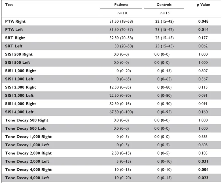
The number of the patients above 65 years old was 10 (29.41%) compared to 15 (33.33%) in the control group. The audiological test results of the two groups are compared in Table 5.
The PTA and tone decay scores in high frequencies were statistically higher in the older patient group.
Discussion
The concept that ET is a more complex and heterogeneous degenerative disease rather than a single modality is gradually becoming widespread.14However, certain accompanying neurological signs have recently been reported in many ET patients, such as
cogwheeling, difficulty in tandem gait, hypermetria, and vestibulo-ocular disturbances.15
In this study, we aimed to measure the prevalence of sensorineural hearing loss (accepted as a part of the disease) in ET patients without depression.
Audiologic tests are the gold standard to detect hearing loss, but the question ‘‘Do you have hearing loss?’’ was also shown to be quite sensitive and specific for the detection of hearing loss when compared with PTA results.16 In our study, 17 out of 34 (50.0%) ET patients reported hearing loss compared to just one (2.2%) subject in the control group, which confirmed that ET patients declared hearing loss even without performing audiologic testing.
PTA is a subjective test that determines the minimum tone frequency at which both ears can hear at various frequencies. In our study, no significant difference was found between the patient and control groups. The SRT measures subjects’ abilities to hear and understand speech.17If the difference from the pure tone average is more than 10 dB, retrocochlear pathologies or hearing loss that is not organic should be suspected. In our study, the difference between the pure tone average values and SRT values was not more than 7 dB for both ears in both groups. Furthermore, no significant difference was
found between the groups’ mean values. Although both tests indicate the presence of hearing loss, they do not discriminate whether it originates from cochlear or retrocochlear areas.
SISI test scores tend to be low at 250 and 500 Hz frequencies (0– 20%); suspicious at 1,000 Hz (40–60%); and very high at 2,000, 3,000, and 4,000 Hz frequencies (80–100%). Thus, the SISI test is quite successful for indicating cochlear injury, particularly at high frequen-cies.18We did not observe a significant difference between the SISI test scores of the two groups, even at the highest frequency.
The tone decay test measures the hearing adaptation phenomenon, which indicates variability in the sensitivity of the hearing system in response to a constant stimulus.19Tone decay values vary largely in subjects with cochlear loss and may reach 30 dB.20 Patients with normal or mild hearing loss at low frequencies may have high tone decay values at high frequencies.21Consistent with the literature, we found that the pure tone averages showed mild hearing loss, and the tone decay test values were found to be significantly higher for the right ears of the patients at 4,000 Hz compared to the control group. When young patients were excluded, there was a significant difference between the PTA scores in subjects older than 65 in both groups. In addition, there was a significant difference of tone decay test Table 1. Comparison of the Right and Left Ear PTA and SRT Results in Patients and Controls
Test Ear Patients Controls p Value
PTA Right 21 (3–58) 20 (7–42) 0.466
Left 19 (5–57) 18 (5–42) 0.644
SRT Right 22.5 (10–58) 20 (5–45) 0.397
Left 20 (10–58) 20 (5–45) 0.397
The scores are expressed as median score (minimum–maximum). Abbreviations: PTA, Pure Tone Audiometry; SRT, Speech Recognition Threshold.
Table 2. Comparison of the Right and Left Ear SISI Test Averages in Patients and Controls
Test Ear Patients Controls p Value
SISI 500 Right 0.0 (0–0) 0.0 (0–0) 1.000 Left 0.0 (0–0) 0.0 (0–0) 1.000 SISI 1,000 Right 0 (0–20) 0 (0–45) 0.446 Left 0 (0–65) 0 (0–65) 0.197 SISI 2,000 Right 0 (0–85) 0 (0–80) 0.210 Left 0 (0–85) 0 (0–80) 0.210 SISI 4,000 Right 0 (0–100) 0 (0–90) 0.090 Left 0 (0–100) 0 (0–95) 0.148
The scores are expressed as median score (minimum–maximum). Abbreviation: SISI, Short Increment Sensitivity Index.
scores of the right ear at 2,000 Hz and both ears at 4,000 Hz. As a result, the hearing test scores of older ET patients were significantly worse, which is consistent with a previous study.4
OAEs are present in 100% of subjects with normal hearing, and they indicate normal cochlear function.22In particular, the external ciliary cells (amplifier of the cochlea) must work regularly for OAE emergence.23The lack of OAE indicates the presence of sensorineural hearing loss. We could not obtain OAE in the right and left ears of 38.2% and 32.3% of patients and 13.3% and 15.5% of control subjects. In conclusion, the percentage of the patients in whom OAE could not be obtained in the right ear was significantly higher than in the control group. These results support the hypothesis of an increased prevalence of sensorineural hearing loss in ET patients compared to controls.
Although the pathogenesis of ET is controversial, the cerebellum is known to play an important role.24Recent postmortem studies have indicated a reduced number of Purkinje cells in the cerebella of ET patients without Lewy bodies.25Moreover, cerebello-thalamo-cortical pathway impairment is hypothesized in ET.26 One of the proposed explanations regarding the relationship between ET and hearing loss involves the passage of sound stimuli through the thalamus.4 An
abnormal stimulus of the ventral thalamus is also suspected in ET. Ondo et al. proposed that physiological perturbation in the ventral thalamus could account for both tremor and hearing loss.4
In our study, tone decay tests at high frequencies were significantly higher in ET patients compared to the control subjects. In addition, the number of subjects in whom OAEs could not be obtained was significantly higher for ET patients. Collectively, these findings support cochlear pathologies. Therefore, another possibility underlying the coexistence of ET and hearing loss is related to connexin proteins. Connexin genes encode the subunits of gap junction proteins, which enable rapid transport of ions, secondary messengers, and small metabolites for fast intercellular communication.27A study of patients who had Charcot-Marie-Tooth disease, an X-linked inherited condition associated with connexin 32 mutation, reported that additional findings such as sensorineural hearing loss and tremor could accompany the clinical manifestations.28 While the relationship between cochlear hearing loss and ET could be associated with connexin mutations, more studies are needed to clearly demonstrate this link.
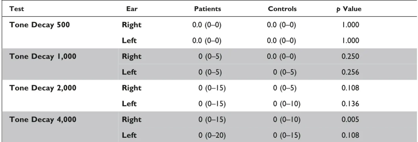
ET is accepted to be a neurodegenerative cerebellar disorder.29A few studies have reported on the relationship between hearing loss and Table 3. Comparison of the Right and Left Ear Tone Decay Test Averages in Patients and Controls
Test Ear Patients Controls p Value
Tone Decay 500 Right 0.0 (0–0) 0.0 (0–0) 1.000
Left 0.0 (0–0) 0.0 (0–0) 1.000
Tone Decay 1,000 Right 0 (0–5) 0.0 (0–0) 0.250
Left 0 (0–5) 0 (0–5) 0.256
Tone Decay 2,000 Right 0 (0–15) 0 (0–5) 0.108
Left 0 (0–15) 0 (0–10) 0.136
Tone Decay 4,000 Right 0 (0–15) 0 (0–10) 0.005
Left 0 (0–20) 0 (0–15) 0.108
The scores are expressed as median score (minimum–maximum).
Table 4. Comparison of the Right and Left Ear OAE Results in Patients and Controls
Patients Controls p Value
n (%) n (%)
Right OAE + 21 (61.3%) 39 (86.7%) 0.022
2 13 (38.7%) 6 (13.3%)
Left OAE + 23 (67.7%) 38 (84.4%) 0.136
2 11 (32.3%) 7 (15.6%)
other cerebellar degenerative disorders. Ikeda et al. reported that sensorineural hearing loss in spinocerebellar ataxia (SCA) 36 was due to inner ear or peripheral auditory system pathology and described a positive correlation between PTA values and disease duration.30
In conclusion, we performed SISI, tone decay, and OAE tests to assess cochlear pathology in both ET patient and control groups. Depressed subjects were excluded to avoid possible negative effects on ET findings and hearing test results. The significantly higher mean values of the tone decay test in high frequencies and the higher number of patients without OAEs indicate the presence of cochlear pathology in subjects with ET and support the findings of previous studies. Although our results were only modestly abnormal when compared to controls, this may be due to the relatively mild tremor
and young age of the ET population. Collectively, these results indicate that there may be a common factor in the pathogeneses of ET and sensorineural hearing loss, and further molecular genetic studies will shed light on the relationship between these two diseases.
References
1. Louis ED, Ottman R, Hauser WA. How common is the most common adult movement disorder? Estimates of the prevalence of essential tremor throughout the world. Mov Disord 1998;13:5–10, doi: http://dx.doi.org/10. 1002/mds.870130105.
2. Benito-Leo´n J, Louis ED. Essential tremor: emerging views of a common disorder. Nat Clin Pract Neurol 2006;2:666–677, doi: http://dx.doi.org/10.1038/ ncpneuro0347.
Table 5.Comparison of the Audiological Test Results in Patients and Controls Older than 65
Test Patients Controls p Value
n510 n515 PTA Right 31.50 (18–58) 22 (15–42) 0.048 PTA Left 31.50 (20–57) 23 (15–42) 0.014 SRT Right 32.50 (20–58) 25 (15–45) 0.177 SRT Left 30 (20–58) 25 (15–45) 0.062 SISI 500 Right 0.0 (0–0) 0.0 (0–0) 1.000 SISI 500 Left 0.0 (0–0) 0.0 (0–0) 1.000 SISI 1,000 Right 0 (0–20) 0 (0–45) 0.807 SISI 1,000 Left 0 (0–65) 0 (0–65) 0.367 SISI 2,000 Right 12.50 (0–85) 0 (0–80) 0.115 SISI 2,000 Left 22.50 (0–90) 0 (0–80) 0.091 SISI 4,000 Right 82.50 (0–95) 0 (0–90) 0.091 SISI 4,000 Left 67.50 (0–100) 0 (0–95) 0.160
Tone Decay 500 Right 0.0 (0–0) 0.0 (0–0) 1.000
Tone Decay 500 Left 0.0 (0–0) 0.0 (0–0) 1.000
Tone Decay 1,000 Right 0 (0–5) 0.0 (0–0) 0.683
Tone Decay 1,000 Left 0 (0–5) 0 (0–5) 0.605
Tone Decay 2,000 Right 2.50 (0–15) 0 (0–5) 0.103
Tone Decay 2,000 Left 5 (0–15) 0 (0–10) 0.031
Tone Decay 4,000 Right 10 (0–15) 0 (0–10) 0.004
Tone Decay 4,000 Left 10 (0–20) 0 (0–15) 0.023
The scores are expressed as median score (minimum–maximum). Abbreviations: PTA, Pure Tone Audiometry; SISI, Short Increment Sensitivity Index; SRT, Speech Recognition Threshold.
3. Cruickshanks KJ, Wiley TL, Tweed TS, et al. Prevalence of hearing loss in older adults in Beaver Dam, Wisconsin. The epidemiology of hearing loss study. Am J Epidemiol 1998;148:879–886, doi: http://dx.doi.org/10.1093/ oxfordjournals.aje.a009713.
4. Ondo WG, Sutton L, Dat Vuong K, Lai D, Jankovic J. Hearing impairment in essential tremor. Neurology 2003;61:1093–1097, doi: http://dx. doi.org/10.1212/01.WNL.0000086376.40750.AF.
5. Benito-Leo´n J, Louis ED, Bermejo-Pareja F, Neurological Disorders in Central Spain (NEDICES) Study Group. Reported hearing impairment in essential tremor: a population-based case-control study. Neuroepidemiology 2007; 29:213–217, doi: http://dx.doi.org/10.1159/000112463.
6. Balaban H, Altuntas EE, Uysal IO, Senturk IA, Topaktas S. Audio-vestibular evaluation in patients with essential tremor. Eur Arch Otorhinolaryngol 2012;269:1577–1581, doi: http://dx.doi.org/10.1007/s00405-011-1801-x.
7. Mulrow CD, Aguilair C, Endicott JE, et al. Quality-of-life changes and hearing impairment. A randomized trial. Ann Intern Med 1990;113:188–194, doi: http://dx.doi.org/10.7326/0003-4819-113-3-188.
8. Louis ED. Essential tremor as a neuropsychiatric disorder. J Neurol Sci 2010;289:144–148, doi: http://dx.doi.org/10.1016/j.jns.2009.08.029.
9. Louis ED, Huey ED, Gerbin M, Viner AS. Depressive traits in essential tremor: impact on disability, quality of life, and medication adherence. Eur J Neurol 2012;19:1349–1354, doi: http://dx.doi.org/10.1111/j.1468-1331.2012. 03774.x.
10. Chouinard S, Louis ED, Fahn S. Agreement among movement disorder specialists on the clinical diagnosis of essential tremor. Mov Disord 1997;12:973– 976, doi: http://dx.doi.org/10.1002/mds.870120621.
11. Fahn S, Tolosa E, Marin C. Clinical rating scale for tremor. In Jankovic J, Tolosa E (eds) Parkinson’s disease and movement disorders. Baltimore, Urban & Schwarzenberg, 1988:225–234.
12. Bain PG, Findley LJ, Atchison P, et al. Assessing tremor severity. J Neurol Neurosurg Psychiatry 1993;56:868–873, doi: http://dx.doi.org/10.1136/jnnp.56. 8.868.
13. Hamilton M. A rating scale for depression. J Neurol Neurosurg Psychiatry 1960;23:56–62, doi: http://dx.doi.org/10.1136/jnnp.23.1.56.
14. Deuschl G, Elble R. Essential tremor-neurodegenerative or nondegen-erative disease towards a working definition of ET. Mov Disord 2009;24:2033– 2041, doi: http://dx.doi.org/10.1002/mds.22755.
15. Benbir G, Ozekmekci S, Oguz S, Kenangil G, Ertan S, Akalan E. Quantitative analysis of reduced arm swing frequency in essential tremor. Eur Neurol 2010;63:302–306, doi: http://dx.doi.org/10.1159/000304194.
16. Sindhusake D, Mitchell P, Smith W, et al. Validation of self-reported hearing loss. The Blue Mountains Hearing Study. Int J Epidemiol 2001;30:1371– 1378, doi: http://dx.doi.org/10.1093/ije/30.6.1371.
17. Saunders AZ, Stein AV, Shuster NL. Audiometry. In Walker HK, Hall WD, Hurst JW (eds). Clinical methods: The history, physical and laboratory examinations. 3rd ed. Boston: Butterworths, 1990.
18. Buus S, Florentine M, Redden RB. The SISI test: a review. Part I. Audiology 1982;21:273–293, doi: http://dx.doi.org/10.3109/00206098209072744.
19. Bhatia PL, Sinha A, Abrol BM. The tone decay test-a simple and reliable audiological test. Laryngoscope 1969;79:1879–1890, doi: http://dx.doi.org/10. 1288/00005537-196911000-00002.
20. Gelfand SA. Behavioral tests for audiological diagnosis. In Hiscock T, Ip I, Caputo GR (eds) Essentials of audiology. 3rd ed. New York: Thieme Medical Publishers, 2009:302–231.
21. Huss M, Moore BCJ. Tone decay for hearing-impaired listeners with and without dead regions in the cochlea. J Acoust Soc Am 2003;114:3283–3294, doi: http://dx.doi.org/10.1121/1.1624063.
22. Zimatore G, Hatzopaulos S, Giuliani A, Martini A, Colosimo A. Comparison of transient otoacoustic emission responses from neonatal and adult ears. J Appl Physiol 2002;92:2521–2528.
23. Sliwinska-Kowalska M, Kotylo P. Otoacoustic emissions in industrial hearing loss assessment. Noise Health 2001;3:75–84.
24. Jenkins IH, Bain PG, Colebatch JG, et al. A positron emission tomography study of essential tremor: evidence for overactivity of cerebellar connections. Ann Neurol 1993;34:82–90, doi: http://dx.doi.org/10.1002/ana. 410340115.
25. Axelrad JE, Louis ED, Honig LS, et al. Reduced Purkinje cell number in essential tremor: a postmortem study. Arch Neurol 2008;65:101–107, doi: http://dx.doi.org/10.1001/archneurol.2007.8.
26. Tro¨ster AI, Woods SP, Fields JA, et al. Neuropsychological deficits in essential tremor: an expression of cerebello-thalamo-cortical pathophysiology? Eur J Neurol 2002;9:143–151, doi: http://dx.doi.org/10.1046/j.1468-1331. 2002.00341.x.
27. Steel KP. A new era in the genetics of deafness. N Engl J Med 1998;339: 1545–1547, doi: http://dx.doi.org/10.1056/NEJM199811193392110.
28. Yiu EM, Geevasinga N, Nicholson GA, Fagan ER, Ryan MM, Ouvrier RA. A retrospective review of X-linked Charcot-Marie-Tooth disease in childhood. Neurology 2011;76:461–466, doi: http://dx.doi.org/10.1212/WNL. 0b013e31820a0ceb.
29. Louis ED. Essential tremor: evolving clinicopathological concepts in an era of intensive post-mortem enquiry. Lancet Neurol 2010;9:613–622, doi: http:// dx.doi.org/10.1016/S1474-4422(10)70090-9.
30. Ikeda Y, Ohta Y, Kurata T, Shiro Y, Takao Y, Abe K. Acoustic impairment is a distinguishable clinical feature of Asidan/SCA36. J Neurol Sci 2013;32:109–112, doi: http://dx.doi.org/10.1016/j.jns.2012.10.013.