E-mail: [email protected] www.wjgnet.com Copyright © 2004 by The WJG Press ISSN 1007-9327
•
CLINICAL RESEARCH
•
Expression of bcl-2 family of genes during resection induced
liver regeneration: Comparison between hepatectomized and
sham groups
Kamil Can Akcali, Aydin Dalgic, Ahmet Ucar,Khemaeis Ben Haj, Dilek Guvenc Kamil Can Akcali, Ahmet Ucar,Khemaeis Ben Haj, Department
of Molecular Biology and Genetics, Bilkent University, Ankara, Turkey, 06800
Dilek Guvenc, Department of Mathematics, Bilkent University, Ankara, Turkey, 06800
Aydin Dalgic, Department of Surgery, Gazi University Medical School, Ankara, Turkey, 06510
Supported by Bilkent University Faculty Development Grant and Bilkent University Research Grant
Correspondence to: Dr. Kamil Can Akcali, Department of Molecular Biology and Genetics, Bilkent University, Bilkent, Ankara, Turkey, 06800. [email protected]
Telephone: +90-312-2902418 Fax: +90-312-2665097 Received: 2003-09-06 Accepted: 2003-10-12
Abstract
AIM: During liver regeneration cellular proliferation and apoptosis result in tissue remodeling to restore normal hepatic mass and structure. Main regulators of the apoptotic machinery are the Bcl-2 family proteins but their roles are not well defined throughout the liver regeneration. We aimed to analyze the expression levels of bcl-2 gene family members during resection induced liver regeneration.
METHODS: We performed semi-quantitative RT-PCR to examine the expression level of bak, bax, bcl-2 and bcl-xL in
the 70% hepatectomized rat livers during the whole regeneration process and compared to that of the sham and normal groups.
RESULTS: The expression of bak and bax were decreased whereas bcl-2 and bcl-xL were increased in hepatectomized
animals compared to normal liver at most time points. We also reported for the first time that sham group of animals had statistically significant higher expression of bak and bax compared to hepatectomized animals. In addition, the area under the curve (AUC) values of these genes were more in sham groups than the hepatectomized groups.
CONCLUSION: We conclude that the expressional changes of bak, bax, bcl-2 and bcl-xL genes were altered not only
due to regeneration, but also due to the effects of surgical operations.
Akcali KC, Dalgic A, Ucar A, Ben Haj K, Guvenc D. Expression of
bcl-2 family of genes during resection induced liver regeneration:
Comparison between hepatectomized and sham groups. World
J Gastroenterol 2004; 10(2): 279-283
http://www.wjgnet.com/1007-9327/10/279.asp
INTRODUCTION
Liver regeneration is a complex physiological response that takes place after the loss of hepatocytes caused by toxic or
viral injury or secondary to liver resection[1,2]. During regeneration, series of reactions take place to maintain the homeostasis and virtually all of the surviving hepatocytes undergo mitosis[3]. Experimentally partial hepatectomy (PH) has been a useful model to study the cellular mechanisms of hepatic regeneration.
Post-hepatectomy-induced proliferative response led to full restoration of the hepatic mass in rats in 14 days[4,5]. Several converging lines of evidence from recent works have established that growth factors and cytokines including hepatocyte growth factor (HGF), tumor necrosis factor-α (TNF-α), interleukin-6 (IL-6), epidermal growth factor (EGF), transforming growth factor-α (TGF-α), fibroblast growth factor (FGF), vascular endothelial growth factor (VEGF), transforming growth factor-β1 (TGF-β1) and hormones such as insulin, glucagon, sex hormones, thyroid hormone, norepinephrine, nitric oxide and vasopressin are important components of liver regeneration[5-9]. Multiple signaling pathways are then activated by these components[10,11]. During liver regeneration, apoptosis occurs as a response to eliminate the defective cells that appear due to rapid cellular divisions after PH, resulting in fine-tuning of the liver size and tissue remodeling[5,12-14]. Therefore, control of apoptosis plays a crucial role in liver regeneration. Among the regulators of apoptosis, the Bcl-2 family of proteins determines the life-or-death of a cell by controlling the releases of mitochondrial apoptogenic factors, cytochrome C and apoptosis inducing factors (AIF), which activate the downstream executional phases, including the activation of the caspases[15,16]. Bcl-2 family of genes consists of both pro- and anti-apoptotic genes and by forming dimers, they exert their function[17]. Since Bcl-2 family of proteins is the most important and critical regulators of apoptosis[18], they should be tightly regulated during regeneration in a time-dependent manner. Existing data suggests the involvement of these proteins in regeneration process during the initial stages of this process[19-23].These studies investigated the expression of these genes during the first four days after hepatectomy, however the expression pattern of the Bcl-2 family members were not examined throughout the regeneration process. Therefore, the purpose of the present study was to quantitate the expression levels of some members of bcl-2 family of genes (bcl-2, bcl-xL, bax,
bak) by using semi-quantitative RT-PCR within a time
spectrum that extended to 14 days after hepatectomy, which was needed for the completion of the regeneration process. We also compared the expression levels of these genes with the levels of the corresponding sham group of animals. For this purpose, we used the “area under the curve” method as used in pharmacodynamic studies and other liver injury and stress models.
MATERIALS AND METHODS Animals
Nine weeks old and 200-250 grams male Sprague-Dawley rats were used. They were housed under controlled environmental
conditions (22 ) with a 12-hour light and 12 hour dark cycle in the animal holding facility of Bilkent University, Turkey. All the animals received care according to the criteria outlined in the “Guide for Care and Use of Laboratory Animals” prepared by the National Academy of Science and this study protocol complied with Bilkent University’s guidelines on humane care and use of laboratory animals.
Experimental groups, partial hepatectomy and sham groups
Three randomly selected three animals were used for each time point. After injecting Ketamine (Ketalar, Park Davis) subcutaneously at a dose of 30 mg/kg, liver resections consisting of 70% of the liver mass were performed in PH group[24]. Sham group of animals underwent the same per operative anesthesia with the PH group. All the surgical operations were done the same as PH, but the liver lobes were not resected. All the operations were performed between 8:00 AM and 12:00 PM to minimize diurnal effects. After the completion of the procedure, the animals were placed under a lamp to prevent the hypotermy and then put into cages (one animal per cage) with continuous supply of food and water. The animals in the PH and corresponding sham groups were sacrificed at 0.5, 2, 8, 18, 36 hours, 3 days, 7 days and 14 days after the operation. The group of animals in which no surgery was performed, as used as normal liver group and mentioned time “0” in quantitated graphs. After sacrifying the animal by cervical dislocation, the remnant liver lobes were excised and washed in DMEM medium, then immediately frozen in liquid nitrogen.
Total RNA isolation and reverse transcription
The RNAs were isolated from all the liver samples using Tripure solution (Roche- Boehringer, Mannheim) according to the manufacturer’s protocol. The integrity of the isolated RNA samples was determined by denaturing- (formaldehyde-) agarose gel electrophoresis. The cDNA samples were synthesized from the total RNA samples with the RevertAid First Strand cDNA Synthesis Kit (MBI Fermentas) by using the manufacturer’s protocol.
Primer design and semi-quantitative PCR
We designed primers for bcl-2, bcl-xL and bax by using the
cDNA sequences of rat homologues of these genes (GenBank accession numbers of rat homologues of bcl-2 is NM_016993,
bcl-xL is U34963, and bax is S76511). In the case of bak, the
cDNA sequences of mouse (Y13231) and human (U23765) were aligned by Blast (NCBI), and primers were chosen from the longest conserved regions by selecting the mouse sequences in the regions of mismatches. As a housekeeping gene, we used cyclophilin, and primers were designed by using the rat cDNA sequence (GenBank accession number: M19533). The primer pairs used for each gene were as follows: cyclophilin: GGGAAGGTGAAAGAAGGCAT and GAGAGCAGAG ATTACAGGGT; bcl-2: CCTGGCATCTTCTCCTTC and TGCTGACCTCACTTGTGG; bcl-xL: TCAATGGCAACC
CTTCCTGG and ATCCGACTCACCAATACCTG; bax: ACGCATCCACCAAGAAGC and GAAGTCCAGAGTC CAGCC; bak: CCGGAATTCCAGGACACAGAGGA and CCAAGCTTGCCCAACAGAACCAC.
In all the reactions, the negative control group was done by using ddH2O instead of cDNA. For each gene, we determined the cycle number of PCR reactions in which the PCR reaction was not saturated. Based on this, we used the following PCR conditions: The initial denaturation step was at 95 , followed by 18 (cyclophilin), 33 (bcl-2), 37 (bcl-xL), 28 (bax), 29 (bak)
cycles of denaturation for 30 seconds at 95 , annealing for 30 seconds at either 55 (cyclophilin, bcl-2, bak) or 52
(bcl-xL), or 60 (bax), and extension for 30 seconds at 72 .
A final extension at 72 for 10 minutes was applied to all the reactions and the PCR products were electrophoresed on a 1.2% agarose gel. Each PCR reaction was replicated three times. The quantitated values for the expression of bcl-2 family members were normalized with the quantitated values for the
cyclophilin for each sample respectively by comparing with
the expression level of cyclophilin in normal liver. The normalized values were then analyzed using Multi-Analyst software and the graphs were drawn.
Calculation of AUC
Area under the curve (AUC) calculations was performed as shown by Tygstrup et al[25]. Results of the expression levels of the bcl-2 family genes by RT-PCR are given as mean ± SEM. As a measure of the change in expression level during the experimental period, the area of the expression level/time curve (AUC: cDNA level x time) was calculated as the sum of the area of the intervals between the samplings for PH and sham groups. Since the time intervals were different, in order to standardize the calculations, we used hour as the unit of X-axis i.e. between 7 days and 14 days, we multiplied with 168 (24x7) to calculate the area under the curve. To determine the hourly changes at the AUC, we divided the AUC values by the hour difference between each two-time points; i.e. between 2 and 8 hour groups, AUC value was calculated and divided by 6.
Statistical tests
For each group of three animals the mean expression level was calculated at the given time points. The means of PH and sham groups were compared using Mann-Whitney test. Since sham group animals seemed to have higher levels of expression of these genes than the hepatectomized animals, one-sided significance level (P value) of 0.05 was used. The null hypothesis of no difference between the expression of bax, bak, bcl-2, and bcl-xL in PH and sham groups was tested versus the research
hypothesis that PH group had lower level of expression than the sham group.
RESULTS
Since the transcriptional control of bcl-2 gene family has been known to be important, we performed semi-quantitative RT-PCR to examine the changes in the expression levels of the transcripts of bcl-2, bcl-xL, bax and bak in the livers of rats
that were subjected to either 70% PH, or sham operation at different time points (Figures 1-4). In all the experiments, the expression at “time 0” was the quantitated expression of each gene in normal livers and accepted as “1” in order to make a comparison with the subsequent time groups.
Expression pattern of pro-apoptotic genes
Two hours after hepatectomy, in 70% PH groups, we observed that the transcript level of bak was decreased by twofold compared with that in normal liver (Figure 1, “time 0”). Although bak expression levels reached the levels observed in normal liver at 8 hours and 7 days, bak was expressed less in comparison with that in the normal liver at other time points (Figure 1, solid circles). On the other hand, in sham group of animals, the expression of bak mRNA was higher than that in the normal liver and hepatectomized group at every time point (Figure 1, open circles). The expression of bak was significantly higher in sham group than in PH group (P<0.001).
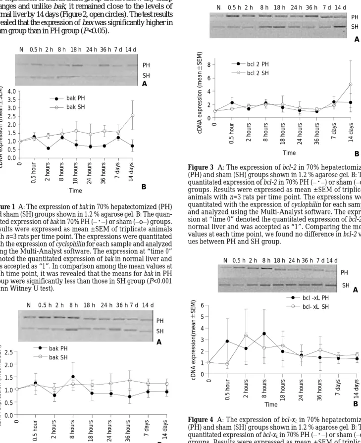
Another pro-apoptotic gene, bax mRNA expression, like
bak, was increased at 0.5 hour after hepatectomy and then
decreased to below the level in normal liver by 2 hours (Figure 2, solid circles). During the 14-day period except the increase
at 8 hours after hepatectomy, bax expression showed a steady pattern and by 14 days after hepatectomy, it reached almost the expression level of the normal liver (Figure 2, solid circles). The expression of bax in sham group did not show any sharp changes and unlike bak, it remained close to the levels of normal liver by 14 days (Figure 2, open circles). The test results revealed that the expression of bax was significantly higher in sham group than in PH group (P<0.05).
Figure 1 A: The expression of bak in 70% hepatectomized (PH) and sham (SH) groups shown in 1.2 % agarose gel. B: The quan-titated expression of bak in 70% PH (__
·
__) or sham (__o__) groups.Results were expressed as mean ± SEM of triplicate animals
with n=3 rats per time point. The expressions were quantitated with the expression of cyclophilin for each sample and analyzed using the Multi-Analyst software. The expression at “time 0” denoted the quantitated expression of bak in normal liver and was accepted as “1”. In comparison among the mean values at each time point, it was revealed that the means for bak in PH group were significantly less than those in SH group (P<0.001 Mann Witney U test).
Figure 2 A: The expression of bax in 70% hepatectomized (PH) and sham (SH) groups shown in 1.2% agarose gel. B: The quan-titated expression of bax in 70% PH (__
·
__) or sham (__o__) groups.Results were expressed as mean ± SEM of triplicate animals
with n=3 rats per time point. The expressions were quantitated with the expression of cyclophilin for each sample and analyzed using the Multi-Analyst software. The expression at “time 0” denoted the quantitated expression of bax in normal liver and was accepted as “1”. In comparison among the mean values at
each time point, it was revealed that the bax values in PH group were less than those in SH group (one-tailed P<0.05, Mann Witney U test).
Figure 3 A: The expression of bcl-2 in 70% hepatectomized (PH) and sham (SH) groups shown in 1.2 % agarose gel. B: The quantitated expression of bcl-2 in 70% PH (__
·
__) or sham (__o__)groups. Results were expressed as mean ± SEM of triplicate
animals with n=3 rats per time point. The expressions were quantitated with the expression of cyclophilin for each sample and analyzed using the Multi-Analyst software. The expres-sion at “time 0” denoted the quantitated expresexpres-sion of bcl-2 in normal liver and was accepted as “1”. Comparing the mean values at each time point, we found no difference in bcl-2 val-ues between PH and SH group.
Figure 4 A: The expression of bcl-xL in 70% hepatectomized (PH) and sham (SH) groups shown in 1.2 % agarose gel. B: The quantitated expression of bcl-xL in 70% PH (__
·
__) or sham (__o__) groups. Results were expressed as mean ± SEM of triplicateanimals with n=3 rats per time point. The expressions were quantitated with the expression of cyclophilin for each sample and analyzed using the Multi-Analyst software. The expres-sion at “time 0” denoted the quantitated expresexpres-sion of bcl-xL in normal liver and was accepted as “1”. Comparing the mean values at each time points, no difference in bcl-xL values be-tween PH and SH groups was found.
Expression pattern of anti-apoptotic genes
Amongst the anti-apoptotic members of 2 gene family,
bcl-N 0.5 h 2 h 8 h 18 h 24 h 36 h 7 d 14 d PH SH 4.0 3.5 3.0 2.5 2.0 1.5 1.0 0.5 0.0 0
0.5 hour 2 hours 8 hours 18 hours 24 hours 36 hours 7 days 14 days
cDNA expression (mean
SEM) bak PH bak SH Time A B N 0.5 h 2 h 8 h 18 h 24 h 36 h 7 d 14 d 0
0.5 hour 2 hours 8 hours 18 hours 24 hours 36 hours 7 days 14 days
PH SH 2.5 2.0 1.5 1.0 0.5 0.0
cDNA expression (mean
SEM) Time bak PH bak SH A B N 0.5 h 2 h 8 h 18 h 24 h 36 h 7 d 14 d PH SH 0
0.5 hour 2 hours 8 hours 18 hours 24 hours 36 hours 7 days 14 days
bcl 2 PH bcl 2 SH Time 8 6 4 2 0
cDNA expression (mean
SEM)
A
B
N 0.5 h 2 h 8 h 18 h 24 h 36 h 7 d 14 d
0
0.5 hour 2 hours 8 hours 18 hours 24 hours 36 hours 7 days 14 days
bcl -xL PH bcl- xL SH 6 5 4 3 2 1 0 cDNA expression(mean SEM) Time A B PH SH
2 mRNA expression did not decrease to below the level in
normal liver both in PH and sham groups. Its level was increased 0.5 hour after hepatectomy and peaked at 8 hours and 7 days after hepatectomy, more than 2-fold that of the normal liver (Figure 3, solid circles). In the sham group of animals, the expression of this gene also increased by 0.5 hour after surgery and then followed a steady level until day 14. The expression of bcl-2 in PH and sham group showed a similar pattern except at 14 day. At this time point, bcl-2 expression was more than two-fold in sham group compared to PH group (Figure 3, open circles).
The expression of another anti-apoptotic gene bcl-xL showed
more than 3-fold increase by 8 hours after hepatectomy whereas at the same time interval, the sham group had almost two-fold increase compared to normal liver. The amount of bcl-xL
expression decreased afterwards both in PH and sham group (Figure 4). However, in both groups, the expression level was always above the levels in normal liver at all time points.
AUC calculation
In order to better compare the expression levels of bak, bax,
bcl-2 and bcl-xL during the whole regeneration process, we
calculated the AUC values for partially hepatectomized and sham groups, i.e.; the area between the lines connecting cDNA levels of different time and zero. It is important to emphasize that AUC is an arbitrary value but provides a comparison between the total expression of these genes in PH and sham groups. The AUC values for PH and sham groups for each gene analyzed are shown in Table 1. AUC values of all the genes were higher in sham group compared with those in the PH group. Interestingly however, the difference was more obvious for pro-apoptotic genes (bak and bax) than for anti-apoptotic genes (bcl-2 and bcl-xL). We detected an 87% and
64% increase in expression levels of pro-apoptotic genes (bak and bax respectively) in sham group compared to PH group, whereas, this difference was 25% in bcl-2 and 21% in bcl-xL .
Since the time intervals ranged from 0.5 hour to 7 days, we calculated hourly changes of AUC values in our experiments for each of bcl-2 family of genes in hepatectomized and sham group of animals. We measured AUC value for each time group and divided this value by the hours of that particular group (Table 2). The values for bak and bax were found to be significantly higher in sham group than in hepatectomized group (P<0.05). No significant difference was found for bcl-2 and bcl-xL values.
Table 1 AUC of cDNA expression for members of the bcl-2 family of genes
AUC (70% PH) AUC (SH) Change
bak 324 609 87%
bax 245 402 64%
bcl-2 666 837 25%
bcl-xL 494 601 21%
DISCUSSION
During liver regeneration, apoptosis allows hepatocytes to die without provoking a potentially harmful inflammatory response. The harmony amongst these complex biological responses is crucial since abnormal regeneration may result in fulminant liver failure, cirrhosis and primary liver cancers[26]. Since programmed cell death is a major component of hepatic regeneration mechanism, and the bcl-2 family members are the main regulators of this cellular death pathway, we concentrated on the expression level of these genes throughout the regeneration of liver induced by partial hepatectomy. For all of the members of this family, it has been shown that the transcriptional activation or suppression is critical[27,28].Existing data in literature suggests the involvement of these genes in regeneration process however, the expression of these genes has been shown for only 4 days after hepatectomy[19-23]. Liver regeneration is a long lasting event but there have been no studies regarding the expression of these genes during the later stages of liver regeneration. Since apoptosis is a homeostatic mechanism, the expression of the genes that regulate apoptosis should be important during the whole regeneration process. Therefore, in this study, we quantitated the expression levels of bak, bax, bcl-2 and bcl-xL up to 14 days post-hepatectomy,
which is the time needed for the completion of regeneration process. In addition, we also examined the expression of these genes in corresponding sham group of animals.
Our data regarding the expression of bak, bax and bcl-xL
during early stages of regeneration (within the first four days) were consistent with previous studies[19-23]. However contradictory data have been published about the expression of bcl-2 in normal liver prior to hepatectomy. It has been reported that by using Northern blot Tzung et al[29] did not find any bcl-2 expression, whereas Kren et al[21] did. Similar to our results Masson et al[22] also found a basal expression of
bcl-2 by using RT-PCR. Since the source of bcl-2 is
non-parenchymal cells, a more sensitive method such as RT-PCR is a better choice of analyzing its expression.
According to our results, the expression levels of bax and
bak in PH group were lower than those in the normal liver
except 0.5 and 8 hours after 70% hepatectomy. On the other hand, the anti-apoptotic genes, bcl-2 and bcl-xL had higher
expression levels than those in the normal liver at most of the time points. Amongst them, the expression of bcl-xL in
hepatectomized animalswas higher than that of normal liver at all the time points. We observed an overall decrease in the pro-apoptotic genes (bax and bak) and an increase in the anti-apoptotic genes (bcl-2 and bcl-xL) expression compared to that
in normal liver. Recently, Locker et al has also shown increased antiapoptotic regulators and down-regulated proapoptotic regulators by using microarray technology during liver regeneration in mouse[30]. Thus, the increase in the ratio of
anti-vs. pro-apoptotic gene expression was in favor of survival of
the liver tissue throughout the regeneration process.
In our study, we also compared the total expression of these Table 2 Hourly changes of AUC values for the members of the bcl-2 family of genes
bak PHa bak SHa bax PHa bax SHa bcl-2 PH bcl-2 SH bcl-xL PH bcl-xL SH
0.5-2 h 0.94 1.25 1.12 2.36 1.81 1.56 2.53 2.13 2-8 h 0.91 1.37 0.8 1.12 1.76 1.88 2.86 2.82 8-18 h 1.01 1.5 1.15 1.19 1.86 2 2.73 2.36 18-24 h 0.75 1.53 0.8 1.19 1.33 1.73 1.78 1.99 24-36 h 0.7 1.52 0.75 1.03 1 1.28 1.53 1.82 36-168 h 0.97 1.58 0.45 1.29 1.65 1.53 1.41 1.87 168-33 6h 0.98 2.1 0.9 1.2 2.35 3.42 1.37 1.64
genes between sham and partially hepatectomized groups by calculating AUC as used by Tygstrup et al[25]. In their study AUC calculation was used to show and compare the mRNA profiles of a set of liver function related and repair related genes in different liver injury and stress models. By comparing AUC values, they have shown that the expressions of several genes related to liver function were reduced[25]. By calculating AUC values, we found an interesting phenomenon that had not been reported in previous studies. The AUC values of all the bcl-2 family of genes were higher in sham group than that of PH group, suggesting the overall expression levels of apoptotic regulator genes in sham group were increased more than those in the hepatectomized group. Since the sham group of animals had undergone the same period of anesthesia and were sacrificed at the same time, the effect of circadian rhythms on the expression of these genes could be ruled out. In addition, when we measured the hourly changes of the expression of these genes, we found an increase in all the genes that we studied in the sham group compared to PH group except for the first 18 hours in bcl-xL. The increase in the case of bak and
bax were statistically significant. This indicated that the
expressions of bak and bax were up regulated in sham group compared to those in hepatectomized group. Therefore, many of the previous findings of expressional changes in transcript levels of bcl-2, bcl-xL, bax and bak might be due to not only
regeneration, but also the effects of surgical operations. The reduction in the expression of proapoptotic bak and
bax in PH group compared to sham group may be related to
the enhanced effects of some growth factors on liver proliferation to prevent apoptosis and to ensure the survival of the tissue. It has also been reported that the majority of the rats that had undergone PH, later developed tumors in their remnant liver[31]. Therefore it is tempting to speculate that decreased expression of proapoptotic genes might be responsible for this outcome. However, the cause for the reduction in the expression of anti-apoptotic bcl-2 family of genes in PH group compared to sham group has been unclear. In the case of bcl-2, since it is expressed exclusively by non-parenchymal cells and especially cholangiocytes, removal of a large portion of the liver might be the explanation for this reduction. Thus, it could be speculated that upon removal of a large portion of the tissue, the liver conferred priority to the expression of vitally most important genes.
REFERENCES
1 Sell S. Is there a liver stem cell? Cancer Res 1990; 50: 3811-3815
2 Fausto N. Hepatology- A Textbook of Liver Disease, 2nd ed.,
Philadelphia: Saunders 1990: 49-64
3 Fausto N, Webber EM. Liver Regeneration In: Arias IM, Boyer
JL, Fausto N, Jakoby WB, Schachter DA, Shafritz DA, eds. The Liver: Biology and Pathobiology. New York: Raven 1994: 1059–1084
4 Fausto N, Thomson NL, Braun L. Cell separation: Methods and
selected applications. Orlando: Academic Press 1986: 45-75
5 Fausto N. Liver regeneration. J Hepatol 2000; 32(1 Suppl): 19-31
6 Bucher NLR. Liver regeneration then and now In: Jirtle RL, ed.
Liver regeneration and carcinogenesis, Molecular and Cellular Mechanisms. San Diego: Academic Press 1995: 1-25
7 Michalopoulos G, Defrances M. Liver regeneration. Science 1997;
276: 60-66
8 Xia M, Xue SB, Xu CS. Shedding of TNFR1 in regenerative liver
can be induced with TNF alpha and PMA. World J Gastroenterol
2002; 8: 1129-1133
9 Li YC, Xu CS, Zhu WL, Li WQ. Isolation and analysis of a novel
gene over-expressed during liver regeneration. World J
Gastroenterol 2003; 9: 1282-1286
10 Brenner DA. Signal transduction during liver regeneration. J
Gastroentorol Hepatol 1998; 13: S93-95
11 Stolz DB, Mars WM, Petersen BE, Kim TH, Michalopoulos GK.
Growth factor signal transduction immediately after two-thirds partial hepatectomy in the rat. Cancer Res 1999; 59: 3954-3960
12 Fan G, Kren BT, Steer CJ. Regulation of apoptosis associated genes
in the regenerating liver. Semin Liver Dis 1998; 18: 123-140
13 Kren BT, Trembley JH, Fan G, Steer CJ. Molecular regulation of
liver regeneration. Ann N Y Acad Sci 1997; 831: 361-381
14 Sakamoto T, Liu Z, Murase N, Ezure T, Yokomuro S, Poli V,
Demetris AJ. Mitosis and apoptosis in the liver of interleukin-6-deficient mice after partial hepatectomy. Hepatology 1999; 29: 403-411
15 Martinou JC, Green DR. Breaking the mitochondrial barrier. Nat
Rev Mol Cell Biol 2001; 2: 63-67
16 Zamzami N, Kroemer G. The mitochondrion in apoptosis: how
Pandora’s box opens. Nat Rev Mol Cell Biol 2001; 2: 67-71
17 Adams JM, Cory S. The Bcl-2 protein family: Arbiters of cell
survival. Science 1998; 281: 1322-1326
18 Cory S, Adams JM. The Bcl-2 family: regulators of the cellular
life-or-death switch. Nat Rev Cancer 2002; 2: 647-656
19 Kamimukai N, Togo S, Hasegawa S, Kubota T, Kurosawa H, Li
XK, Suzuki S, Shimada H. Expression of Bcl-2 family reduces apoptotic hepatocytes after excessive hepatectomy. Eur Surg Res 2001; 33: 8-15
20 Taira K, Hiroyasu S, Shiraishi M, Muto Y, Koji T. Role of the Fas
system in liver regeneration after a partial hepatectomy in rats.
Eur Surg Res 2001; 33: 334-341
21 Kren BT, Trembly JH, Krajewski J, Behren TW, Reed JC, Steer CJ.
Modulation of apoptosis-associated genes bcl-2, bcl-x, and bax during rat liver regeneration. Cell Growth Differ 1996; 7: 1633-1642
22 Masson S, Scotte M, Garnier S, Francois A, Hiron M, Teniere P,
Fallu J, Salier JP, Daveau M. Differential expression of apoptosis-associated genes post-hepatectomy in cirrhotic vs normal rats.
Apoptosis 2000; 5: 173-179
23 Karavias DD, Tsamandas AC, Tepetes K, Kritikos N, Kourelis T,
Ravazoula P, Vagenas K, Siasos N, Mirra N, Bonikos DS. BCL-2 and BAX expression and cell proliferation, after partial hepatec-tomy with and without ischemia, on cholestatic liver in rats: an experimental study. J Surg Res 2003; 110: 399-408
24 Wang X, Parsson H, Anderson R, Soltesz V, Johansson K,
Bengmark S. Bacterial translocation, intestinal ultrastructure and cell membrane permeability early after major liver resection in the rat. Br J Surg 1994; 81: 579-584
25 Tygstrup N, Bangert K, Ott P, Bisgaard HC. Messenger RNA
pro-files in liver injury and stress: A comparison of lethal and nonle-thal rat models. Biochem Biophys Res Comm 2002; 290: 518-525
26 Diehl AM. Liver generation. Front Biosci 2002; 7: e301-314
27 Gross A, McDonnell JM, Korsmeyer SJ. Bcl-2 family members
and the mitochondria in apoptosis. Genes Dev 1999; 13: 1899-1911
28 Tsujimoto Y, Shimizu S. Bcl-2 family: Life-or-death switch. FEBS
Lett 2000; 466: 6-10
29 Tzung SP, Fausto N, Hockenberry DM. Expression of Bcl-2
fam-ily during liver regeneration and identification of bcl-x as a de-layed early response gene. Am J Pathol 1997; 150: 1985-1995
30 Locker J, Tian J, Carver R, Concas D, Cossu C, Ledda-Columbano
GM, Columbano A. A common set of immediate-early response genes in liver regeneration and hyperplasia. Hepatology 2003; 38: 314-325
31 Picardo A, Karpoff HM, Ng B, Lee J, Brennan MF, Fong Y.
Par-tial hepatectomy accelerates local tumor growth: potenPar-tial roles of local cytokine activation. Surgery 1998; 124: 57-64