THE ROLE OF SUPEROXIDE DISMUTASE (SOD) ACTIVITY AND
MALONDIALDEHYDE (MDA) LEVELS IN THE DIFFERENTIATION
OF BENIGN-MALIGN PLEURAL EFFUSION
Tansu ULUKAVAK ÇİFTÇİ
1, Yıldız GÜNEY
2, Ayşe BİLGİHAN², Filiz ÇİMEN
31
Department of Chest Diseases, Gazi University Hospital, Ankara, Turkey 2
Department of Radiation Oncology, Ankara University Hospital, Ankara, Turkey 3
Atatürk Chest Diseases and Chest Surgery Hospital, Ankara, Turkey
ABSTRACT
The Role Of Superoxide Dismutase (SOD) Activity And Malondialdehyde (MDA) Levels In The Differentiation Of Benign-Malign Pleural Effusion
Introduction: Free radicals are highly reactive oxygen-containing molecular species that have been implicated in the pathogenesis of many diseases. There is a critical balance between free radical generation and antioxidant defenses. It has been suggested that oxidative stress may be associated with malignancy. Pleural effusions (PE) complicating bronchogenic malignancy is not uncommon but many diseases that cause PE are also present. The aim of our study was to assess the level of the malondialdehide (MDA) and antioxidant enzyme superoxide dismutase (SOD), in pleural fluids of malign and benign patients.
Material and Methods: Forty-eight patients were enrolled into the study. Group I had 24 cases with malign pleural effusion and group II had 24 cases with benign pleural effusion. The MDA levels and SOD activities in pleural fluid were quantified.
Results: There was a significant increase in MDA levels in group I (135,15 ± 18,95 nmol/ml) than in group II (67,35 ± 13,02 nmol/ml) (p<0,001). The activities of SOD is found to be increased significantly in group I when compared to group II (40,33 ± 7,1 U/ml vs 33,2 ± 6,2 U/ml) (p< 0,05).
Discussion: These findings may support the previous findings showing that reactive oxygen species increased in malignant pleural fluid with increased activities of SOD. These parameters might be helpful in differation of malign from benign effusion and could be used as non-spesific diagnostic markers.
Keywords: Pleural effusion, SOD, MDA ÖZET
Giriş:Serbest radikaller birçok hastalığın patogenezinde yer alan yüksek oranda oksijen içeren moleküllerdir. Serbest radikal oluşumu ile antioksidan savunma sistemi arasında kritik bir denge vardır. Oksidatif stresin malignensi ile birlikte olduğu bilinmektedir. Pleural effüzyon (PE) bronkojenik malignensi komplikasyonu olarak nadir değildir fakat diğer bazı hastalıklarda da PE görülebilir. Bu çalışmanın amacı malign ve benign hastalarda pleural sıvıdaki malondialdehide (MDA) seviyesi ve antioksidant enzim süperoksid dismutaz (SOD) aktivitesinin arasında herhangi bir fark olup olmadığını saptamaktır.
Gereç ve Yöntem:Bu çalışmaya kırksekiz hasta dahil edildi. Grup I’de 24 malign pleural effüzyonlu hasta ve grup II’de ise 24 benign pleural effüzyonlu hasta vardı. MDA seviyesi ve SOD aktivitesi pleural sıvıda incelendi.
Sonuçlar:MDA seviyesi Grup I’de (135,15 ± 18,95 nmol/ml) group II’ ye göre belirgin olarak yüksektir (67,35 ± 13,02 nmol/ml) (p<0,001). SOD aktivitesi ise grup I’de grup II ile karşılaştırıldığında anlamlı
olarak yüksek bulunmuştur. (40,33 ± 7,1 U/ml vs 33,2 ± 6,2 U/ml) (p< 0,05).
Tartışma: Bu sonuçlarımız reaktif oksijen türlerinin malign pleural sıvıda SOD aktivitesi ile birlikte arttığını göstermektedir. Bu parametreler malign efüzyonu bening efüzyondan ayırmak için non-spesifik bir diagnostik markır olabilir.
Anahtar Kelimeler: Pleural efüzyon, SOD, MDA
Introduction
Experimental and epidemiological studies implicate the involvement of oxygen derived radicals in the pathogenesis of cancer development in the last years (1). In healthy conditions at the cellular level, a subtle balance exists between the free radical generation and the antioxidant defense. If the balance tilts towards free radical generation could lead to oxidative damage (2). Especially, in the presence of metal ions, oxygen derived radicals may cause an oxidative damage by reacting with macromolecules including lipids, proteins and DNA in the cell (3). The accumulation of DNA damages has been suggested to contribute to carcinogenesis (4). The cells protect themselves against oxidative injury by enzymatic and nonenzymatic antioxidant systems (1,5).
Superoxide dismutase (SOD) primary enzymatic defense system, catalyses dismutation of superoxide radicals to hydrogen peroxide and protects aerobic organism against the potential damage of superoxide radicals (6).
Lipid peroxidation is a chain reaction providing a continuous supply of free radicals as it involves the oxidation of polyunsaturated fatty acids in membranes and it shows the oxidative cell damage. MDA is formed as an end product of lipid peroxidation and it is an indicator of lipid peroxidation (7,8).
The diagnosis of a malignant pleural effusion is established by demonstrating malignant cells in the pleural fluid, needle biopsy of the pleura, or thoracoscopy. However many articles have been devoted to the biochemical differentiation of malign from non-malign pleural effusion. This study was undertaken to examine the MDA levels and
SOD activities in benign and malign pleural effusions.
Method
Patients who applied to Atatürk Chest Diseases and Chest Surgery Hospital in December 2000 and February 2001 with the diagnosis of their pleural effusion that have been confirmed either malign or benign were included in the study. Investigation were classified into two groups as follows; Group I Malign pleural effusion (24 subjects), Group II Benign pleural effusion (24 subjects). None of these patients was receiving known antioxidants such as vitamin E and vitamin C patients with malignancies had not yet received cytotoxic drugs.
The study was approved by the ethical committee of Atatürk Chest Diseases and Chest Surgery Hospital. All patients in this study gave their informed can sent before entering the study.
The pleural fluid sample was received fresh after thoracentesis. Samples were stored at –20ºC until the analyze was performed.
Lipid peroxidation products were quantified by the TBA method (9). MDA is formed as an end product of lipid peroxidation. In this reaction, the pink color which was obtained by TBA with MDA was determined at 532 nm. The results were given as nmol MDA / ml. Tetramethoxypropane (Sigma T9889) was used as an external standart .
SOD activity was assayed according to the method of Yi-Sun (10). This method employs xanthine and xanthine oxidase to generate superoxide anion which reacts with nitroblue tetrazolium (NBT) and then measured by the degree of inhibition of the reaction one unit of enzyme provides 50% inhibition of NBT reduction. Results were expressed as U/ml.
Statistics
Data were expressed as the mean ± S.D. significance of the difference was calculated by Mann-Whitney U test. Values of p< 0,005 was considered significant.
Results
All pleural fluids obtained from patients were classified into two groups. Malign pleural
fluids were collected in group I which consisted of 24 patients (17 males, 7 females) with a mean age of 62,3 ± 11,3 years. Benign pleural fluids were included in group II which contained 24 patients (15 males, 9 females) with a mean age of 58,3±9,7 years. The classification of pleural fluids acconding to their etyology were summarized in Table I.
Table 1: The classification of pleural fluids according to their etyology
Malign (Group 1) n
Non Small Cell Cancer 16 Small Cell Cancer 2
Lymphoma 1
Malign Mesotelioma 4 Metastatic Lung Cancer 1 Non Malign (Group 2)
Congestive Heart Failure 7 Tuberculosis Pleuresy 14 Benign Asbestosis 2
Hidatic cyst 1
Fig I presents MDA levels from group I and group II. There was a significant increase in MDA levels in group I (mean 135,15 ± 18,95 nmol/ml) than in group II (mean 67,350 ± 13,02 nmol/ml) (p<0,001).
Figure 1: MDA levels from group I and group II. There was a significant increase in MDA levels in the group I (mean 135,15 ± 18,95 nmol/ml) than in group II (mean
67,350 ± 13,02 nmol/ml) (p<0,001).
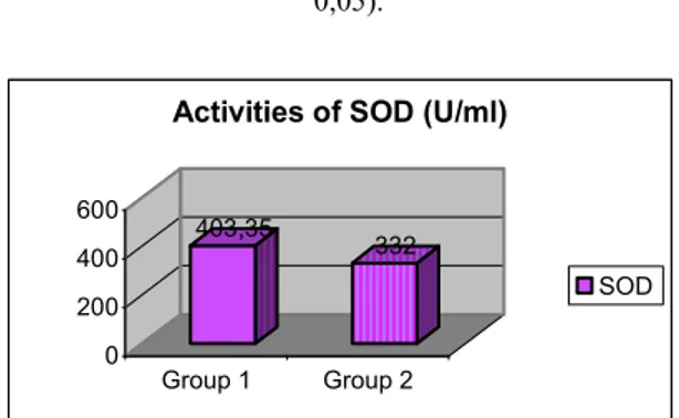
Figure 2: Activities of SOD wich is found to be increased significantly in group I when compared to group II (mean 40,33 ± 7,1 U/ml vs 33,2 ± 6,2 U/ml; p<
0,05).
Fig II represents the activities of SOD which is found to be significantly higher in group I when compared to group II. (mean 40,33 ± 7,1 U/ml vs 33,2 ± 6,2 U/ml; p<0,05).
Discussion
Free radicals have been implicated in the pathogenesis of many diseases (2,11). Reactive oxygen species (ROS) can initiate lipid peroxidation and DNA damage leading to carcinogenesis and cell death if the antioxidant system is impaired (3,12).
Lipid peroxidation (LP) is a chain reaction that involves the oxidation of fatty acids. MDA is a product of lipid peroxidation and used as an indicator of this chain reaction. Products of lipid peroxidation may cause DNA damage (13). It has been reported that lipid peroxidation may also play an indirect role in the conversion of procarcinogenesis to the last carcinogenesis (14,15).
Increased lipid peroxide products in abnormally proliferating cells due to the oxidative damage are thought to be released into the systemic circulation resulting in increased lipid peroxide levels in serum or pleural fluids of cancer patients (16).
In the present study, MDA content in malign pleural fluids was higher than in benign pleural fluids. Some previous observations reported higher MDA concentrations in human cell lines and solid tumors (1,17). Earlier reports described elevated plasma MDA levels in malignant pleural exudates when compared to those of non-malignant effusions (18,19). 135,15 67,35 0 50 100 150 Group 1 Group 2
MDA levels (nmol/ml)
MDA 403,35 332 0 200 400 600 Group 1 Group 2
Activities of SOD (U/ml)
Free radicals like superoxide anion are highly reactive and can cause both morphological and functional damage in the cell. Also an accumulation of superoxide anion in tumor cells was reported (8). The close relationship between free radical function and malignancy has been well documented (18,20). The cells protect themselves against oxidative damage by enzymatic and nonenzymatic antioxidant system. Superoxide dismutase is the major primary defense enzyme in the cell (21). This scavenging enzyme plays an important role in the protection of cell against the potentially harmful effects of superoxide anion generated by a wide variety of biological processes (22). Since SOD is known to help the protect epithelial cells against malign transformation in vitro. Also it was shown that antioxidant enzymes like SOD have the ability to inhibit tumor promotion and initiation in vivo and in vitro (23).
We have found that total SOD activitiy in malignant pleural exudates were significantly higher than in benign pleural fluids. The increase in SOD activity may be due to the higher enzyme expression in tumor cells or activation of the enzymes. The increase in SOD activity in malign pleural fluids may be related with an increase production of superoxide anion. Studies on antioxidant enzyme activities showed significant increased red cell Cu-Zn SOD and GSH-Px activities in leukemia patients (8). In another study, increased Mn-SOD activity was also determined in serum from patients with ovarian cancer and concerned as a tumor marker (24).
In a previous study (18) the levels of antioxidants were found to be decreased in lung malignant pleural exudates when compared to those of non-malign effusion. These lower levels of antioxidant enzymes might have caused the accumulation of free
radicals in pleural fluid of lung cancer patients. It has been observed that free radicals attack proteins, also enzymes (25). In lung cancers due to elevated MDA levels, it is assumed that the decrease in the activities of antioxidant enzymes in malignant pleural fluids when compared to the control group (18). This was contrary to our results.
The consequence of increased free radical formation and imbalances in antioxidant defence is oxidative stress which leads to oxidative damage and causing in increased lipid peroxide levels. Lipid peroxide levels may be changed by mild, moderate and transient oxidative stress. The different alterations observed in antioxidant enzymes could be primarily due to the lipid peroxide levels.
Concerning the lung cancer, Lizutci et al reported that Mn-SOD activity was significantly elevated in cancerous lung tissues when compared with those of normal uninvolved tissues (26). Similarly, Durak et al determined higher serum and pleural fluid SOD enzyme activities in patients with lung cancer than in those of healthy controls (27).
In our study, we have found elevated MDA levels and increased activities of SOD enzymes in malign pleural fluids when compared to the benign pleural fluids. These results suggest that, in spite of an increase in the activity of SOD, there is an increase in lipid peroxidation products which could reflect the fact that oxidative stress was in excess of the capacity of the antioxidants enzymes in pleural fluid. It is concluded that there is a significant association of lipid peroxidation of pleural effussion with increase activities of SOD in malign group. This could be suggested that these parameteres in pleural fluid might be helpful in differentiation of malign from malign effusions and could be used as non-specific diagnostic markers.
REFERENCES
1. Portakal O, Özkaya Ö, İnal ME et al. Coenzyme Q10 concentrations and antioxidant status in tissues of breast cancer patients. Clinical Biochemistry 2000; 33(4): 279-284.
2. Timothy RR, Sharma HM. Free radicals in health and disease. Indian J Clin Pract 1991; 2: 15-27. 3. Halliwell B. Reactive oxygen species in living systems: Source, biochemistry and role in human disease. Am J Med 1991; 91: 14-21.
4. Halliwell B, Gutterage JMC. Role of free radicals and catalytic metal ions in human disease: an overview. Methods Enzymol 1989; 186: 1-85.
5. Galleotti T, Masotti L, Borrello S. Oxy-radical metabolism and control of tumour growth. Xenobiotica 1991; 21: 1041-1451.
6. Yamaguchi S, Sakurada S, Nagumo M. Role of intracellular SOD in protecting human leukaemic and cancer cells against superoxide and radiation. Free Radical Biol Med 1994; 17: 389-395.
7. Gavino VC, Miller JS, Ikharebha SO et al. Effects of polyunsaturated fatty acids and antioxidants on lipid peroxidation in tissue cultures. J Lipid Res 1981; 22: 763-769.
8. Devi GS, Prasad MH, Saraswathi I et al. Free radicals antioxidant enzymes and lipid peroxidation indifferent types of leukemias. Clinica Chimica Acta 2000; 293: 53-62.
9. Yoshioka T, Kawada K, Shimoda T, Muri M. Lipid peroxidation in maternal and cord blood and protective mechanisms against activated oxygen toxicity in the blood. Am J Obstet Gynecol 1979; 135: 372-376.
10. Yi-Sun, Larry W. Oberley R. Superoxide dismutase. Clin Chem 1988; 3413: 497-500.
11. Kakkar R, Mantha SV, Kalra S et al. Time course study of oxidative stress in aorta and heart of diabetic rat. Clinical Science 1996; 91: 441-448.
12. Jaruga P, Zastawny TH, Skoleowski S et al. Oxidation DNA base damage and antioxidant enzyme activities in human lung cancer. FEBS Letters 1994; 341:59-64.
13. O’Brien PJ. Antioxidants and cancer. Molecular mechanism. In: Armstrong D, Ed. Free radicals in diagnostic medicine. New York; Plenum Press, 1994: 215-239.
14. Floyd RA, Soung LM, Walker RN. Lipid hydroperoxide of N-hydroxyl-N-acetylaminofluorane via free radical route. Cancer Res 1976; 36: 2761-2766.
15. Cercitti PA. Proxidant states and tumor promotion. Science 1985; 227: 375-381.
16. Scott MD, Eaton JW, Frans A. Enhancement of erythrocyte superoxide dismutase activity. Effects on cellular oxidant defense. Blood 1989; 74: 2542-2549.
17. Diplock AT, Rice-Evans K, Burdan RH. Is there a significant role for lipid peroxidation in the aqusation of malignancy and for antioxidants. Cancer Prevent 1993; 54: 1952-1956.
18. Vanisree AJ, Syamaladevi CS. Status of lipid peroxidation and antioxidant enzymes in malignant (bronchogenic carcinoma) and non-malignant pleural effusions. Indian Journal of Cancer 1999; 36: 127-134.
19. Hammouda MA, Khalil MM, Salem A. Lipid peroxidation products in pleural fluid for seperation of transudates and exudates. Clinical Chemistry 1995; 41(9): 1314-1315.
20. Dormandy TL. Free-Radical oxidation and antioxidants. Lancet 1978; 1: 647-653.
21. Galleotti T, Masotti L, Borrello S. Oxy-radical metabolism and control of tumour growth Xenobiotica 1991; 21: 1041-1051.
22. Bhuvarahamurthy V, Balasubramaniant M, Govindasamy S. Effect of radiotherapy and chemoradiatherapy on circulating antioxidant system of human uterine cervical carcinoma. Molecular and Cellular Biochemistry 1996 158; 17-23
23. Guyton KZ, Kensler TW. Oxidative mechanism in carcinogenesis. Br Med Bull 1993; 49: 523-544. 24. Taniguchi N. Superoxide dismutase significances in aging, diabetes, ischemia and cancer. Rinshe Boyri 1990; 38: 376-381.
25. Comporti M. Biology of disease: lipid peroxidation and cellular damage in toxic liver injury. Lab Invest 1985; 53: 599-623.
26. Lizuka S. Human manganase-containing superoxide dismutase; immunoassay and contents in lung cancer. Hokkoido Igatu Zasshi 1984; 59: 739-749.
27. Durak İ, Canbolat O, Kavutçu M et al. Activities of total, cytoplasmic and mitochondrial superoxide dismutase enzymes in sera and pleural fluids from patients with lung cancer. Journal of Clinical Laboratory Analysis 1996; 10:17-20.