BASIC RESEARCH
Comparison of ultrastructural changes and the anticarcinogenic effects of
thymol and carvacrol on ovarian cancer cells: which is more effective?
Hulya Elbea, Gurkan Yigitturka, Turker Cavusoglub,c, Tuba Baygard, Melike Ozgul Onala, and Feral Ozturka aFaculty of Medicine, Department of Histology and Embryology, Mugla Sıtkı Kocman University, Mugla, Turkey;bFaculty of Medicine,
Department of Histology and Embryology, Ege University, Izmir, Turkey;cCell and Tissue Research and Application Centre, Ege University &
Cord Blood, Izmir, Turkey;dResearch Laboratories Center, Material Research Laboratory, Mugla Sıtkı Kocman University, Mugla, Turkey
ABSTRACT
Ovarian cancer is the seventh most common cancer worldwide in women. Many anticancer drugs are currently used clinically have been isolated from plant species or are based on such sub-stances. Thymol (5-methyl-2-isopropylphenol) and carvacrol are oxygenated aromatic compounds from the monoterpene group. They are the main constituents of thyme essential oil and show antiproliferative, antioxidant, and antiseptic properties. The aim of this study is to compare the antiproliferative and apoptotic effects of thymol and carvacrol on SKOV-3 ovarian cancer cell line. The cancer cells were treated with different concentrations of thymol and carvacrol (100, 200, 400, 600 µM) at 24 h and 48 h durations. The cell viability was investigated by MTT assay and analysis of apoptosis with annexin V assay was determined. The study show that thymol and carvacrol significantly induced apoptosis in all groups as dose and time-dependent (p < .05). The data in the present study demonstrated that thymol and carvacrol have apoptotic and antiproliferative properties in a concentration-dependent manner toward ovarian cancer cells. SKOV-3 cancer cell line was much more sensitive to the toxic effect of thymol than carvacrol.
ARTICLE HISTORY Received 13 November 2019 Revised 2 March 2020 Accepted 5 March 2020 KEYWORDS Apoptosis; antiproliferative; carvacrol; ovarian cancer; ultrastructure; thymol
Introduction
Ovarian cancer is the seventh most common can-cer worldwide in women. It is the second most common malignancy after breast cancer in women over the age of 40, and the fifth leading cause of cancer-related death in women and the most fatal gynecologic cancer particularly in developed countries.1 Ovarian cancer is rare in young women, particularly under the age of 30; risk increases with age, with the occurrence spiking drastically after the age of 50, the maximum inci-dence occurs in the 80- to 84-year old and average diagnosis between the ages of 50 and 70 years.1,2 Epithelial ovarian cancer (EOC) is a relatively common one.3 Although the cause of EOC is not known, it is thought to occur as a result of the accumulation of genetic damage at the cellular level, but the reason for the occurrence of this damage is not fully defined.4 While the role of some factors, such as parity, is well defined, the role of others, such as the exogenous administra-tion of gonadotrophins, remains far more
controversial. Treatment of EOC was mainly by surgery, combined with chemotherapy.3
Several agents including life habits, exposure to chemical agents, and diet have been correlated with the risk of cancer development.5Besides, pharmaco-logical or nutritional intervention can significantly affect patients’ quality of life by delaying cancer progression.6 Therefore, the role of dietary compo-nents in the prevention of the onset and progression of cancer is an area of scientific and clinical interest.7 The plant-derived products are expected to induce lesser side effects compared to synthetic drugs.8 Many anticancer drugs currently used clinically have been isolated from plant species based on their substances.9 A plant-derived compound, essential oils are one among of the most valuable plant pro-ducts used in medicine and complementary treatment strategies.8
Extensive researches about biologically active com-pounds from essential oils have proven to be potential antibacterial, antifungal and antioxidant agents.10 Accumulating data has revealed the anticarcinogenic
CONTACTHulya Elbe h_elbe@hotmail.com Department of Histology and Embryology, Mugla Sıtkı Kocman University Faculty of Medicine, Mugla, Turkey 2020, VOL. 44, NO. 2, 193–202
https://doi.org/10.1080/01913123.2020.1740366
activity of plant-derived monoterpenes.8,9 Thymol (2-isopropyl-5-methylphenol) is a major phenolic compound that is present in the essential oils of various plants, including Thymus vulgaris (Lamiaceae).11,12 Several biological properties were reported for thymol that it has antiinflammatory, antibacterial, antispasmodic, wound healing and anti-oxidant effects. It is also an active compound for the inhibition of cancer cells.11–14Thymol is a major phe-nolic compound present in the essential oil of Thymus vulgaris.11,15
Carvacrol is a natural-bioactive monoterpenoid phenol, which is found in essential oils of the family Lamiaceae, including the genera Origanum and Thymus.16–18 Previous studies have demonstrated that carvacrol has antiangiogenic, analgesic, antioxi-dant, antimicrobial, and anti-inflammatory properties.18,19,20 Furthermore, carvacrol was reported to have an antiproliferative effect on liver, lung, colon, and breast cancer cell lines.10,20–23
In this study, we compared the anticarcinogenic and apoptotic effects of thymol and carvacrol on SKOV-3 ovarian cancer cell line. We also expected to reveal that the ultrastructural morphology of the cell surface of SKOV-3 cancer cells after exposure to these monoterpene phenols and investigated the potential underlying mechanisms involved in these effects.
Materials and methods
Ovarian epithelial adenocarcinoma cell line (SKOV-3, ATCC® HTB-77TM) were purchased from American Type Culture Collection (ATCC) (Rockville, Maryland, USA). Cells were cultured in RPMI 1640 (Lonza, Basel, Switzerland) culture medium contain-ing 10% heat-inactivated fetal bovine serum (Gibco, Invitrogen Life Technologies, Paisley, UK), and 1% penicillin/streptomycin (Sigma-Aldrich, St Louis, MO, USA). Cells were cultured in 25 cm2polystyrene flasks (Corning Life Sciences, UK) and maintained in an incubator at 37°C in a humidified atmosphere in the presence of 5% CO2. Growth and morphology
were checked microscopically daily to ensure cell health. Cells were passaged when they had reached approximately 80% confluency. Cells were harvested using 0.05% trypsin-EDTA (Sigma-Aldrich) and cen-trifuged (Nuve NF200; Laboratory and Sterilization Technology, Ankara, Turkey) after the addition of
RPMI 1640 for trypsin inactivation. After centrifuga-tion, they were resuspended in a culture medium. Thymol (Sigma-Aldrich, 16254) and carvacrol (Sigma-Aldrich, 282197) were prepared as a 4 mM stock solution in dimethyl sulfoxide (DMSO). The DMSO concentration in the assay did not exceed 0.1% and was not cytotoxic to the cancer cells.
Cell viability assay
The viability of the cells was evaluated with the MTT 3-(4,5-Dimethylthiazol-2-yl)-2,5-Diphenyl tetrazolium Bromide) assay. Briefly, cells were seeded in triplicate in 96-well plates at a density of 2 × 10⁴ cells/well. Cells were incubated for 24 h before to treat with thymol and carvacrol to main-tain the adhesion and then 100 μM, 200 μM, 400 μM ve 600 μM doses of each compound was applied to determine IC50(half maximal inhibitory
concentration) values. Then, plates were incubated at 37°C in a 5% CO2incubator for 24 h and 48 h.
For the preparing MTT solution, 5 mg MTT (pow-der) was dissolved in 5 mL PBS (phosphate buffer saline). For each well of 96-well plates the media were changed with 100 μl fresh RPMI-1640 (non-containing FBS), then 10 μl MTT solution was added into each well and plates were incubated at 37°C for 4 h. DMSO was added to all wells and mixed thoroughly to dissolve the dark blue crys-tals. After a few minutes at room temperature to ensure that all crystals were dissolved, the plates were read on a microplate reader (Bio-Rad), using a test wavelength of 570 nm. IC50 values were
determined for thymol and carvacrol by evaluating the results of MTT and these doses were used in the later stages of the study.
Cell death analysis with Annexin V
The apoptotic cell profile was determined using the Muse™ Annexin V & Dead Cell kit (Merck) according to the manufacturer’s instructions. Briefly, after treatment with thymol and carvacrol, SKOV-3 cancer cells were incubated for 24 h, and collected and diluted with PBS as a dilution buffer to a concentration of 7 × 10⁵ cells/ml. 100 μL of cell suspension and 100 μL of Annexin V/dead reagent were mixed and incubated 20 min at room temperature. Then, cells were analyzed
with Muse™ Cell Analyzer (Merck Millipore). The apoptotic profile was determined by the identifica-tion of four populaidentifica-tions: (i) non-apoptotic cells, not undergoing detectable apoptosis: Annexin V (−) and 7-amino-actinomycin D (AAD) (−), (ii) early apoptotic cells, Annexin V (+) and 7-AAD (−), (iii) late apoptotic cells, Annexin V (+) and 7-AAD (+), (iv) cells that have died through non-apoptotic pathway: Annexin V (−) and 7-AAD (+).
Morphological analysis with hematoxylin and eosin (H-E)
After fixation in 96% ethanol for 12 h, the SKOV-3 ovarian cancer cells were stained for 12 min with hematoxylin, washed with phosphate-buffered sal-ine (PBS) for development of the blue color for 3 min, and then incubated with eosin for another 3 min. During the next stage, the cells were washed with PBS and dehydrated using a graded series of increasing concentrations of ethanol (50%; 70%; 80%; 90%; 96%; 99.6%). The slides were left in a solution of ethanol: xylene (50:50) in the tank for at last 1 min and then they were exposed to pure xylene for 1 min. The mounted slides were visually assessing with a Nikon Eclipse 80i image analyze system.
Ultrastructural analysis with scanning electron microscopy (SEM)
Effects of thymol and carvacrol on the morphology of SKOV-3 ovarian cancer cell line were observed by SEM. Sterile circle glass coverslips (13 mm) were placed into the wells of the cell culture plates. 3 × 10⁵ cells were plated in each well. IC50 values
of thymol and carvacrol determined by the MTT method were applied to cells. After 24 h incubation, the coverslips were gently rinsed with PBS (pH 7.4) and fixed with 2.5% glutaraldehyde at 4°C for 1 h. After glutaraldehyde fixation, the coverslips were washed again with PBS and dehydrated by increas-ing concentrations of ethanol (50%, 70%, 90%, and 96%). Specimens were air-dried and coated by gold (Emmitech K550, UK) before examination by SEM (JSM-7600 F, JEOL Ltd., Tokyo, Japan).
Statistical analysis
Statistical analysis was carried out using the SPSS 17.0 statistical program (SPSS Inc., Chicago, Ill., USA). All experiments were carried out in tripli-cate, and presented as mean±SD. Statistical analy-sis was performed by using one-way analyanaly-sis of variance, followed by Tukey’s or Dunnett’s post hoc test. p < .05 was considered to indicate a sta-tistically significant difference.
Results
Effects of thymol and carvacrol on cell viability
The cell viability decreased in dose and time-dependent manner in SKOV-3 ovarian cancer cell line. These cells were much more sensitive to the toxic effect of thymol than carvacrol. The IC50
values of thymol and carvacrol were calculated as 316.08, 322.50 µM at 24 h and 258.38, 289.54 µM at 48 h, respectively (Figure 1).
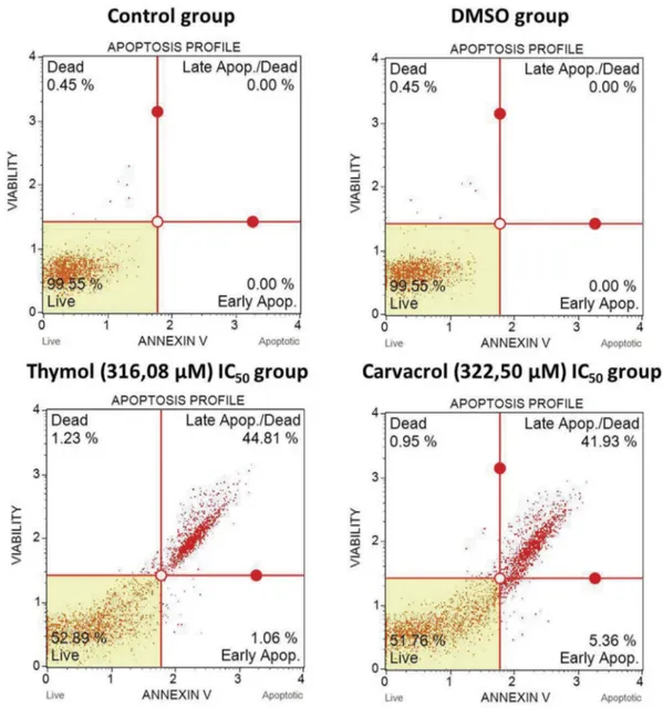
Effects of thymol and carvacrol on apoptosis
Thymol and carvacrol significantly induced apoptosis in SKOV-3 ovarian cancer cell line as dose and time-dependent. Statistical analyzes showed a significant difference between thymol and carvacrol-treated cells compared to non-treated cells (p < .05). At the IC50
value of thymol, 44.81% of the cells were in the late apoptosis phase and 1.06% were in the early apoptosis phase. In the IC50 value of carvacrol, 41.93% of the
cells were in the late apoptosis phase and 5.36% were in the early apoptosis phase. There was no significant difference between elevated thymol concentration and apoptotic cells (p > .05). As the concentration of carvacrol increased, early apoptotic cells (p < .001) and late apoptotic cells (p < .0001) were found to increase at high rates (Figure 2).
Light microscopic changes of ovarian cancer cells
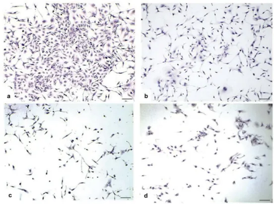
When the hematoxylin-eosin (H-E) staining of the control group (untreated) SKOV-3 ovarian cancer cell line was examined, three different cell morpholo-gies, epithelial, round and spindle-shaped, were deter-mined. The number of epithelial-shaped (+++) cells were higher than spindle-shaped cells; spindle-shaped cells were found to be more than round (+) shaped
Figure 1.Effects of thymol and carvacrol on cell viability by MTT analysis. Thymol and carvacrol suppressed the cell proliferation of ovarian cancer cells. As shown in graph, time and dose-dependent decrease in the growth of SKOV-3 ovarian cancer cells was observed with increasing concentrations of (a) thymol and (b) carvacrol. The percent viable cells were calculated in comparison to untreated cells taken as 100%. Data were expressed as mean±SD (p < .05).
Figure 2.Effects of thymol and carvacrol on apoptosis by Annexin-V analysis. Cell death analysis with Annexin V/PI staining of SKOV-3 ovarian cancer line. The non-stained population (bottom left) represents viable cells. Thymol and carvacrol induced apoptosis in ovarian cancer cells. Cells were incubated for 24 h. Data were expressed as mean±SD (p < .05).
cells. In the DMSO group, the cell morphology is similar to the control group. The concentration of DMSO used as the solvent did not appear to cause cytotoxic effects on cells. In the thymol group, although the number of cells with three different morphologies was observed to decrease, the decrease in the number of epithelial (+) shaped cells was more prominent. In this group, the cell population consists mainly of the spindle (++) and round (+) shaped cells. In the carvacrol group, there was a decrease in the number of cells with three different morphologies. Although not as much as the thymol group, the decrease number in epithelial (++) shaped cells in the carvacrol group is remarkable. Spindle (++) and round (+) shaped cells also decreased (Figure 3).
Ultrastructural changes of ovarian cancer cells
Control group cells without any treatment were found to be abundant and spreading on the substrate sur-face. On the other hand, thymol and carvacrol treated
group cells were found to have lower amounts. The number of cells was found to be decreased. Control group cells displayed epithelial morphology with mul-tipolar shape. Thymol and carvacrol treated group cells were mostly elongated and were bipolar/spindle-like shaped. Control group cells had clear nuclei and relatively higher amounts of cytoplasm. The nuclei of the thymol and carvacrol treated group cells were less distinct and the cells had a lower amount of cyto-plasm. Control group cells appear more grouped and closer to each other. Thymol and carvacrol treated cells were observed to be spread onto the substrate surface independently (Figure 4).
Discussion
Today, chemotherapy is one of the main methods of modern cancer treatment.24Epithelial ovarian cancer treatment was mainly performed with cytoreductive surgery in combination with cisplatin and paclitaxel chemotherapy, most of the patients’ clinical
Figure 3.Light microscopic changes of SKOV-3 ovarian cancer line. When the control group (untreated) hematoxylin-eosin (H&E) staining of the SKOV-3 ovarian cancer cells was examined, three different cell morphologies, epithelial, round and spindle shaped, were determined. Cells were incubated for 24 h. (a) Control group X10; H-E. In the DMSO group, the cell morphology is similar to that of the control group. (b) DMSO group X10; H-E. In the thymol group, although the number of cells with three different morphologies was observed to decrease, the decrease in the number of epithelial (+) shaped cells was more prominent. (c) Thymol group X10; H-E. In the carvacrol group, there was a decrease in the number of cells with three different morphologies. (d) Carvacrol group X10; H-E.
symptoms improved, but most patients with complete remission were prone to recurrent tumors, and some patients developed resistance.3 However, most che-motherapeutic agents have several important short-and long-term side effects.24 Recently, researchers have focused on the biologically active derivatives of medicinal plants which have been considered for the development of novel potential nontoxic drugs and for the prevention and treatment of certain types of cancer.10,24 The medicinal plants of the Lamiaceae family have been used by humans for thousands of years and are known for their therapeutic properties.25,26Particular attention has been given to the in vitro anticarcinogenic effects of thyme essential oils.25
In recent years, anticarcinogenic effects of thymol and carvacrol have been investigated; however, its effect on cancer has not yet been fully elucidated. Several studies have been performed with extracts of Thymus spp., and some studies have evaluated the therapeutic effects of thymol and carvacrol. These studies have shown that thymol and carvacrol have cytotoxic effects on cancer cells. Ferraz et al.27 evalu-ated the cytotoxic activity of essential oil of L. gracilis is chemically characterized by the presence of thymol, as major constituent on K562 (human chronic mye-logenous leukemia), HepG2 (human hepatocellular
carcinoma), B16-F10 (mouse melanoma), and normal peripheral blood mononuclear (PBMC) cell lines. Three tumor cell lines were treated with increasing concentrations of essential oil and thymol for 72 h. Thymol showed cytotoxicity only for the B16-F10 melanoma cell line at IC50value of 18.23 µg/ml. The
essential oil was cytotoxic on both cancer cells and normal PBMC cells. But, thymol did not show cyto-toxicity to normal cells at the tested concentrations.27 Similarly, Deb et al.28 investigated the anticancer activity of thymol on PBMC and HL-60 (human acute promyelotic leukemia) cells. In this study, thy-mol demonstrated dose-dependent cytotoxic effects on HL-60 cells after 24 h of exposure. However, thymol did not show any cytotoxic effect in human normal PBMC cell line like the previous study.28Calo et al.29treated 1–8 g/mL thymol to the NCTC 2544 (human keratinocyte) cell line. Cell viability of sam-ples exposed to thymol never decreased under 80%.29 In the study of Khan et al.30carvacrol nanoemulsion displayed no cytotoxicity up to 100 µg/ml against normal bronchial epithelium cells (BEAS-2B).30 Koparal et al.21reported that carvacrol has no signifi-cant effects on HFL1 (Human Lung fibroblast) cells. Literature studies show that the cytotoxic effect of thymol and carvacrol on normal cells is relatively very low.21 On the other hand, Mastelić et al.31 Figure 4.Ultrastructural changes of SKOV-3 ovarian cancer line by SEM. Cells of control group (untreated) were found to be abundant and spreading on the substrate surface. Cells were incubated for 24 h. (a) Control group X250. Cells of thymol and carvacrol treated groups were found to have lower amounts. (b) Thymol group IC50X250, (c) Carvacrol group IC50X250. Control cells displayed epithelial morphology with multipolar shape. Cells of thymol and carvacrol treated groups were mostly elongated and were bipolar/spindle-like shaped. (d) Control group X1000, (e) Thymol group X1000, (f) Carvacrol group X1000.
reported that thymol had dose-dependent (0.1–-10 mM) antiproliferative effects on HeLa (human epithelial cervical cancer) cell line.31Stammati et al.32 reported that thymol has been shown to induce non-apoptotic cell death in human laryngeal carcinoma Hep-2 cells at IC50value of 700μM.32Previous studies
reported that thymol has cytotoxic and apoptotic effects on human gastric carcinoma cells, on P815 mastocytoma cells, on Caco-2 human colon adeno-carcinoma cells, and HepG2 human hepatoma cells.11,33,34The different results in these studies may be due to different metabolic activities of the cells and the methods used to measure cytotoxic activity. Yeh et al.35reported that thymol (100–900 μM) was cyto-toxic to PC-3 cells in a concentration-dependent manner. Thymol also induced cell death in PC-3 cells.35 In a comprehensive study, Abed36 have ana-lyzed the effect of thymol on two cancer (HeLa, Hep) cell lines at five concentrations (15, 30.5, 61, 122, 244 ng/ml). They observed a dose-dependent decrease in survival of the two tumor cell lines. Thymol exhib-ited stronger cytotoxicity at concentrations of 30.5 ng/ ml toward HeLa (human epithelial cervical cancer) and Hep (Human larynx epidermoid carcinoma) cell lines.36 In our study, we also detected that thymol reduced cell viability in SKOV-3 ovarian cancer cells. IC50 values of thymol were 316.08 µM at 24 h and
258.38 at 48 h. The exact mechanisms underlying the cytotoxic effects of thymol on cancer cells are not fully understood.
Arunasree10and Abid et al.20reported that carva-crol inhibits cell growth and induces apoptosis on human breast cancer cell lines.10,20 Fan et al.22 reported that carvacrol has anticarcinogenic effects on HCT116 and LoVo colon cancer cell lines.22 Yin et al.23reported that carvacrol has a cytotoxic effect on HepG2 human hepatocellular carcinoma cell line.23 Koparal and Zeytinoglu21 reported that carvacrol inhibits dose-dependent cell growth on A549 non-small cell lung cancer cell lines at the concentrations of 250, 500 and 1000 M for 24 h.21Esmaeili-Mahani et al.24reported that Thymus caramanicus extract, the major constituents of essential oil is carvacrol and thymol, has a potential apoptotic and antiproliferative property against MCF-7 human breast cancer cells and the combination with vincristine, a chemother-apeutic agent, may effectively induce cell death and be a potent modality to treat this type of cancer.24 Karkabounas et al.37 showed that the anticancer
effects of carvacrol in MDA-MB-231 human meta-static breast cancer cells were based on the activation of the classical apoptosis response, including decrease in mitochondrial membrane potential and increase in cytochrome-c release from mitochondria, increase in caspase activity, decrease in Bcl-2/Bax ratio, and clea-vage of PARP and fragmentation of DNA, which belong to the mitochondrial pathway of the apoptosis.37 Kahn et al.30, Mehdi et al.38 and Liang et al.39 found that carvacrol has cytotoxic effects to different cancer cells including human lung adeno-carcinoma A549 cells, servical cancer cells and human glioblastoma cells, respectively.30,38,39In our study, we detected that carvacrol also reduced cell viability in SKOV-3 ovarian cancer cell line. IC50values of
carva-crol were 322.50 µM at 24 h and 289.54 at 48 h. When we compared the IC50values of thymol and carvacrol,
thymol was much more effective on ovarian cancer cells than carvacrol.
Apoptosis is a physiological process that leads to cell death.21 Excessive cell proliferation and loss of apoptosis control lead to the onset and progression of cancer.28 So, apoptosis is an important phenom-enon in cytotoxicity induced by antitumor agents.40 In recent years, the induction of apoptosis has become a targeted strategy for antitumor drug discovery.21 Hence, natural products causing apoptosis in the can-cer cells are valuable resources in cancan-cer suppression.8 Deb et al.28reported that thymol-induced apoptosis in HL-60 cells involves both caspase-dependent and cas-pase independent pathways.28 Koparal et al.21 reported that A549 lung cancer cells treated with 100 M carvacrol did not show any apoptotic morpho-logical changes for 24 h. The cells were treated with 500 and 1000 M carvacrol showed some apoptotic characteristics as well as morphological changes.21 Our results showed that thymol and carvacrol acti-vated apoptosis in a dose-dependent manner. Although the utility of thymol and carvacrol in the treatment of malignancy has started to be understood, the mechanism of apoptosis induced by these mole-cules is unknown. Perhaps both thymol and carvacrol may affect the cell membrane and the cytoplasm. The hydrophobic properties appear to be responsible for the deterioration of cancer cell structures that cause increased cytoplasmic membrane permeability. From these data, the cytotoxic effects of thymol and carva-crol on SKOV-3 ovarian cancer cell line was found to be associated with apoptotic cell death.
The literature regarding ultrastructural features of thymol or carvacrol-treated ovarian cancer cells is scant. Few studies refer to the ultrastructural morphol-ogy of the ovarian cancer cells.41–44 Bailey et al.41 reported that for epithelial ovarian cancer cells, there is an abundance of microvilli that appear as dense microscopic protrusions on the cell surface in the SEM images. The LPA-treated ovarian cancer cells have a bare, smooth cell surface. This finding suggests that the loss or reconstruction of microvilli and cell surface proteins correspond to the cellular sheddings observed and newly formed larger protrusions.41 Gilloteaux et al.42,45) reported that MDAH 2774 human ovarian cancer cells are oblong to round in shape in SEM images. Mitotically active cells display bulging, spheroidal shapes during the cell cycle pre-ceding metaphase. In general, cell surfaces do not dis-play blisters; instead, they are smooth and flat except for the bulge around the nucleus. Numerous long, delicate, branching filopodia or microspikes and large, flattened lamellipodia emanate from the cell surface and extend to adjacent cells or overlap each other. On the other hand, SEM views reveal that Vitamin-C treated MDAH 2774 human ovarian can-cer cells are oblong to polygonal in shape. In addition, Vitamin-K3-treated ovarian cancer cells show a profound alteration in shape, which suggests a large degree of intracellular defects in SEM views. These cells display large, thick, cytoplasmic extensions as well as spherical blisters or blebs.42,44 Gilloteaux et al.45,46) reported that combined treatment with ascorbate and menadione exacerbated human bladder carcinoma cell damage caused by individual vitamins and accelerated cell death primarily through the induction of a new cell death called autoschizis.45,46 This mode of induced cancer cells is different that apoptosis as it is not programmed. Autoschizis exhi-bits a unique set of morphological alterations.43,44This morphological characterization of autoschizic cell death confirms and extends the previous studies and demonstrates that this cell death is distinct from apoptosis.42–47Tumor cells undergo a progressive ser-ies of profound cellular changes such as membranous, nuclear chromatin decondensation, and nucleolar changes.45Our study revealed that both thymol and carvacrol-treated cells have similar ultrastructural fea-tures; by SEM. Thymol and carvacrol treated cancer cells were found to have lower amounts. The number of SKOV-3 cancer cells was found to be decreased.
Untreated control cells displayed epithelial morphol-ogy with multipolar shape and had clear nuclei and relatively higher amounts of cytoplasm. Thymol and carvacrol-treated cells were mostly elongated and were bipolar/spindle-like shaped and nuclei of the thymol and carvacrol-treated cells were less distinct and the cells had a lower amount of cytoplasm.
Conclusion
In conclusion, this is the first study about ultrastruc-tural changes in SKOV-3 ovarian cancer cells under the treatment of monoterpene phenols. Traditional treatment of ovarian cancer has been proven to be effective but there are many highly undesirable side effects. Thus, alternative agents are needed which have similar efficacy of conventional chemotherapy with minimal side effects. Our study showed that thymol and carvacrol were cytotoxic to SKOV-3 ovar-ian cancer cell line based on dose and time-dependant manner. When we compared the IC50values of
thy-mol and carvacrol, thythy-mol was much more effective on ovarian cancer cells than carvacrol. Ultrastructural changes of cells under the effects of thymol and car-vacrol were similar. Our observations suggested that thymol is highly efficacious in reducing cancer cell number and that the growth inhibitory effect of thy-mol is time and dose-dependent. These findings sug-gest that thymol and carvacrol may be a potential chemopreventive agents in cancer, and further experi-ments to investigate these possibilities are required.
Acknowledgments
The authors extend their sincere gratitude to the Department of Histology and Embryology, Faculty of Medicine, Mugla Sıtkı Kocman University, Mugla, Turkey, for their support. The authors thanks Mugla Sıtkı Kocman University, Research Laboratories Center, Material Research Laboratory for the access of the SEM facility.
Declaration of interest
The authors declare no conflicts of interest in this work.
Ethical approval
This article does not contain any studies with human parti-cipants or animals performed by any of the authors.
Funding
This study has been granted by the Mugla Sıtkı Kocman University Research Projects Coordination Office through Project Grant Number: (17/062). The funders had no role in study design, data collection and analysis, decision to publish, or preparation of the manuscript
References
1. Stewart C, Ralyea C, Lockwood S. Ovarian cancer: an integrated review. Semin Oncol Nurs. 2019;35 (2):151–156. doi:10.1016/j.soncn.2019.02.001.
2. Edmondson RJ, Monoghan JM. The epidemiology of ovarian cancer. Int J Gynecol Cancer. 2001;11 (6):423–429. doi:10.1046/j.1525-1438.2001.01053.x. 3. Sun ZL, Tang YJ, Wu WG, et al. AZD1480 can inhibit
the biological behavior of ovarian cancer SKOV-3 cells
in vitro. Asian Pac J Cancer Prev. 2013;14
(8):4823–4827.doi:10.7314/apjcp.2013.14.8.4823. 4. Baker TR, Piver MS. Epidemiology and etiology of
ovarian cancer. Semin Surg Oncol. 1994;10
(4):242–248. doi:10.1002/ssu.2980100403.
5. da Silva HB, Amaral EP, Nolasco EL. et al. Dissecting major signaling pathways throughout the development of prostate cancer. Prostate Cancer.2013;2013:920612. doi:10.1155/2013/920612.
6. Syed DN, Khan N, Afaq F, et al. Chemoprevention of prostate cancer through dietary agents: progress and promise. Cancer Epidemiol Biomarkers Prev. 2007;16 (11):2193–2203.doi:10.1158/1055-9965.EPI-06-0942. 7. Jackson CL, Dreaden TM, Theobald LK, et al. Pectin
induces apoptosis in human prostate cancer cells: correla-tion of apoptotic funccorrela-tion with pectin structure. Glycobiology. 2007;17(8):805–819.doi:10.1093/glycob/ cwm054.
8. Gautam N, Mantha AK, Mittal S. Essential oils and their constituents as anticancer agents: a mechanistic view. Biomed Res Int. 2014;2014:154106. doi:10.1155/2014/ 154106.
9. Sobral MV, Xavier AL, Lima TC. et al. Antitumor activity of monoterpenes found in essential oils. Sci World J.2014;2014:953451. doi:10.1155/2014/953451. 10. Arunasree KM. Antiproliferative effects of carvacrol on
a human metastatic breast cancer cell line,
MDA-MB231. Phytomedicine. 2010;17(8–9):581–588. doi:10.1016/j.phymed.2009.12.008.
11. Kang SH, Kim YS, Kim EK, et al. Anticancer effect of thymol on AGS human gastric carcinoma cells. J Microbiol Biotechnol. 2016;26(1):28–37.doi:10.4014/ jmb.1506.06073.
12. Nagoor Meeran MF, Javed H, Al Taee H. et al. Pharmacological properties and molecular mechanisms of thymol: prospects for its therapeutic potential and pharmaceutical development. Front Pharmacol. 2017;8:380. doi:10.3389/fphar.2017.00380.
13. Arab HA, Fathi M, Mortezai E, et al. Chemoprotective effect of thymol against genotoxicity induced by bleo-mycin in human lymphocytes. Pharm Biomed Res. 2015;1(1):26–31.doi:10.18869/acadpub.pbr.1.1.26. 14. Lee KP, Kim JE, Park WH, et al. Regulation of C6
glioma cell migration by thymol. Oncol Lett. 2016;11 (4):2619–2624.doi:10.3892/ol.2016.4237.
15. Aeschbach R, Löliger J, Scott BC, et al. Antioxidant actions of thymol, carvacrol, 6-gingerol, zingerone and hydroxytyrosol. Food Chem Toxicol.1994;32(1):31–36. doi:10.1016/0278-6915(84)90033-4.
16. Lee SY, Jin HH. Inhibitory activity of natural antimi-crobial compounds alone or in combination with nisin against Enterobacter sakazakii Journal compilation. Lett Appl Microbiol. 2008;47(4):315–321. doi:10.1111/ j.1472-765x.2008.02432.x.
17. Luo Y, Wu JY, Lu MH. et al. Carvacrol alleviates prostate cancer cell proliferation, migration, and inva-sion through regulation of PI3k/AKT and MAPK
sig-naling pathways. Oxid Med Cell Longev.
2016;2016:1469693. doi:10.1155/2016/1469693. 18. Dai W, Sun C, Huang S. et al. Carvacrol suppresses
proliferation and invasion in human oral squamous cell carcinoma. Onco Targets Ther.2016;9:2297–2304. doi:10.2147/OTT.S98875.
19. Liolios CC, Gortzi O, Lalas S, et al. Liposomal incor-poration of carvacrol and thymol isolated from the essential oil of Origanum dictamnus L. and in vitro antimicrobial activity. Food Chem.2009;112(1):77–83. doi:10.1016/j.foodchem.2008.05.060.
20. Abid AT, Al-Kafaji B, Karbel H, et al. Damper effect of carvacrol on T47-D, a human breast cancer cell line. Med J Babylon. 2014 ;11(4):792–798.doi:10.1812-156X-11-4.
21. Koparal AT, Zeytinoglu M. Effects of carvacrol on a human non-small cell lung cancer (NSCLC) cell line, A549. Cytotechnology. 2003;43(1–3):149–154. doi:10.1023/B:CYTO.0000039917.60348.45.
22. Fan K, Li X, Cao Y, et al. Carvacrol inhibits prolifera-tion and induces apoptosis in human colon cancer cells. Anticancer Drugs. 2015;26(8):813–823. doi:10.1097/CAD.0000000000000263.
23. Yin QH, Yan FX, Zu XY, et al. Anti-proliferative and pro-apoptotic effect of carvacrol on human hepatocel-lular carcinoma cell line HepG-2. Cytotechnology. 2012;64(1):43–51.doi:10.1007/s10616-011-9389-y.
24. Esmaeili-Mahani S, Falahi F, Yaghoobi MM.
Proapoptotic and antiproliferative effects of Thymus caramanicus on human breast cancer cell line (MCF-7) and its interaction with anticancer drug vincristine. Evid Based Complement Alternat Med. 2014;2014:893247. doi:10.1155/2014/893247.
25. Alexa E, Sumalan RM, Danciu C, et al. Synergistic antifungal, allelopatic and anti-proliferative potential of Salvia officinalis L., and Thymus vulgaris L. essential oils. Molecules.2018;23(1):185.doi:10.3390/ molecules23010185.
26. Palabiyik SS, Karakus E, Halici Z, et al. The protective effects of carvacrol and thymol against paracetamol– induced toxicity on human hepatocellular carcinoma cell lines (HepG2). Hum Exp Toxicol. 2016;35 (12):1252–1263.doi:10.1177/0960327115627688. 27. Ferraz RP, Bomfim DS, Carvalho NC, et al. Cytotoxic
effect of leaf essential oil of Lippia gracilis Schauer (Verbenaceae). Phytomedicine. 2013;20(7):615–621. doi:10.1016/j.phymed.2013.01.015.
28. Deb DD, Parimala G, Devi SS, et al. Effect of thymol on peripheral blood mononuclear cell PBMC and acute promyelotic cancer cell line HL-60. Chem Biol Interact. 2011;193(1):97–106.doi:10.1016/j.cbi.2011.0 5.009.
29. Calò R, Visone CM, Marabini L. Thymol and
ThymusvulgarisL. activity against UVA- and
UVB-induced damage in NCTC2544 cell line. Mutat Res Genet Toxicol Environ Mutagen. 2015;791:30–37. doi:10.1016/j.mrgentox.2015.07.009.
30. Khan I, Bahuguna A, Kumar P, et al. In vitro and in vivo antitumor potential of carvacrol nanoemulsion against human lung adenocarcinoma A549 cells via mitochondrial mediated apoptosis. Sci Rep. 2018;8 (1):144.doi:10.1038/s41598-017-18644-9.
31. Mastelić J, Jerković I, Blazević I, et al. Comparative study on the antioxidant and biological activities of carvacrol, thymol, and eugenol derivatives. J Agric Food Chem.2008;56(11):3989–3996.doi:10.1021/jf0732 72v.
32. Stammati A, Bonsi P, Zucco F, et al. Toxicity of selected plant volatiles in microbial and mammalian short-term assays. Food Chem Toxicol. 1999;37 (8):813–823.doi:10.1016/s0278-6915(99)00075-7. 33. Jaafari A, Mouse HA, Rakib EM, et al. Chemical
composition and antitumor activity of different wild varieties of Moroccan thyme. Braz J Pharmacogn. 2007;17(4):477–491.doi:10.1590/S0102-695X20070004 00002.
34. Slamenová D, Horváthová E, Sramková M, et al. DNA-protective effects of two components of essential plant oils carvacrol and thymol on mammalian cells cultured in vitro. Neoplasma.2007;54(2):108–112.
35. Yeh JH, Chou CT, Chen IS, et al. Effect of thymol on Ca2+homeostasis and viability in PC3 human prostate cancer cells. Chin J Physiol. 2017;60(1):32–40. doi:10.4077/CJP.2017.BAF447.
36. Abed RM. Cytotoxic, cytogenetics and immunomodula-tory effects of thymol from Thymus vulgaris on cancer and normal cell lines in vitro and in vivo. Al-Mustansiriyah J Sci.2011;22:41–53.
37. Karkabounas S, Kostoula OK, Daskalou T, et al. Anticarcinogenic and antiplatelet effects of carvacrol. Exp Oncol.2006;28(2):121–125.
38. Mehdi SJ, Ahmad A, Irshad M, et al. Cytotoxic effect of Carvacrol on human cervical cancer cells. Biol Med. 2011;3(2):307–312.doi:10.4172/0974-8369.10000119. 39. Liang WZ, Lu CH. Carvacrol-induced [Ca2+]i rise and
apoptosis in human glioblastoma cells. Life Sci. 2012 15;90(17–18):703–711. doi:10.1016/j.lfs.2012.03.027. 40. Fatimah SAS, Wimardhani YS. The ultrastructural
sur-face morphology of oral cancer cells and keratinocytes after exposure to chitosan. J Phys Conf Series.2017;884 (1):012064. doi:10.1088/1742-6596/884/1/012064. 41. Bailey KA, Klymenko Y, Feist PE, et al. Chemical
analysis of morphological changes in lysophosphatidic acid-treated ovarian cancer cells. Sci Rep. 2017;7 (1):15295.doi:10.1038/s41598-017-15547-7.
42. Gilloteaux J, Jamison JM, Arnold D, et al. Autoschizis of human ovarian carcinoma cells: scanning electron and light microscopy of a new cell death induced by sodium ascorbate: menadione treatment. Scanning. 2003;25(3):137–149.doi:10.1002/sca.4950250306. 43. Gilloteaux J, Jamison JM, Lorimer HE, et al.
Autoschizis: a new form of cell death for human ovar-ian carcinoma cells following ascorbate: menadione-treatment. Nuclear and DNA degradation. Tissue Cell. 2004;36(3):197–209. doi:10.1016/j.tice.2004.01.006. 44. Gilloteaux J, Lau L, Gourari I, et al. Apatone® induces
endometrioid ovarian carcinoma (MDAH 2774) cells to undergo karyolysis and cell death by autoschizis: A potent and safe anticancer treatment. Transl Res Anat. 2015;1:25–39.
45. Gilloteaux J, Jamison JM, Arnold D, et al.
Ultrastructural aspects of autoschizis: a new cancer cell death induced by the synergistic action of ascor-bate/menadione on human bladder carcinoma cells. Ultrastruct Pathol. 2001;25(3):183–192. doi:10.1080/ 019131201300343810.
46. Gilloteaux J, Jamison JM, Arnold D, et al. Cancer cell necrosis by autoschizis. Synergism of antitumor activity of vitamin C:Vitamin K3 on human bladder carcinoma T24 cells. Scanning. 1998;20:564–575. doi:10.1002/ sca.4950200805.
47. Gilloteaux J, Jamison JM, Arnold D, et al. Autoschizis: A mode of cell death of cancer cells ınduced by a prooxidant treatment ın vitro and ın vivo. In: Radosevich J, ed. Apoptosis and Beyond: The Many Ways Cells Die. New York: J Wiley;2018. Chapter 28, pp. 583–694. ISBN:9781119432463. doi:10.1002/ 9781119432463.