DOI 10.1007/s00776-014-0632-1
ORIGINAL ARTICLE
Reconstruction of intercalary bone defects following bone tumor
resection with segmental bone transport using an Ilizarov circular
external fixator
Bahtiyar Demiralp · Tolga Ege · Ozkan Kose · Yuksel Yurttas · Mustafa Basbozkurt
Received: 12 May 2014 / Accepted: 4 August 2014 / Published online: 22 August 2014 © The Japanese Orthopaedic Association 2014
the SF-36 and BQUILI indexes was scored as 104 (88–150) and 4 (0–13), respectively.
Conclusion From the long-term follow-up results, recon-struction with distraction osteogenesis seems to be an efficient method in patients with long life expectancies. However, a long external fixation time is a disadvantage of this technique. Problems in patient compliance and pos-sible complications such as nonunion should be managed promptly.
Introduction
Recent advances in the management of bone tumors have led to a significant increase in the survival rates of patients with malignant bone tumors [1–5]. Therefore, limb salvage surgery has gained importance for preserving limb func-tion and providing a better quality of life. Reconstrucfunc-tion of large bone defects following en bloc resection of malig-nant or aggressive benign bone tumors is a problem in orthopedic surgery. Several biological and non-biological options are available for limb reconstruction after benign or malignant tumor resection, such as non-vascularized or vascularized autologous osseous grafts, allografts, spac-ers, tumor prostheses and bone transport with distraction osteogenesis [5, 6]. However, it is difficult to determine the best treatment strategy for a particular patient. In gen-eral, reconstructive procedures have been chosen consider-ing several factors such as the site and involvement of the tumor, pathology and biological behavior of the tumor, life expectancy and predicted function of the limb [7].
An ideal skeletal reconstruction should be biologi-cal and resistant to infection and should have sufficient biomechanical strength and durability particularly in the weight-bearing lower extremities [8]. The bone transport
Abstract
Aim The purpose of this retrospective study was to report the long-term follow-up results of the reconstruction of bony defects with Ilizarov distraction osteogenesis using the bone transport method following en bloc resection of bone tumors.
Materials and methods En bloc resection was performed in 13 patients with bone tumors between October 1991 and December 2010 in our clinic. The mean age of the patients was 19.46 years (range 7–42 years) at the time of surgery. Histological diagnosis was osteosarcoma in seven cases, Ewing’s sarcoma in three cases, giant cell tumor in one case, osteoblastoma in one case and fibrous dysplasia in one case. In all cases either the femur or tibia was involved.
Results The average follow-up period was 157.23 months (range 32–288 months), and the bone defect after resection was 14.61 cm ± (9–24 cm). The mean Musculoskeletal Tumor Society score of the patients was 89.46 (83–96) at the final follow-up. The mean Knee Society Scale scores of patients in whom reconstruction was performed around the knee joint were 74.3 (51–84). The mean foot and ankle dis-ability index of patients with a tumor around the ankle joint was 81 (73–95). Quality of life of the patients according to
B. Demiralp
Department of Orthopedics and Traumatology, Medipol University, Istanbul, Turkey
T. Ege (*) · Y. Yurttas · M. Basbozkurt
Department of Orthopedics and Traumatology, Gulhane Military Medical Academy, Ankara, Turkey
e-mail: [email protected] O. Kose
Department of Orthopedics and Traumatology, Antalya Education and Research Hospital, Antalya, Turkey
technique using an Ilizarov circular external fixator (CEF) and distraction osteogenesis have so far been successfully utilized for skeletal defects after resection of infected bone segments or traumatic bone loss [9–11]. However, this technique has not been discussed much in the current lit-erature for the reconstruction of defects remaining after the resection of bone tumors. The purpose of this study was to report the long-term results of biological limb reconstruc-tion with the bone transport technique after en bloc resec-tion of primary bone tumors in the lower extremities.
Materials and methods
This study was carried out according to the Declaration of Helsinki, and the Institutional Review Board at our institu-tion approved the study protocol (IRB no. 10840098-70). Patients
A retrospective review was made of 13 patients with vari-ous primary bone tumors who underwent limb-sparing sur-gery and skeletal reconstruction with bone transport using an Ilizarov CEF between 1991 and 2010 in our institution. The patients comprised ten males and three females with a mean age of 19.46 years (range 7–42 years) at the time of surgery. All of the tumors were localized on the lower extremity on either the femur or tibia. Eight tumors were localized on the tibia: three distal, four diaphyseal and one proximal. The five tumors on the femur were all in the dis-tal region. All patients underwent routine imaging workup including direct radiography, bone scintigraphy, comput-erized tomography (CT) and magnetic resonance imaging (MRI). Definitive diagnosis was made through open inci-sional biopsy in all cases. Histological diagnosis was con-ventional osteosarcoma in seven cases, Ewing’s sarcoma in three cases, giant cell tumor in one case, osteoblastoma in one case and fibrous dysplasia in one case. The tumor was staged according to the Musculoskeletal Tumor Soci-ety (MSTS) Staging System [12]. No patients had distant metastasis at the time of surgery. The patients with malig-nant bone tumors (conventional osteosarcoma and Ewing’s sarcoma) received neoadjuvant and postoperative chemo-therapy. The demographic and clinical characteristics of the patients are presented in Table 1.
Surgical technique
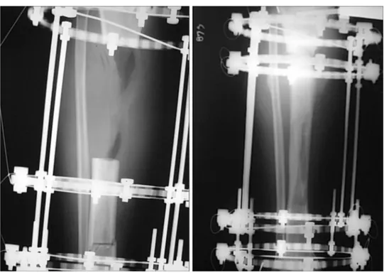
After wide/marginal resection of the tumor, the Ilizarov frame was applied to the extremity. For each case, the Ilizarov frame was constructed according to the skeletal defect. After tumor resection and application of the Ilizarov frame, corticotomy was performed on the proximal or
distal metaphysis of the bone defect (Figs. 1, 2, 3, 4, 5, 6). In all cases at least 1 cm or more of tumor-free epiphysis was preserved. An intramedullary guide wire was used to improve the alignment in three cases. Distraction was started on days 5–9 at a rate of 4 × 0.25 mm per day. Pro-phylactic antibiotics were administered to all patients for 5 days postoperatively. The patients underwent a rehabilita-tion program immediately. Knee and quadriceps exercises were started, and weight bearing was allowed on the 2nd postoperative day. After surgery, patients with a malig-nant bone tumor were referred to the oncology clinic, and postoperative chemotherapy was started. The distraction procedure continued throughout the chemotherapy period. The CEFs were applied until sufficient cortex development had been observed, and they were removed after adequate consolidation. Thereafter, a cast or protective splint was applied for 4–6 weeks.
Outcome measures
Three indices were used to evaluate the results: an
exter-nal fixation index calculated by dividing the entire dura-tion of external fixadura-tion by the length of bone regeneradura-tion; a distraction index calculated by dividing the duration of distraction by the length of bone regeneration; a
matura-tion index calculated by dividing the duration of exter-nal fixation, measured from the completion of distrac-tion to the removal of external fixadistrac-tion, by the length of
Table 1 Patient demographics
Case no. Age Sex Diagnosis Site of involve-ment MSTS stage 1 14 F Parosteal osteo-sarcoma Tibial shaft G1T1M0 2 15 F Conventional osteosarcoma Tibial shaft G1T1M0 3 24 M Osteosarcoma Distal femur G1T1M0 4 21 M Giant cell tumor Proximal tibia G0T1M0 5 20 M Fibrous dysplasiaTibial shaft G0T0M0
6 42 M Aggressive
osteoblastoma
Tibial shaft G0M0T0 7 7 M Ewing’s sarcoma Distal femur G1T1M0 8 10 M Ewing’s sarcoma Distal femur G2T1M0 9 19 M Conventional
osteosarcoma
Distal femur G1T1M0 10 10 F Ewing’s sarcoma Distal femur G1T1M0 11 33 M Conventional osteosarcoma Distal tibia G1T1M0 12 17 M Conventional osteosarcoma Distal tibia G1T1M0 13 21 M Conventional osteosarcoma Distal tibia G1T1M0
bone regeneration. The function of the affected limb was assessed according to the revised 30-point functional clas-sification system established by the International Society of Limb Salvage and the Musculo-skeletal Tumor Society [12]. Patients were evaluated with physical examination and radiologically throughout the follow-up period. All patients were called to the hospital for a final follow-up examination, at which the quality of life and general health status were evaluated using Short-Form 36 (SF-36) and the Body Image Quality of Life Inventory (BIQLI) [13]. In patients where the tumor was close to either the knee or the ankle joint, the Knee Society Score [14] and Foot and Ankle Disability Index (FADI) were also used [15]. All complications and secondary surgical interventions were screened and recorded during the treatment period.
Results
The average follow-up period was 157.23 months (range 32–288 months). The external fixation device was removed at an average of 18.1 months (range 4–19 months) when sufficient bone consolidation was observed on radiographs. The bone defect after resection was a mean of 14.61 cm (range 9–24 cm). While the mean external fixation index was 28.30 day/cm (range 18.75–40), the mean distraction index and maturation indexes were 11.08 days/cm (range 10.4–11.7) and 16.8 days/cm (range 7.5–32.7), respec-tively. The MSTS score of the patients was a mean of 89.46 (range 83–96) at the final follow-up. The Knee Society Scale score of patients for whom reconstruction was around the knee joint was a mean of 74.3 (range 51–84). The Foot
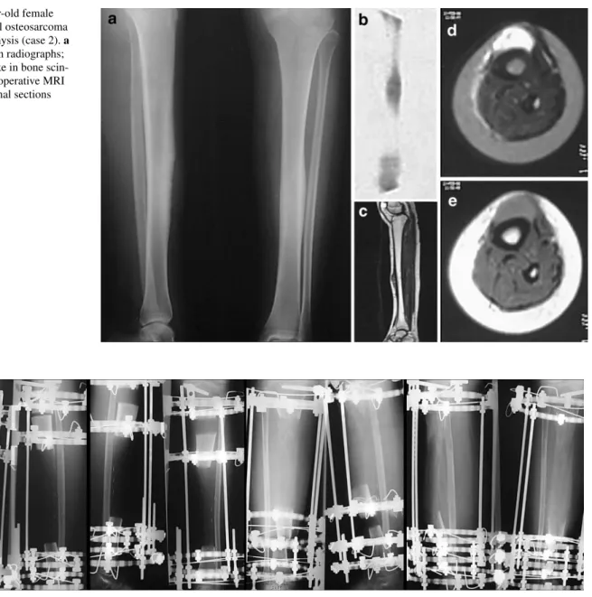
Fig. 1 A 15-year-old female
with conventional osteosarcoma in the tibial diaphysis (case 2). a Preoperative plain radiographs;
b increased uptake in bone
scin-tigraphy. c–e Preoperative MRI in axial and coronal sections
and Ankle Disability Index (FADI) of patients with a tumor around the ankle joint was a mean of 81 (range 73–95). The mean scores of the SF-36 and BQUILI indexes were 104 (range 88–150) and 4 (range 0–13), respectively. A sum-mary of the results is presented in Table 2.
Complications and secondary surgical interventions
Pin tract infection and reduced range of motion (ROM) were the most common complications during the distrac-tion period. All pin tract infecdistrac-tions were successfully man-aged with local care and oral antibiotics without the need for wire removal. Case 2 had a supracondylar femur frac-ture during the consolidation period of the tibial defect despite no major complications in the tibia. The fracture was successfully treated with open reduction and inter-nal fixation with a dynamic condylar screw. However, the patient died of leukemia in the 7th year of follow-up. Five patients with restricted movement in the ankle and knee joints were managed with aggressive physiotherapy, but a residual limitation of ROM was observed at the final fol-low-up in all of these patients. Delayed consolidation and union problems at the docking site were other common complications, which were seen in five patients. In case 9, the Ilizarov frame was removed, and intramedullary fixa-tion was performed twice because of nonunion and severe infection. In the 59th month of follow-up, a locking plate and grafting were applied because of non-union. On the last visit, sufficient union was not observed, and a 10-cm limb length inequality was detected. The patient was not compli-ant and rejected the Ilizarov method. Docking site problems were encountered in four cases. Union was achieved with the reapplication of the Ilizarov CEF in one case, grafting of the docking site in two cases and distraction compres-sion maneuvers in one case. In case 3, plastic deforma-tion in the distracdeforma-tion zone developed 2 months after the CEF had been removed because of skin invagination. The deformity was corrected by Ilizarov surgery, and union was achieved 6 months after surgery. In the 56th month, a refracture developed on the osteotomy line, and it was operated on with plate and screw fixation. Knee ROM was
Fig. 3 Clinical appearance and final plain radiographs after consolidation of the resected bone segment
Fig. 4 A 42-year-old male patient (case 6) with aggressive
osteoblas-toma in the tibia diaphysis. Preoperative plain radiographs and MRI findings
significantly limited in the short-term follow-up (extension was full but flexion was limited at 19°), and long-term fol-low-up revealed full extension and limited flexion at 33°. Data pertaining to the patients’ follow-up examinations are summarized in Table 2.
Discussion
In this study, the aim was to assess the safety and efficacy of limb reconstruction using distraction osteogenesis and the Ilizarov method on long-term follow-up after treat-ment of benign and malignant bone tumors. The results of the patients in the study were also compared with the results in the current literature and different treatment modalities. The current literature emphasizes the indica-tion of using distracindica-tion osteogenesis in bone tumors in a lesion <15 cm in length with at least 0.5 cm of subchon-dral bone and sufficient metaphyseal cortex retained after the excision of tumors that have responded well to chemo-therapy [1]. In all of the cases in the current study, the same rules were strictly followed when choosing the optimal treatment method for both benign and malignant tumors. However in four cases (cases 2, 3, 8 and 11) with malig-nant bone tumors, the resected bone segment was >15 cm, but despite this, the functional MSTS scores and SF 36 scores were good. Distraction osteogenesis was applied to ten patients who were undergoing chemotherapy after the operation. While this can be expected to delay bone regen-eration initially and to increase the distraction and matura-tion indexes, callus was successfully formed and matured. At the final follow-up, the mean functional MSTS, SF-36 and BQUILI scores were satisfactory and similar to the results in current reports [1, 16, 17]. In addition, none of the patients required amputation or lost functional ability. Despite the satisfactory results, frequent complications were experienced such as union problems (docking site) and pin site infections in the majority of our patients. All
Fig. 5 A 13-cm bone segment
was resected, and the distraction period was 145 days
Fig. 6 Plain radiographs obtained after consolidation of the bone
complications were promptly managed. We strongly rec-ommend autologous grafting, bone marrow injections, application of percutaneous demineralized bone matrix and hyperbaric oxygen alone or with open reduction inter-nal fixation in case of union problems at the docking site. Pin site infections are generally overcome with appropriate antibiotics and hyperbaric oxygen. Malalignment after the distraction period, especially in patients with a larger bone defect, is another problem. In the current study, the use of intramedullary guide wires to obtain alignment during dis-traction period was preferred to overcome this complica-tion as stated in our surgical technique. Finally, restricted ROM around the ankle and knee joints during the distrac-tion period in five patients was managed appropriately with aggressive physiotherapy and resulted in only a slight ROM restriction. These complications were generally similar to those reported in recent studies for distraction osteogenesis in bone tumors [1, 6, 8, 16, 17]. Although the complication rate was quite high in the current study, all were appropri-ately managed, and this series emphasizes the efficacy and reliability of applying the Ilizarov method to reconstruc-tions of large bone deficits arising from either benign or malignant tumors with good results in long-term follow-up. However, this treatment modality should not be recom-mended for patients with a poorer prognosis and short life expectancy because of the frequent complications.
The use of distraction osteogenesis and the Ilizarov method in bone defects after resection of benign or malig-nant bone tumors has been rarely reported, and the results are often conflicting. In most of the studies, distraction osteogenesis has been widely accepted as a basic treatment method despite frequent complications similar to those experienced in the current study. There has been agree-ment that this treatagree-ment modality should be preferred for bone tumors in children in particular and patients with a long life expectancy. Kapukaya et al. [17] reported excel-lent results in four, good in three and fair in two of nine patients treated with distraction osteogenesis for malig-nant bone tumors during a 22-month follow-up period. Similarly, in a larger patient group, Tsuchiya et al. [8] reported excellent results in 12, good in 5 and fair in 2 of 19 patients, and distraction osteogenesis was found to be beneficial in patients with an expectation of long-term sur-vival and in growing children. Watanabe et al. [1] reported excellent results over a 10-year period in a larger patient group with various bone tumors, and it was stated that although this technique requires both time and effort, it can provide excellent long-term outcomes, resulting in a stable reconstruction that functionally restores the natu-ral limb. McCoy et al. [16] also reported good results in bone tumors treated with the Ilizarov method from a mean follow-up period of 81.7 months (range 26–131 months).
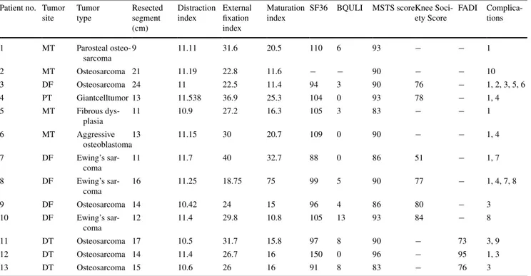
Table 2 Patient details and treatment results
MT tibial diaphysis, DT distal tibia, PT proximal tibia
Complications: 1, pin tract infection; 2, skin invagination; 3, restriction of ROM; 4, docking site; 5, malunion; 6, bowing at the transported bone; 7, delayed consolidation of the medial cortex; 8, leg length discrepancy; 9, delayed consolidation; 10, exitus with leukemia
Patient no. Tumor site Tumor type Resected segment (cm) Distraction index External fixation index Maturation index
SF36 BQULI MSTS scoreKnee Soci-ety Score FADI Complica-tions 1 MT Parosteal osteo-sarcoma 9 11.11 31.6 20.5 110 6 93 − − 1 2 MT Osteosarcoma 21 11.19 22.8 11.6 − − 90 − − 10 3 DF Osteosarcoma 24 11 22.5 11.4 94 3 90 76 − 1, 2, 3, 5, 6 4 PT Giantcelltumor 13 11.538 36.9 25.3 104 0 93 78 − 1, 4 5 MT Fibrous dys-plasia 11 10.9 27.2 16.3 105 3 83 − − 1 6 MT Aggressive osteoblastoma 13 11.15 30 20.7 109 0 90 − − 1, 4 7 DF Ewing’s sar-coma 11 11.7 40 32.7 88 0 86 51 − 1, 7 8 DF Ewing’s sar-coma 16 11.25 18.75 75 99 5 90 77 − 1, 4, 7, 8 9 DF Osteosarcoma 14 10.42 24 15 96 4 86 80 − 3 10 DF Ewing’s sar-coma 12 11.4 29.8 10.8 105 13 93 84 − 8 11 DT Osteosarcoma 17 10.5 31.7 15.8 97 8 90 − 73 3, 9 12 DT Osteosarcoma 14 11.4 26.7 16 150 0 96 − 95 1, 3 13 DT Osteosarcoma 15 10.6 26 16 91 8 83 − 76 3
No patient in that study was simultaneously undergoing chemotherapy during the distraction period, so no com-ment could be made on the use of the Ilizarov method with these treatments. In most of the published reports, general complications have been reported, such as pin site infec-tions, docking site and consolidation problems. Most of these problems can usually be successfully managed with conservative treatment or additional surgery. Contrary to the these favorable reports, Ozaki et al. [18] stated that this method cannot be recommended for patients with bone sarcoma who have a poor prognosis, as it has an unacceptably high complication rate, although their study was limited to 5 patients with a short follow-up period of 48 months. Technical errors were also reported such as resection of too much soft tissue around the tumor, which reduces the blood supply, and incorrect settings of the magnitude of the transported bone segment. In the current study, the complication rate was significantly lower than that in the Ozaki et al. study, and the mean MSTS scores from long-term follow-up were similar to those in current the literature [1].
In malignant or benign bone tumors, there are several alternative treatment methods instead of distraction osteo-genesis, and each technique has some advantages and disadvantages. The advantages of allografts are that they allow some bony host-to-allograft incorporation, preserva-tion of bone stock, facilitating future joint revision surgery, and provision of soft tissue attachments for the host limb and joint reconstruction [5, 19]. On the other hand, the most important disadvantages are the lack of growth, joint degeneration, graft resorption, joint instability, pathological fractures and high infection rates [20, 21]. In addition, the complication rates are even higher in children and patients receiving chemotherapy. Nonvascularized fibular grafting is a simpler and less expensive procedure in reconstruction after the resection of bone tumors [22], although it has the drawbacks of the absence of biological activity, resorption and the risk of non-union [22, 23]. Vascularized grafting has a lower infection rate, high rate of bone union and good functional outcomes. It is recommended in the reconstruc-tion of large, complex bone defects resulting from tumor extirpation. The disadvantage to this is that sufficient fibu-lar thickness to allow full weight bearing is achieved sev-eral years after the vascularized fibular grafting; therefore, patients have to use crutches for long periods. Furthermore, the same amount of bone ossification as seen in distraction osteogenesis during reconstruction has not been observed in vascularized fibulae. Non-union, stress fracture, infec-tion and malunion are also other complicainfec-tions [5, 24]. Autoclaved allografts conform to the defect and use the patient’s own tissue for reconstruction [25]. This eliminates the need for bone banking and the risk of disease transmis-sion associated with allografts [25]. However, autoclaved
bone has a long healing time and limited autogenic poten-tial, and there is the likelihood of resorption of the grafted bone or fracture of the graft. Endoprosthetic replacement has many advantages, such as early stability, mobilization and weight bearing, a high level of emotional acceptance by the patient and rapid restoration of function with a good functional outcome, but infection, mechanical failure and aseptic loosening may limit the long-term survival of the prosthesis, and the risk of revision increases with time, par-ticularly in skeletally immature patients [5, 6]. Reconstruc-tion of an extremity with a tumor with the use of autografts treated with liquid nitrogen is another alternative method that may resemble autoclaved bone grafting. Several stud-ies have reported favorable outcomes [26–28], although major complications of infection, fracture and local recur-rence have also been observed with this technique [26–28]. The current study has some limitations. The number of patients was relatively small with a large spectrum of tumors from benign to malignant with different sites on the bone. This technique is more demanding than other treatment methods, and further studies with larger patient groups are needed. Those patients took adjuvant chemo-therapy during the distraction period, which may have contributed to some complications, although the effect of chemotherapy on complications in distraction osteogen-esis has not yet been fully clarified. The major strength of this study is that it had a relatively longer follow-up time, which supports the advantage of distraction osteogenesis for various bone tumors.
In conclusion, it is suggested that, due to the high com-plication rates in the short-term follow-up, tumor recon-struction surgery with the distraction method is not appro-priate. However, in patients with long life expectancies, reconstruction with distraction osteogenesis, which is a biological reconstruction, seems to be an efficient method in the long-term follow-up, on the condition that complica-tions are promptly managed.
Conflict of interest The authors declare that they have no conflict
of interest.
References
1. Watanabe K, Tsuchiya H, Yamamoto N, Shirai T, Nishida H, Hayashi K, Takeuchi A, Matsubara H, Nomura I. Over 10 year follow up of functional outcome in patients with bone tumors reconstructed using distraction osteogenesis. J Orthop Sci. 2013;18(1):101–9.
2. Messerschmitt PJ, Garcia RM, Abdul-Karim FW, Green-field EM, Getty PJ. Osteosarcoma. J Am Acad Orthop Surg. 2009;17(8):515–27.
3. Grimer RJ. Surgical options for children with osteosarcoma. Lan-cet Oncol. 2005;6(2):85–92.
4. Heare T, Hensley MA. Dell’Orfano S. Bone tumors: osteosarcoma and Ewing’s sarcoma. Curr Opin Pediatr. 2009;21(3):365–72.
5. Kunz P, Bernd L. Methods of biological reconstruction for bone sarcoma: indications and limits. Recent Results Cancer Res. 2009;179:113–40.
6. Erler K, Yildiz C, Baykal B, Atesalp AS, Ozdemir MT, Basboz-kurt M. Reconstruction of defects following bone tumor resec-tions by distraction osteogenesis. Arch Orthop Trauma Surg. 2005;125(3):177–83.
7. Aldlyami E, Abudu A, Grimer RJ, Carter SR, Tillman RM. Endo-prosthetic replacement of diaphyseal bone defects. Long-term results. Int Orthop. 2005;29(1):25–9.
8. Tsuchiya H, Tomita K, Minematsu K, Mori Y, Asada N, Kitano S. Limb salvage using distraction osteogenesis. A classification of the technique. J Bone Joint Surg Br. 1997;79(3):403–11.
9. Aronson J, Johnson E, Harp JH. Local bone transportation for treatment of intercalary defects by the Ilizarov technique. Bio-mechanical and clinical considerations. Clin Orthop Relat Res. 1989;243:71–9.
10. Atesalp AS, Yildiz C, Basbozkurt M, Gur E. Treatment of type IIIa open fractures with Ilizarov fixation and delayed pri-mary closure in high-velocity gunshot wounds. Mil Med. 2002;167(1):56–62.
11. Li Z, Zhang X, Duan L, Chen X. Distraction osteogenesis tech-nique using an intramedullary nail and a monolateral external fixator in the reconstruction of massive postosteomyelitis skeletal defects of the femur. Can J Surg. 2009;52(2):103–11.
12. Enneking WF, Dunham W, Gebhardt MC, Malawar M, Pritchard DJ. A system for the functional evaluation of reconstructive pro-cedures after surgical treatment of tumors of the musculoskeletal system. Clin Orthop Relat Res. 1993;286:241–6.
13. Cash TF, Fleming EC. The impact of body image experiences: development of the body image quality of life inventory. Int J Eat Disord. 2002;31(4):455–60.
14. Liow RY, Walker K, Wajid MA, Bedi G, Lennox CM. The reli-ability of the American Knee Society Score. Acta Orthop Scand. 2000;71(6):603–8.
15. Martin RL, Burdett RG, Irrgang JJ. Development of the Foot and Ankle Disability Index (FADI). J Orthop Sports Phys Ther. 1999;29:A32–3.
16. McCoy TH Jr, Kim HJ, Cross MB, Fragomen AT, Healey JH, Athanasian EA, Rozbruch SR. Bone tumor reconstruction with the Ilizarov method. J Surg Oncol. 2013;107(4):343–52.
17. Kapukaya A, Subas¸i M, Kandiya E, Ozates¸ M, Yilmaz F. Limb reconstruction with the callus distraction method after bone tumor resection. Arch Orthop Trauma Surg. 2000;120(3–4):215–8.
18. Ozaki T, Nakatsuka Y, Kunisada T, Kawai A, Dan’ura T, Naito N, Inoue H. High complication rate of reconstruction using Ilizarov bone transport method in patients with bone sarcomas. Arch Orthop Trauma Surg. 1998;118(3):136–9.
19. Fox EJ, Hau MA, Gebhardt MC, Hornicek FJ, Tomford WW, Mankin HJ. Long-term followup of proximal femoral allografts. Clin Orthop Relat Res. 2002;397:106–13.
20. Gebhardt MC, Flugstad DI, Springfield DS, Mankin HJ. The use of bone allografts for limb salvage in high-grade extremity osteo-sarcoma. Clin Orthop Relat Res. 1991;270:181–96.
21. Ortiz-Cruz E, Gebhardt MC, Jennings LC, Springfield DS, Mankin HJ. The results of transplantation of intercalary allografts after resection of tumors. A long-term follow up study. J Bone Joint Surg Am. 1997;79(1):97–106.
22. Krieg AH, Hefti F. Reconstruction with non-vascularised fibu-lar grafts after resection of bone tumours. J Bone Joint Surg Br. 2007;89:215–21.
23. Chen Z, Chen Z, Zhang G. Fibula grafting for treatment of aggressive benign bone tumor and malignant bone tumor of extremities. Chin Med J (Engl). 1997;110(2):125–8.
24. Chen CM, Disa JJ, Lee HY, Mehrara BJ, Hu QY, Nathan S, Boland P, Healey J, Cordeiro PG. Reconstruction of extremity long bone defects after sarcoma resection with vascularized fibula flaps: a 10 year review. Plast Reconstr Surg. 2007;119(3):915–24 (discussion 925–6).
25. Khattak MJ, Umer M, Haroon UR, Umar M. Autoclaved tumor bone for reconstruction: an alternative in developing countries. Clin Orthop Relat Res. 2006;447:138–44.
26. Tsuchiya H, Wan SL, Sakayama K, Yamamoto N, Nishida H, Tomita K. Reconstruction using an autograft contain-ing tumour treated by liquid nitrogen. J Bone Joint Surg Br. 2005;87(2):218–25.
27. Igarashi K, Yamamoto N, Shirai T, Hayashi K, Nishida H, Kimura H, Takeuchi A, Tsuchiya H. The long-term outcome fol-lowing the use of frozen autograft treated with liquid nitrogen in the management of bone and soft-tissue sarcomas. Bone Joint J. 2014;96-B(4):555–61.
28. Paholpak P, Sirichativapee W, Wisanuyotin T, Kosuwon W, Jeer-avipoolvarn P. Clinical results of primary malignant musculo-skeletal tumor treated by wide resection and recycling autograft reconstruction using liquid nitrogen. Asia Pac J Clin Oncol. 2014 Jun 3.