catalytically competent
non-transforming H-RAS
G12P
Mutant
provides insight into Molecular
Switch function and
GAp-independent Gtpase Activity of
RAS
Metehan ilter
1& ozge Sensoy
2RAS mutants have been extensively studied as they are associated with development of cancer; however, H-RASG12P mutant has remained untouched since it does not lead to transformation in the cell. To the best of our knowledge, this is the first study where structural/dynamical properties of H-RASG12P have been investigated -in comparison to H-RASWt, H-RASG12D, RAF-RBD-bound and GAP-bound H-RASWt- using molecular dynamics simulations (total of 9 μs). We observed remarkable differences in dynamics of Y32. Specifically, it is located far from the nucleotide binding pocket in the catalytically-active GAp-bound H-RASWt, whereas it makes close interaction with the nucleotide in signaling-active systems (H-RASG12D, KRAS4BG12D, RAF-RBD-bound H-RASWt) and H-RASWt. The accessibility of Y32 in wild type protein is achieved upon GAp binding. interestingly; however, it is intrinsically accessible in H-RASG12P. Considering the fact that incomplete opening of Y32 is associated with cancer, we propose that Y32 can be targeted by means of small therapeutics that can displace it from the nucleotide binding site, thus introducing intrinsic Gtpase activity to RAS mutants, which cannot bind to GAp. therefore, mimicking properties of H-RASG12P in RAS-centered drug discovery studies has the potential of improving success rates since it acts as a molecular switch per se.
RAS proteins are guanine nucleotide-dependent molecular switches which participate in cell proliferation, dif-ferentiation, growth, and survival1–6. Three human RAS oncogenes (H-RAS, K-RAS, and N-RAS) encode four
proteins, namely H-RAS, N-RAS, K-RAS4A and K-RAS4B, which are comprised of 188- or 189 amino acids, having a sequence identity of 90% or more among them7–9. The activity of this protein family is modulated by the
phosphorylation status of the nucleotide. That is to say, when RAS is bound to guanosine triphosphate (GTP), it is activated and can initiate several cellular signalling pathways such as Raf/MEK/ERK or PI3K/Akt10–12. Upon
hydrolysis of GTP to guanosine diphosphate (GDP), which is mediated by GTPase activating proteins (GAPs), RAS is inactivated and signaling is terminated13.
Since RAS protein family is involved in crucial cell processes as mentioned above, mutations are closely asso-ciated with development of various cancer types such as lung, bladder, pancreas, and colon14,15. They are generally
seen in the 12th, 13th, and 61st residues8,15. In particular, the mutation that occurs in the 12th residue is the most
frequent mutation in K-RAS and H-RAS subtypes. Moreover, the G12D mutation is the most prevalent one among three frequent G12 mutations (G12C, G12D and G12V)8. They cause loss of intrinsic or GAP-mediated
GTPase activity of the protein. Therefore, the mutant RAS is locked in the signaling-active state and cannot terminate signal. The underlying structural cause of this functional change has been revealed by elucidation of
1Istanbul Medipol University, The School of Engineering and Natural Sciences, Department of Biomedical
Engineering, Istanbul, 34810, Turkey. 2Istanbul Medipol University, The School of Engineering and Natural Sciences,
Department of Computer Engineering, Istanbul, 34810, Turkey. Correspondence and requests for materials should be addressed to O.S. (email: [email protected])
Received: 17 February 2019 Accepted: 15 July 2019 Published: xx xx xxxx
In spite of close association of RAS mutants with development of cancer, H-RAS mutant does not lead to cancer in the cell (non-transforming mutant). Similar to transforming RAS mutants, H-RASG12P mutant also
cannot bind to GAP since pyrrolidine ring of P12 points outward and prevents interaction between RAS and GAP effector8,16,26–29; however, it can still hydrolyze GTP using its intrinsic GTPase activity. Structurally, transforming
H-RASG12D (PDB ID: 1AGP)16 and non-transforming H-RASG12P mutant (PDB ID: 1JAH)17 are very similar as the
backbone RMSD between them is measured as 0.44 Å and increases up only to 1.4 Å when considering the side chain atoms as well. Consequently, this suggests that non-transforming mutant displays different dynamics that provides intrinsic GTPase activity to the protein, hence can be used as a model system to understand molecular mechanism of GAP-independent GTP hydrolysis.
In this study, we performed comparative analysis of structural and dynamical properties among H-RASG12D,
H-RASG12P and H-RASWT by means of long atomistic molecular dynamics simulations (total of 9 μs). Trajectories
of GAP-bound and RAF-RBD-bound H-RASWT were used to represent the catalytically- and signaling-active
states of the protein, respectively. We showed that non-transforming H-RASG12P mutant displays remarkable
dif-ferences in terms of dynamics of the system. Specifically, Switch I and II are more stable in the non-transforming mutant which leads to have a more compact nucleotide binding pocket than the transforming mutant. Considering that Switch II is responsible for interaction with nucleotide exchange factors (GEF) the stability of this region increases the strength of interaction between RAS and GEF, thus expediting the rate of nucleo-tide exchange as in agreement with experimental data16. Interestingly, Y32, which has been shown to stimulate
GTPase activity of RAS30, displays different dynamics in signaling- and catalytically-active states. It is positioned
closer to the nucleotide in the former and in wild type RAS, whereas it is located far from the nucleotide in the latter. Moreover, the accessibility of Y32 in wild type protein increased upon GAP-binding. On the other hand, Y32 samples both of these states in the GTP-bound non-transforming mutant per se, and the accessibility of the residue decreases in the GDP-bound state, which agrees with experimental data30. Considering i) the role of Y32
in intrinsic GTPase activity of RAS31,32, ii) the relation between incomplete opening of Y32 and the onset of
can-cer33, and iii) undraggibility of RAS34–36 we propose that Y32 can be used as an alternative site that can be targeted
by means of small therapeutic molecules to displace it from the nucleotide binding pocket, thus introducing intrinsic GTPase activity to transforming RAS mutants, which cannot bind to GAP.
Results
In this study, we carried out comparative analyses of local and global structural/dynamic properties of H-RASWT/H-RASG12D/H-RASG12P (wild-type/transforming mutant/non-transforming mutant), GAP-bound
and RAF-RBD-bound H-RASWT by means of atomistic molecular dynamics simulations to provide mechanistic
insight into transforming activities of these systems. For H-RASWT and H-RASG12P, we studied GDP-bound states
as well. RAF-RBD-bound and GAP-bound systems were used to represent signaling- and catalytically-active states of the protein, respectively.
Comparison of root-mean-square fluctuation (RMSF) profiles reveals significant differences in
residues with functional relevance.
Comparative RMSF analysis of the systems shows remarkable dif-ferences in fluctuation patterns of mutant RAS proteins. Specifically, the mutation of glycine to proline residue increases stability more in H-RASG12P than H-RASG12D. In particular, T35 and G60 which coordinate γ-phosphateof the nucleotide, and Q61 that participates in the catalytic activity of RAS less fluctuate in H-RASG12P, whereas
they relatively more fluctuate in the transforming mutant. (Compare red and green in Fig. 1 and see Table S1). It is also interesting that G60 and Q61 residues are more mobile in wild-type protein than in H-RASG12P mutant
despite the fact that both systems do not cause transformation in the cell (See Table S1). Moreover, residues 62–69, which are known to mediate binding of RAS to nucleotide exchange factor, namely GEF37, also relatively
less fluctuate in the non-transforming mutant compared to wild-type RAS (See Table S2), which might explain why H-RASG12P has lower dissociation constant for GDP16. Consequently, tight binding between GEF and RAS
might trigger rapid nucleotide exchange in the non-transforming mutant than wild type protein. In addition, these residues fluctuate more in GDP-bound H-RASWT than in GDP-bound H-RASG12P mutant suggesting that
the nucleotide binding pocket might be intrinsically more stable in non-transforming mutant independent of the phosphorylation status of the nucleotide (See Fig. S1). Besides mutants, we also compared fluctuation patterns of H-RASWT and GAP-bound H-RASWT and showed that the presence of the effector further stabilizes T35, G60
and Q61 residues. Interestingly, however, Y32 residue highly fluctuates in H-RASG12P mutant and in GAP-bound
RAS than the other systems.
the catalytically important residues are positioned closer to the nucleotide in the
non-transforming mutant independent of the phosphorylation state of the nucleotide.
In a recent 31PNMR study38,39, GppNHp-bound H-RASWT has shown to be found in a mixture of differentby complete interaction between T35, G60, and γ-phosphate of GTP. The inactive state is further grouped into three sub-states, namely, inactive state 1, 2 and 3. In the first one, interaction between nucleotide, T35 and G60 is completely lost, whereas in the second one, interaction between G60 and nucleotide is maintained but the one formed between T35 and the nucleotide is lost. The inactive state 3 is discovered in a computational study which was conducted by Nussinov et al., where interaction between γ-phosphate of GTP and T35 is maintained, but the one formed between G60 and the nucleotide is lost8,40.
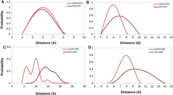
In this part, we investigated conformational state of the nucleotide binding pocket of the systems using the same atom pair (T35-G60) given above. To do so, we calculated probability distributions of two atom-pairs dis-tances between side chain oxygen of T35 and GTP Pβ atom; namely Distance 1, and between backbone amide of G60 and GTP Pβ atom; namely Distance 2. For comparison, we utilized corresponding distances in crystal struc-tures of GppNHp-bound H-RASWT (PDB ID:5P21)41 complex, where Distance 1 and Distance 2 were measured
as 5.5 Å and 6.2 Å, respectively. In general, the nucleotide is tightly coordinated by T35 and G60 residues in all of the systems, except H-RASG12D, as evident from sampling relatively longer distance values in Distance 1 and 2 (See
Fig. 2). Similar coordination is also observed for K-RAS4BG12D mutant8,40. The transforming mutant resembles
inactive State 1 with an open nucleotide binding pocket (longer Distance 1 & 2 values) (See Fig. 3C), whereas non-transforming mutant resembles State2 (shorter Distance 1 & 2 values) (See Fig. 3A), which corresponds to the active state with a compact nucleotide binding pocket. In addition, RAF-RBD-bound system resembles State 2, whereas GAP-bound system resembles a mixture of State 2 and Inactive State 3 (shorter Distance 1 and longer Distance 2 values), albeit with low probability, where interaction between Pβ atom of the nucleotide and back-bone amide of G60 is lost but the one formed between T35 and the nucleotide is maintained (See Fig. 3B). We also investigated the impact of phosphorylation status of the nucleotide on the conformational state of the catalytic site in non-transforming mutant and H-RASWT. To do so, we compared probability distributions for the same atom
pairs as given in Fig. 2. We showed that exchange of GDP by GTP causes tight coordination of the nucleotide by both T35 and G60 which is evident by left shifts in Distance 1 & 2 values in both H-RASG12P mutant and H-RASWT
(Compare A–C and B–D in Fig. 4), which is similar to what is observed for K-RAS4BG12D and K-RAS4BG12C
mutants40. Moreover, T35 and G60 sample shorter distances from the nucleotide in both GDP- and GTP-bound
H-RASG12P mutant compared to H-RASWT which might expedite the organization of the catalytically important
residues around the nucleotide binding pocket.
Figure 1. Root-mean-square fluctuations of H-RASG12D (PDB ID: 1AGP), H-RASG12P (PDB ID: 1JAH),
GAP-bound H-RASWT (PDB ID: 1WQ1), RAF-RBD-bound H-RASWT (PDB ID: 4G0N) calculated from molecular
dynamics trajectories of GTP-bound systems.
Figure 2. Probability distributions of two atom-pairs distances in GTP-bound states. (A) Distance between side chain oxygen atom of T35 and Pβ atom of GTP, namely Distance-1 and (B) Distance between backbone amide of G60 and Pβ atom of GTP, namely Distance 2, was measured using MD trajectories of corresponding systems. Green lines correspond to distances which are measured using crystal structure of GppNHp-bound H-RASWT
Dynamics of Q61 gives an insight into transforming capacity of systems.
To have more insight on the catalytic activity of systems, we investigated orientational dynamics of Q61, which is known to contribute to GTPase activity of RAS. To do so, we computed probability distributions of atom-pair distances which are meas-ured between the side chain oxygen atom of Q61 and Pβ atom of the nucleotide as shown in Fig. 5.We showed that Q61 is positioned relatively closer to the nucleotide in GAP-bound H-RASWT, (See yellow
in Fig. 5), which is expected since it represents the catalytically-active state of RAS. On the other hand, the same residue is located farther from the nucleotide binding pocket in signaling-active RAF-RBD-bound system which prevents GTP hydrolysis (See purple in Fig. 5). Q61 samples longer distances from the nucleotide in the trans-forming mutant (See blue in Fig. 5) than GAP-bound system, which explains why it cannot catalyze hydrolysis and causes transformation in the cell. It samples even longer distances from the nucleotide in H-RASG12P than
H-RASG12D, which is similar to RAF-RBD-bound H-RASWT (See red in Fig. 5). This is an interesting observation
because the catalytically important residue is positioned closer to the nucleotide in the transforming mutant than the non-transforming mutant. We also investigated the impact of the phosphorylation status of the nucleotide on dynamics of Q61 by measuring the same distance in both GDP- and GTP-bound protein. We showed that exchange of GDP by GTP increases the coordination of the nucleotide in both H-RASWT and H-RASG12P as
evi-dent from relatively shorter distance values measured for GTP-bound state (compare Fig. 5A and B). Lastly, it is also important to emphasize that Q61 samples shorter distance values in GDP-bound H-RASG12P compared to
GDP-bound H-RASWT (Fig. 5B) as also seen for T35 and G60 residue.
Figure 3. Representative structures that correspond to different states of the nucleotide binding pocket were taken from trajectories of GTP-bound systems. (A) State 2 (closed nucleotide binding pocket), (B) Inactive state 3, and (C) Inactive state 1 (exposed nucleotide binding pocket). Protein is shown in surface whereas GTP is shown in licorice representation. Switch I is shown in green, Switch II is shown in pink and rest of the protein is shown in orange color.
Figure 4. Probability distributions of two atom-pairs distances which were measured between (A,C) side chain oxygen atom of T35 and Pβ atom of the nucleotide (B,D) backbone amide of G60 and Pβ atom of the nucleotide.
Dynamics of Y32 provides mechanistic insight into molecular switch function of H-RAS
G12Pmutant.
As comparative analysis of RMSF plots reveals (See Fig. 1), Y32 highly fluctuates in H-RASG12Pmutant as well as in GAP-bound H-RASWT, whereas it is quite stable in the transforming mutant,
RAF-RBD-bound H-RASWT and H-RASWT. Detailed analysis of the trajectories showed that Y32 forms hydrogen
bond with γ-oxygen of GTP in the transforming mutant and RAF-RBD-bound H-RASWT, so it is positioned
closer to the nucleotide throughout the trajectory as shown in Fig. 6 (See A (blue and green, respectively), D and F). Here, the electrostatic repulsion between negatively charged aspartic acid residue and GTP in H-RASG12D
mutant causes this residue to be repelled from the nucleotide binding site, thus leaving a space for Y32 to interact with GTP. On the other hand, the abovementioned hydrogen bond is not formed in the non-transforming mutant and GAP-bound H-RASWT as it is evident from wider distance distributions which are measured between the side
chain of Y32 and Pβ atom of GTP (See Fig. 6 (See A (red and yellow), B and E). Detailed analysis of the trajectory of non-transforming mutant showed that the proline residue at 12th position in H-RASG12P mutant is closely
posi-tioned to GTP, thus leaving partly no space for Y32 to interact with the nucleotide.
Considering the fact that RAF-RBD-bound H-RASWT represents the signaling-active state of RAS, it is
expected to observe that Y32 makes close interaction with the nucleotide, thus preventing access to GTP for hydrolysis. Similarly, Y32 is not accessible also in the transforming mutant, making close interaction with the GTP, thus preventing access to the nucleotide and locking RAS in the “on” state. On the other hand, Y32 is dom-inantly exposed in the catalytically-active GAP-bound H-RASWT, which is also expected because, in this way,
GTP can be accessed by the nucleophilic water, thus triggering hydrolysis of the nucleotide. Interestingly, in the non-transforming H-RAS, Y32 samples both states (“exposed” and “non-exposed”), albeit with low probability of the “non-exposed” state. However, Y32 is exposed in H-RASWT upon GAP-binding as it is found in the “exposed”
state in GAP-bound H-RASWT but not in H-RASWT trajectories. Moreover, we also showed that dynamics of
Y32 in H-RASG12P mutant is modulated by the phosphorylation status of the nucleotide as Y32 can be closely
positioned to the nucleotide in GDP-bound RAS (See Fig. SI-2). Lastly, it is also important to emphasize that Y32 is positioned closer to the nucleotide in another mutant RAS subtype as well, namely, K-RAS4BG12D (PDB
ID:4DSN)18 as shown in Fig. 7 suggesting that the incomplete opening of Y32 is conserved among different
mutant RAS subtypes.
Local dynamic properties also emerge in dominant global motions of systems.
Beside exploring local structural/dynamics properties of the systems, we also investigated dominant collective motions in trajec-tories by means of essential dynamics analysis. Here, we depicted first three eigenvectors which cumulatively constitute 50% of the overall motion and highlight distinctions in dynamics of the systems as shown in Fig. 8. In general, we observed that local system-specific properties also emerge as dominant global motions in the systems, which are evident from fluctuation profiles obtained from projection of the trajectories along their first three essential eigenvectors (See Fig. 8A). According to that, Switch I and II regions dominate overall motion in trans-forming mutant as evidenced by higher fluctuation in these regions with respect to the nucleotide binding pocket. This is represented by extreme structures that are obtained by projection of trajectories along their first eigenvec-tors (See Fig. 8B). On the other hand, motion of Y32 contributes more to the overall motion in non-transforming mutant and GAP-bound H-RASWT (See red and orange in Fig. 8A).Discussion
To the best of our knowledge, this is the first study, where structural and dynamical properties of non-transforming H-RASG12P mutant have been revealed. Our results showed that glycine to proline mutation introduced stability
to H-RASG12P as evident from lower flexibility of Switch I & II in this non-transforming mutant. Interestingly,
despite the fact that both wild type protein and H-RASG12P do not cause transformation, the latter resembles more
catalytically competent state than the former. In particular, relatively lower fluctuation of the catalytically impor-tant residue Q61 in both GDP- and GTP-bound states of H-RASG12P might expedite proper organization of the
nucleotide binding pocket, thus increasing GTPase activity of non-transforming mutant compared to wild type protein, which agrees with experimental data16.
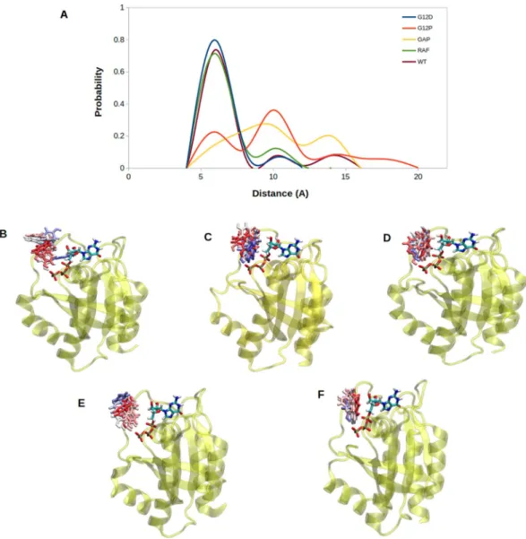
Apart from T35, G60 and Q61, we also observed remarkable differences in dynamics of Y32. Specifically, it is positioned closer to the nucleotide in signaling-active systems, which are represented by RAF-RBD-bound Figure 5. Probability distributions of two atom-pairs distances which were measured between side chain oxygen atom of Q61 and Pβ atom of the nucleotide in (A). GTP-bound H-RASG12D, H-RASG12P, GAP-bound
Figure 6. (A) Probability plots of distance distributions were measured between side chain oxygen atom of Y32 and Pβ atom of GTP by using MD trajectories of the systems. Snapshots show the orientation of Y32 with respect to nucleotide binding pocket in (B) H-RASG12P, (C) H-RASWT, (D) H-RASG12D, (E) GAP- bound
H-RASWT, (F) RAF-RBD-bound H-RASWT. Y32 is colored with respect to the frame number in corresponding
MD trajectories. Y32 and GTP are shown in licorice, whereas protein is shown in yellow and new cartoon representation.
Figure 7. Probability plot shows distance distributions which were measured between side chain oxygen of Y32 and Pβ atom of GTP in H-RASG12D and K-RAS4BG12D mutants.
H-RASWT and H-RASG12D, thus preventing accessibility of GTP for hydrolysis. On the other hand, it is located far
from the nucleotide binding pocket in catalytically-active state, which is represented by GAP-bound H-RASWT
complex. Interestingly, however; Y32 samples both of these orientations in H-RASG12P. Considering the fact
that orientational dynamics of Y32 is crucial for proper function of RAS30,33, understanding simultaneous
sam-pling of “exposed” and “non-exposed” states of Y32 can provide insight into intrinsic molecular switch func-tion of H-RASG12P. Presumably, when Y32 is positioned closer to and Q61 is positioned far from the nucleotide
H-RASG12P can conduct signal; however, when Y32 is exposed and Q61 is positioned closer to the nucleotide,
non-transforming mutant catalyzes hydrolysis of GTP.
In light of these findings, we propose that Y32 can be used as an alternative site which can be targeted by small therapeutic molecules to displace it from the nucleotide binding pocket, thus introducing intrinsic GTPase activity to transforming RAS mutants that cannot bind to GAP. Moreover, we believe that mimicking structural and dynamical properties of H-RASG12P will improve success rates in drug discovery studies since it resembles
catalytically competent state more than H-RASWT.
Methods
System setup for MD simulations.
Simulations of systems were performed as (a) guanosine triphos-phate (GTP-) bound H-RASG12P (PDB ID: 1JAH)17, H-RASG12D (PDB ID: 1AGP)16, H-RASWT (PDB ID: 5P21)41,GAP-bound H-RASWT (PDB ID: 1WQ1)42, and RAF-RBD-bound H-RASWT (PDB ID:4G0N)43 (b) guanosine
diphospate (GDP-) bound H-RASG12P and H-RASWT (PDB ID: 4Q21)44. Corresponding crystal structures were
retrieved from Protein Data Bank (PDB). In order to prepare GTP and GDP-bound H-RASG12P systems,
phos-phomethylphosphonic acid guanylate ester (GCP) was converted to GTP and GDP by substituting C3B with O
and removing γ-phosphate, sequentially. For H-RASG12D and H-RASWT, conversion of phosphoaminophosphonic
acid-guanylate ester (GNP) to GTP was done by interchanging N3B with O. As a side note, GDP bound H-RASWT
does not require any manipulation since it has been resolved with GDP. For GAP-bound H-RASWT, aluminum
fluoride (AlF3) was extracted and GDP was exchanged with GTP. For RAF-bound H-RASWT system, acetate
ion, (2R,3S) -1,4-dimercaptobutane-2.3-diol, and calcium ion were removed from the system, and GNP was substituted with GTP as it has done for both H-RASG12D and H-RASWT. Lastly, GTP-bound K-RAS4BG12D were
prepared by removing 1,2-ethanediol and substituting GCP with GTP in the same way mentioned for H-RASG12P.
Also, crystal waters which are located within 5 Å of nucleotide were kept for all the systems. After these manip-ulations, protonation states of amino acids for all systems were assigned by using PropKa server45 at pH 7.4. By
taking periodic boundary conditions (PBCs) into account, thickness of water layer was adjusted as 9 Å, except GAP- and RAF-RBD-bound H-RASWT. For these systems, it was set as 11 Å and 13 Å, respectively. Furthermore,
TIP3P46 water model was utilized, and systems were neutralized with NaCl.
Simulation protocols.
MD simulations were carried out by using Compute Unified Device Architecture (CUDA) version of Nano Scale Molecular Dynamics (NAMD) with the help of graphical processing units (GPUs) acceleration where CHARMM 36 was used to model proteins, ligands, and ions47,48. Systems were minimizedFigure 8. (A) Comparison profile of fluctuations along first three essential eigenvectors. (B) Extreme structures obtained along eigenvector 1 were shown for H-RASG12D (blue), H-RASG12P (red), H-RASWT (purple),
GAP-bound H-RASWT (orange), and RAF-RBD-bound H-RASWT (green), where thickness of ribbons corresponds to
the magnitude of contributions of residues to overall dynamics. Mg2+ ion is shown in pink and van der Waals
Root-mean-square fluctuation (RMSF).
Root-mean-square fluctuation was computed as following:∑
= − = N X n X RMSF (1/ ) ( ( ) ) n N 1 i iwhere N was duration of simulation, Xi(n) coordinates of backbone atom Xi at time n. Sum of squared difference
of mean coordinate Xi and Xi(n) was computed and divided by the duration of simulation. Lastly, square root of
the result was taken. For this analysis, ‘gmx rmsf’ module of GROMACS was utilized to explore dynamic profiles by assessments of fluctuations of residues49.
principal component analysis (pcA).
Apart from exploring local dynamic and structural properties of the systems, dominant collective motions occurring in the trajectories were demystified by means of PCA. For this, trajectories were aligned with respect to the Cα atoms of reference structure.= 〈 Δ Δ 〉 Cij M r rij i j
where Cij corresponds to covariance matrix. A change in position from time-averaged structure for each
coordi-nates of all atoms i and j was denoted as MijΔriΔrj.
Covariance matrices were generated as shown in above.
δ
= v
Cv 2
Diagonalisation of covariance matrices provided a set of eigenvalues and eigenvectors δ2, and v, respectively.
Computation and diagonalisation of covariance matrices were done by making use of ‘gmx covar’ module of GROMACS while ‘gmx anaeig’ module of GROMACS was used to obtain eigenvectors and eigenvalues from diagonalized covariance matrices49.
References
1. Wiesmüller, L. & Wittinghofer, F. Signal transduction pathways involving ras. Cellular Signalling 6, 247–267, https://doi. org/10.1016/0898-6568(94)90030-2 (1994).
2. Bryant, K. L., Mancias, J. D., Kimmelman, A. C. & Der, C. J. Kras: feeding pancreatic cancer proliferation. Trends in Biochemical
Sciences 39, 91–100, https://doi.org/10.1016/j.tibs.2013.12.004 (2014).
3. Pylayeva-Gupta, Y., Grabocka, E. & Bar-Sagi, D. Ras oncogenes: weaving a tumorigenic web. Nature Reviews Cancer 11, 761–774,
https://doi.org/10.1038/nrc3106 (2011).
4. Cox, A. D. & Der, C. J. Ras history: The saga continues. Small GTPases 1, 2–27, https://doi.org/10.4161/sgtp.1.1.12178 (2010). 5. Drosten, M. et al. Genetic analysis of ras signalling pathways in cell proliferation, migration and survival. EMBO J 29, 1091–1104,
https://doi.org/10.1038/emboj.2010.7 (2010).
6. Crespo, P. & León, J. Ras proteins in the control of the cell cycle and cell differentiation. Cellular and Molecular Life Sciences 57, 1613–1636, https://doi.org/10.1007/pl00000645 (2000).
7. Baines, A. T., Xu, D. & Der, C. J. Inhibition of ras for cancer treatment: the search continues. Future Med Chem 3, 1787–1808, https:// doi.org/10.4155/fmc.11.121 (2011).
8. Lu, S. et al. Ras conformational ensembles, allostery, and signaling. Chemical Reviews 116, 6607–6665, https://doi.org/10.1021/acs. chemrev.5b00542 (2016).
9. Castellano, E. & Santos, E. Functional specificity of ras isoforms: so similar but so different. Genes Cancer 2, 216–231, https://doi. org/10.1177/1947601911408081 (2011).
10. Luca, A. D., Maiello, M. R., Dalessio, A., Pergameno, M. & Normanno, N. The ras/raf/mek/erk and the pi3k/akt signalling pathways: role in cancer pathogenesis and implications for therapeutic approaches. Expert Opinion on Therapeutic Targets 16, https://doi.org/ 10.1517/14728222.2011.639361 (2012).
11. Young, A., Lou, D. & McCormick, F. Oncogenic and wild-type ras play divergent roles in the regulation of mitogen-activated protein kinase signaling. Cancer Discovery 3, 112–123, https://doi.org/10.1158/2159-8290.CD-12-0231 (2013).
12. Knight, T. & Irving, J. A. E. Ras/raf/mek/erk pathway activation in childhood acute lymphoblastic leukemia and its therapeutic targeting. Front Oncol 4, 160–160, https://doi.org/10.3389/fonc.2014.00160 (2014).
13. Wittinghofer, A., Scheffzek, K. & Ahmadian, M. R. The interaction of ras with gtpase-activating proteins. FEBS Letters 410, 63–67,
https://doi.org/10.1016/S0014-5793(97)00321-9 (1997).
14. Holderfield, M., Deuker, M. M., McCormick, F. & McMahon, M. Targeting raf kinases for cancer therapy: Braf-mutated melanoma and beyond. Nature Reviews Cancer 14, 455 EP–, Review Article (2014).
15. Prior, I. A., Lewis, P. D. & Mattos, C. A comprehensive survey of ras mutations in cancer. Cancer Res 72, 2457–2467, https://doi. org/10.1158/0008-5472.CAN-11-2612 (2012).
16. Franken, S. M. et al. Three-dimensional structures and properties of a transforming and a nontransforming glycine-12 mutant of p21h-ras. Biochemistry 32, 8411–8420, https://doi.org/10.1021/bi00084a005 (1993).
17. Schweins, T., Scheffzek, K., Aßheuer, R. & Wittinghofer, A. The role of the metal ion in the p21ras catalysed gtp-hydrolysis: Mn2+versus mg2+11edited by k. nagai. Journal of Molecular Biology 266, 847–856, https://doi.org/10.1006/jmbi.1996.0814
(1997).
18. Maurer, T. et al. Small-molecule ligands bind to a distinct pocket in ras and inhibit sos-mediated nucleotide exchange activity. Proc
19. Ostrem, J. M., Peters, U., Sos, M. L., Wells, J. A. & Shokat, K. M. K-ras (g12c) inhibitors allosterically control gtp affinity and effector interactions. Nature 503, 548–551, https://doi.org/10.1038/nature12796 (2013).
20. Muraoka, S. et al. Crystal structures of the state 1 conformations of the gtp-bound h-ras protein and its oncogenic g12v and q61l mutants. Current neurology and neuroscience reports, https://doi.org/10.1016/j.febslet.2012.04.058 (2012).
21. Hunter, J. C. et al. Biochemical and structural analysis of common cancer-associated kras mutations. Molecular cancer research 13, 1325–1335 (2015).
22. Xu, S. et al. Structural insight into the rearrangement of the switch i region in gtp-bound g12a k-ras. Acta Crystallographica Section
D: Structural Biology 73, 970–984 (2017).
23. Zeng, M. et al. Potent and selective covalent quinazoline inhibitors of kras g12c. Cell chemical biology 24, 1005–1016 (2017). 24. Sayyed-Ahmad, A., Prakash, P. & Gorfe, A. A. Distinct dynamics and interaction patterns in h-and k-ras oncogenic p-loop mutants.
Proteins: Structure, Function, and Bioinformatics 85, 1618–1632 (2017).
25. Vatansever, S., Gümüş, Z. H. & Erman, B. Intrinsic k-ras dynamics: A novel molecular dynamics data analysis method shows causality between residue pair motions. Scientific reports 6, 37012 (2016).
26. Mishra, A. K. & Lambright, D. G. Invited review: Small gtpases and their gaps. Biopolymers 105, 431–448, https://doi.org/10.1002/ bip.22833 (2016).
27. Ahmadian, M. R. M. et al. Guanosine triphosphatase stimulation of oncogenic ras mutants. Proc Natl Acad Sci USA 96, 7065–7070 (1999).
28. Hah, J. H. et al. Hras mutations and resistance to the epidermal growth factor receptor tyrosine kinase inhibitor erlotinib in head and neck squamous cell carcinoma cells. Head & Neck 36, 1547–1554 (2014).
29. Seeburg, P. H., Colby, W. W., Capon, D. J., Goeddel, D. V. & Levinson, A. D. Biological properties of human c-ha-ras1 genes mutated at codon 12. Nature 312, 71–75, https://doi.org/10.1038/312071a0 (1984).
30. Bunda, S. et al. Src promotes gtpase activity of ras via tyrosine 32 phosphorylation. Proceedings of the National Academy of Sciences
111, https://doi.org/10.1073/pnas.1406559111 (2014).
31. Buhrman, G., Wink, G. & Mattos, C. Transformation efficiency of rasq61 mutants linked to structural features of the switch regions in the presence of raf. Structure Article, https://doi.org/10.1016/j.str.2007.10.011 (2007).
32. Buhrman, G., Holzapfel, G., Fetics, S. & Mattos, C. Allosteric modulation of ras positions q61 for a direct role in catalysis. Proceedings
of the National Academy of Sciences 107, 4931–4936 (2010).
33. Gao, C. & Eriksson, L. A. Impact of mutations on k-ras-p120gap interaction. Computational Molecular Bioscience 3, 9 (2013). 34. Wilson, C. Y. & Tolias, P. Recent advances in cancer drug discovery targeting ras. Drug Discovery Today 21, 1915–1919, https://doi.
org/10.1016/j.drudis.2016.08.002 (2016).
35. Han, C. W., Jeong, M. S. & Jang, S. B. Structure, signaling and the drug discovery of the ras oncogene protein. BMB Rep 50, 355–360,
https://doi.org/10.5483/BMBRep.2017.50.7.062 (2017).
36. Keeton, A. B., Salter, E. A. & Piazza, G. A. The ras-effector interaction as a drug target. Cancer Res 77, 221–226, https://doi. org/10.1158/0008-5472.CAN-16-0938 (2017).
37. Quilliam, L. A. et al. Involvement of the switch 2 domain of ras in its interaction with guanine nucleotide exchange factors. Journal
of Biological Chemistry 271, 11076–11082, https://doi.org/10.1074/jbc.271.19.11076 (1996).
38. Shima, F. et al. Structural basis for conformational dynamics of gtp-bound ras protein. J Biol Chem 285, 22696–22705, https://doi. org/10.1074/jbc.M110.125161 (2010).
39. Araki, M. et al. Solution structure of the state 1 conformer of gtp-bound h-ras protein and distinct dynamic properties between the state 1 and state 2 conformers. J Biol Chem 286, 39644–39653, https://doi.org/10.1074/jbc.M111.227074 (2011).
40. Lu, S., Jang, H., Nussinov, R. & Zhang, J. The structural basis of oncogenic mutations g12, g13 and q61 in small gtpase k-ras4b.
Scientific Reports 6, 21949 EP (2016).
41. Pai, E. F. et al. Refined crystal structure of the triphosphate conformation of h-ras p21 at 1.35 a resolution: implications for the mechanism of gtp hydrolysis. EMBO J 9, 2351–2359 (1990).
42. Scheffzek, K. et al. The ras-rasgap complex: Structural basis for gtpase activation and its loss in oncogenic ras mutants. Science 277, 333–339, https://doi.org/10.1126/science.277.5324.333 (1997).
43. Fetics, S. et al. Allosteric effects of the oncogenic rasq61l mutant on raf-rbd. Structure 23, 505–516, https://doi.org/10.1016/j. str.2014.12.017 (2015).
44. Milburn, M. et al. Molecular switch for signal transduction: structural differences between active and inactive forms of proto-oncogenic ras proteins. Science 247, 939–945, https://doi.org/10.1126/science.2406906 (1990).
45. Olsson, M. H. M., Søndergaard, C. R., Rostkowski, M. & Jensen, J. H. Propka3: Consistent treatment of internal and surface residues in empirical pka predictions. Journal of Chemical Theory and Computation 7, 525–537, https://doi.org/10.1021/ ct100578z (2011).
46. Mark, P. & Nilsson, L. Structure and dynamics of the tip3p, spc, and spc/e water models at 298 k. The Journal of Physical Chemistry
A 105, 9954–9960, https://doi.org/10.1021/jp003020w (2001).
47. Huang, J. et al. Charmm36m: an improved force field for folded and intrinsically disordered proteins. Nature Methods 14 (2016). 48. Phillips, J. C. et al. Scalable molecular dynamics with namd. Journal of Computational Chemistry 26, 1781–1802, https://doi.
org/10.1002/jcc.20289 (2005).
49. Abraham, M. J. et al. Gromacs: High performance molecular simulations through multi-level parallelism from laptops to supercomputers. SoftwareX 1–2, 19–25, https://doi.org/10.1016/j.softx.2015.06.001 (2015).
Acknowledgements
Metehan Ilter would like to acknowledge TUBITAK for providing funding in scope of 2209-A Undergraduate Research Support Program, reference number: 1919B011701434. Some of the molecular dynamics simulations were performed at TUBITAK ULAKBIM, High Performance and Grid Computing Center.
Author contributions
Conceived and designed experiments: M.I. and O.S. Performed the experiments: M.I. Analysed the data: M.I. and O.S. Contributed reagents/,materials/analysis tools: M.I. Wrote the paper: M.I. and O.S.
Additional information
Supplementary information accompanies this paper at https://doi.org/10.1038/s41598-019-47481-1. Competing Interests: The authors declare no competing interests.
Publisher’s note: Springer Nature remains neutral with regard to jurisdictional claims in published maps and institutional affiliations.