C L I N I C A L A N D E X P E R I M E N T A L
OPTOMETRY
CLINICAL COMMUNICATION
A case of pigmented, free-
floating vitreous cyst treated with
micropulse diode laser
Clin Exp Optom 2016; 99: 90–92 DOI:10.1111/cxo.12308
Gokhan Gulkilik MD Mahmut Odabasi MD Sevil Karaman ErdurMD Mustafa Ozsutcu MD Mustafa Eliacik MD Goktug Demirci MD Mehmet Selim Kocabora MD
Istanbul Medipol University, Department of Ophthalmology, Istanbul, Turkey E-mail: [email protected]
Submitted: 7 January 2015 Revised: 7 February 2015
Accepted for publication: 16 April 2015 Key words: laser, low vision, retina, vitreous cyst
Vitreous cysts, unattached to the retina, are rare findings that can be congenital or acquired.1Although controversy exists, con-genital vitreous cysts are usually non-pigmented and are believed to originate from the hyaloid vascular system.2In contrast, ac-quired cysts usually have pigmented surfaces and are thought to arise from the iris or ciliary body pigment epithelium, which then dislodges into the vitreous either spontane-ously or after blunt trauma.3These cysts are usually asymptomatic and treatment is unnecessary. Argon laser or neodymium: yttrium aluminium garnet (Nd:YAG) laser photocystotomy and cyst removal with pars plana vitrectomy (PPV) have been described for the treatment of symptomatic cysts.4–6 We describe herein the first reported treatment of a pigmented, free-floating, vitreous cyst using a micropulse diode laser. CASE REPORT
A 32-year-old White man presented with a three-year history of intermittent visual blurring in the left eye. There was no history of intraocular inflammation, surgery or trauma. His visual acuity (VA) was 1.0 R and
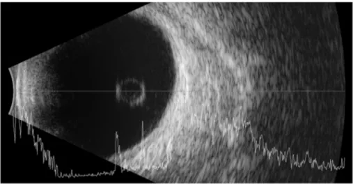
0.6 L on the day of presentation. The patient described an intermittent shadow in front of his left eye. Anterior segment examination was unremarkable. Dilated fundus examina-tion of the left eye using a 90 D lens showed a semi-transparent, oval-shaped, cystic mass with speckled surface pigmentationfloating freely in the vitreous. It obscured the visual axis and foveal details (Figure 1). The optic disc and retina beyond the arcades were normal. The vitreous was partially liquefied but there was no posterior vitreous detach-ment. The patient denied any history of ocular trauma. The ophthalmic examination revealed no signs of ocular trauma. Ultraso-nography (Eye Cubed, Ellex, Adelaide, Australia) revealed a 5.32 mm intra-vitreal cyst that was acoustically hollow internally and hyper-echogenic externally (Figure 2). The cyst was not connected to the optic disc or peripheral retina. Spectral domain optical coherence tomography (SD-OCT) (Avanti RTVue-XR, Optovue, Fremont, California, USA) showed a thin, hyper-reflective cyst wall (Figure 3). Its lumen was divided by numerous hyper-reflective septa forming a multi-lobular structure. The interior of each lobule was hypo-reflective. There was no scolex-like appear-ance inside the cyst and parasite serologies and immunoglobulin E were normal.
Because the patient was symptomatic, photocystotomy was performed using a 577 nm micropulse diode laser (Supra Scan, Quantel Medical, Bozeman, Montana, USA), which has advantages such as minimal collateral damage, less heat conduction and temperature rise in the retinal tissue. Photocystotomy was performed at multiple points to prevent under-treatment of the multi-lobular cyst. Pigmented areas of the cyst were selected to maximise laser absorption. Laser parameters were power at 500 mW for 0.01 seconds. Ten minutes later, the cyst size decreased to 2.04 mm and it no longer blocked the visual axis (Figure 4). One day later, VA improved to 1.0 and the patient was symptom-free except for minimal
floaters. During follow-up, the cyst shrank further and only a small pigmented mem-branous mass could be seen outside the vi-sual axis.
DISCUSSION
Pigmented vitreous cysts, unattached to the retina, are rarely seen and treatment is not indicated unless they block the visual axis or become symptomatic. There is little treatment information in the literature.
Cyst removal via PPV, argon laser photocystotomy and Nd:YAG laser cystotomy has been previously described.4–6 All these methods effectively relieve symptoms but have their own risks. Orellana and colleagues6successfully performed PPV and aspirated the cyst using a 20G canula. Vitrec-tomy has become safer with newer technol-ogy but is still associated with a long list of complications. For instance, cataracts are likely to form in young phakic eyes. We believe that, unless other methods fail to relieve symptoms, PPV should not be a first-line treatment for this disease in young phakic patients. Awan4suggested argon laser photocystotomy as a safe and effective method to treat vitreous cysts. Historically, this treatment has been used for lesions three tofive millimetres in diameter. Recently, De-sai and Saffra7successfully performed argon laser photocystotomy for a 7.1-mm diameter vitreous cyst, emphasising its safety; however, Nork and Milecchia8reported an inadvertent retinal burn that occurred during argon green laser photocystotomy. Because the cyst is transparent, this complication may occur if the cyst is close to the posterior pole and the fovea can be damaged. Lastly, Ruby and Jampol5 described Nd:YAG laser cystotomy to disrupt such cysts. Unlike the thermal ef-fect of argon laser, Nd:YAG laser produces acoustic shock waves, which mechanically breakdown the cyst wall. This technique is also not without complications, as shock waves may be transmitted to the retina.
© 2016 Optometry Australia Clinical and Experimental Optometry 99.1 January 2016
Whereas peripheral vitreous traction may re-sult in a retinal tear and eventual retinal de-tachment, perifoveal contraction can cause a macular hole.9
In the present case, we report a pigmented vitreous cyst unattached to the retina in a 32-year-old man with no pertinent medical history and no secondary cause of the cyst. Lisch and Roschel2suggest that pigmented, vitreous cysts arise from the iris or ciliary pigment epithelium, which then dislodges into the vitreous either traumatically or spontaneously. They state that these cysts are not connected to the retina and that they are transparent with speckled surface pigmentation. We were also unable to dem-onstrate any connection to the optic disc or hyaloidal system by ultrasonography or fundus examination. We believe that Lisch
and Roschel’s theory2for the origin of this type of pigmented, free-floating cyst is correct. Histological examinations demon-strated a monolayer of pigmented cells similar to the pigment epithelium of retina, ciliary body and iris with microvillous pro-cesses toward the vitreous.8This orientation of the cells may causefluid to be pumped from the vitreous cavity into the interior of the cyst and cause the cyst to enlarge or reform after treatment. When we performed
SD-OCT on the cyst, we demonstrated nu-merous septal divisions and multiple cavities. This finding may add new information to the current treatment approach. We suggest that performing laser photocystotomy at a single point on the cyst may be insufficient for complete resolution. Laser treatment at multiple points may be required to increase the success rate but may also increase the rate of laser-related complications, both for Nd: YAG laser and for argon laser (because cysts must be located in front of the macula to be symptomatic).
Recently, 577 nm micropulse diode laser technology was introduced. It has demon-strated effectiveness but causes minimal collateral retinal damage. It dispenses laser energy in micropulses instead of a single pulse; thus, heat conduction and tempera-ture rise in the retinal tissue is limited.10 Because the cyst was just in front of the mac-ula, we decided to perform photocystotomy with micropulse diode laser to decrease the risk of inadvertent retinal burn. We applied laser to multiple spots but we did not see any retinal burns. The cyst shrank signi fi-cantly at 10 minutes and has not recurred during follow-up. In conclusion, pigmented cysts that are not attached to the retina are very rare lesions that may have multi-lobular structures. Symptomatic cysts can safely be treated using photocystotomy with a micropulse diode laser at multiple points, thus, avoiding the risks of conventional argon or Nd:YAG laser cystotomy.
Figure 2. B-scan ultrasonography of the cyst. Its wall was hyperechogenic and the inner part was acoustically hollow. There was no scolex-like formation inside. Any connections to other parts of the eye could not be seen.
Figure 1. A free-floating, pigmented cyst obscuring the visual axis and masking the foveal details. The cyst had no connections to the optic disc or peripheral vitreous.
Figure 3. Spectral domain optical coher-ence tomography image showing a hyper-re-flective, thin-walled cyst. The multilobed cyst interior was formed by multiple hyper-reflective septa.
Figure 4. Coloured photograph of the cyst just after micropulse diode laser treatment. The cyst deflated and moved away from the visual axis. Multiple laser spots can be seen on the cyst surface.
Vitreous cyst treated with micropulse diode laser Gulkilik, Odabasi, Erdur, Ozsutcu, Eliacik, Demirci and Kocabora
© 2016 Optometry Australia Clinical and Experimental Optometry 99.1 January 2016
REFERENCES
1. Cruciani F, Santino G, Salandri AG. Monolateral idiopathic cyst of the vitreous. Acta Ophthalmol Scand 1999; 77: 601–603.
2. Bullock JD. Developmental vitreous cysts. Arch Ophthalmol 1974; 91: 83–84.
3. Lisch W, Rochels R: Pathogenesis of congenital vitre-ous cysts. Klin Monbl Augenheilkd 1989; 195: 375–378. 4. Awan KJ. Biomicroscopy and argon laser photocystotomy of free-floating vitreous cysts. Oph-thalmology 1985; 92: 1710–1711.
5. Ruby AJ, Jampol LM. Nd:YAG treatment of a posterior vitreous cyst. Am J Ophthalmol 1990; 110: 428–429.
6. Orellana J, O’Malley QE, McPherson AR, Font RL. Pigmented free-floating vitreous cysts in two young adults.Electron microscopic observations. Ophthal-mology 1985; 92: 297–302.
7. Desai RU, Saffra NA. Argon laser photocystotomy of a vitreous cyst. Ophthalmic Surg Lasers Imaging 2010; 9: 1–4. 8. Nork TM, Millecchia LL. Treatment and histopa-thology of a congenital vitreous cyst. Ophthalmology 1998; 105: 825–830.
9. Chaudhary R, Sheidow T, Gonder JR, Merchea MM. Macular hole following YAG capsulotomy. Br J Ophthalmol 1999; 83: 755.
10. Sramek CK, Leung LS, Paulus YM, Palanker DV. Therapeutic window of retinal photocoagulation with green (532 nm) and yellow (577 nm) lasers. Ophthalmic Surg Lasers Imaging 2012; 43: 341–347.
Vitreous cyst treated with micropulse diode laser Gulkilik, Odabasi, Erdur, Ozsutcu, Eliacik, Demirci and Kocabora
© 2016 Optometry Australia Clinical and Experimental Optometry 99.1 January 2016