See discussions, stats, and author profiles for this publication at: https://www.researchgate.net/publication/317697620
The relationship of serum Pentraxin-3 with peripheral arterial disease
Article · June 2017 DOI: 10.5152/eajem.2017.06977 CITATIONS 0 READS 104 11 authors, including:Some of the authors of this publication are also working on these related projects:
irisin View project
Author View project Burak Katipoglu Ufuk Üniversitesi 36PUBLICATIONS 13CITATIONS
SEE PROFILE
Ali Aygün
University Education and Research Hospital, 25PUBLICATIONS 30CITATIONS
SEE PROFILE
Selim Demir
Karadeniz Technical University 66PUBLICATIONS 89CITATIONS
SEE PROFILE
Ozgur Tatli
Trabzon KanuniTraining and Research Hospital 30PUBLICATIONS 197CITATIONS
SEE PROFILE
All content following this page was uploaded by Ali Aygün on 04 October 2017.
Relationship of Serum Pentraxin-3 with Peripheral Arterial Disease
Burak Katipoğlu1, Ali Aygün2, Selim Demir3, Özgür Tatlı4, Ahmet Menteşe5, Ümit Menteşe6, Gökalp Altun7, Asım Örem8, Abdulkadir Gündüz4,
Semih Korkut9, Togay Evrin10
1Department of Emergency Medicine, Ankara Training and Research Hospital, Ankara, Turkey
2Department of Emergency Medicine, Ordu University Training and Research Hospital, Ordu, Turkey
3Department of Nutrition and Dietetics, Karadeniz Technical University School of Health Sciences, Trabzon, Turkey
4Department of Emergency Medicine, Karadeniz Technical University School of Medicine, Trabzon, Turkey
5Program of Medical Laboratory Techniques, Karadeniz Technical University Vocational School of Health Sciences, Trabzon, Turkey
6Department of Cardiovascular Surgery, Ahi Evren Thoracic and Cardiovascular Surgery Training and Research Hospital, Trabzon, Turkey
7Department of Cardiovascular Surgery, Karadeniz Technical University School of Medicine, Trabzon, Turkey
8Department of Medical Biochemistry, Karadeniz Technical University School of Medicine, Trabzon, Turkey
9Public Hospitals Association Bakırköy General Secretaria, İstanbul
10Department of Emergency Medicine, Ufuk University School of Medicine, Dr. Rıdvan Ege Training and Research Hospital, Ankara, Turkey
Introduction
Peripheral artery disease (PAD) is a widely occurring condition. The main cause is atherosclerosis, and the causes of atherosclerosis con-stitute the predisposing factors for the disease (1).
Atherosclerosis, the cause of PAD, is generally a condition of advanced age. It develops in association with intimal plaques affecting arterial cir-culation of the vascular system and containing varying proportions of lipids, macrophages, fibroblasts, smooth muscle cells, and extracellular materials. Atherosclerosis is also a chronic inflammatory condition (2).
Correspondence to: Ali Aygün e-mail: [email protected]
Received: 08.04.2017 • Accepted: 06.06.2017 • Available Online Date: xx.xx.xxxx
©Copyright 2017 by Emergency Physicians Association of Turkey - Available online at www.eajem.com DOI: 10.5152/eajem.2017.06977
Abstract
Aim: Since atherosclerosis is a chronic inflammatory process associated with peripheral artery disease (PAD), the inflammatory marker pentraxin (PTX) may
increase in PAD.
Materials and Methods: This cross-sectional clinical study was performed at the tertiary university hospital emergency department and cardiovascular
sur-gery departments in Turkey. The purpose was to determine the value of PTX3 in the diagnosis of PAD. This study was performed on 43 symptomatic patients aged >18 years and diagnosed with PAD.
Results: Median PTX3 value was 1.027 (25–75th percentile: 0.395–2.902) in the control group and 0.585 (25–75th percentiles: 0.406–5.467) in the PAD group (p=0.913). A comparison of PTX3 with ankle brachial index (ABI) values revealed a weak and non-significant correlation (rho: 0.016, p=0.886). Analysis of
PTX3 values with other parameters (age, systolic and diastolic blood pressure, heart rate, respiratory rate, temperature, and SpO2) revealed no significant
correlation with any of the other parameters.
Conclusion: Our findings indicate that PTX3 levels cannot be used as a marker in patients with the chronic process of PAD. Keywords: Atherosclerosis, Inflammation, Pentraxin-3, Peripheral arterial disease
EMERGENCY MEDICINE
Cite this article as: Katipoğlu B, Aygün A, Demir S, Tatlı Ö, Menteşe A, Menteşe Ü, et al. Relationship of Serum Pentraxin-3 with Peripheral
Pentraxins (PTX) are multifunctional protein superfamily that play a role in the inflammatory response. PTX3 is one of the main acute phase reactants, which may increase in circulation 3–5 times above the baseline in inflammatory conditions. PTX3 is produced in the region of inflammation and binds immediately to the endothelium. PTX3 levels are believed to be an independent marker of disease ac-tivity (3).
Since a chronic inflammatory process is involved in atherosclerosis, PTX3 is an inflammatory marker that might be expected to increase in PAD. This study investigated whether PTX3 increases in patients with PAD.
Materials and Methods
Patients with suspected PAD based on symptoms at presentation to the tertiary university hospital emergency department and cardio-vascular surgery departments over a 12-month period were included in this cross-sectional clinical study after receiving of approval from the local ethics committee. The study enrolled 43 patients presenting to the aforementioned units and with suspected PAD and 40 healthy control group volunteers contacted from outside the hospital. Patients aged ≥18 years presenting to the departments with suspect-ed PAD and agreeing to participate were includsuspect-ed. Exclusion criteria were patients with acute coronary syndrome, acute kidney failure, chronic kidney failure, hemorrhagic stroke, cerebrovascular disease, liver failure, acute pulmonary edema, cardiopulmonary arrest, acute mesenteric ischemia, or pulmonary thromboembolism.
Clinical and demographic characteristics, such as symptoms, physi-cal examination findings and Doppler ultrasound, peripheral arterial with contrast computerized tomography, and magnetic resonance angiography details were recorded into a study form.
The control group inclusion criteria included non-pregnant or puer-perant patients, aged ≥18 years with no acute kidney failure, chronic kidney failure, sepsis, ischemic stroke, liver failure, acute pulmonary edema, PAD, deep vein thrombosis, acute coronary syndrome, pul-monary embolism, mesenteric ischemia, cardiopulpul-monary arrest, multi-trauma, Tissue Plasminogen Activator (TPA)-related hemor-rhage or acute trauma.
For PTX3 measurements at the time of presentation, a complete blood count (CBC) was performed by collecting blood samples in the anticoagulant ethylenediaminetetraacetic acid (EDTA) tubes. Plas-ma was separated by centrifugation at 1800× g for 10 min and then stored at −80°C until PTX3 study.
Measurement of plasma pentraxin-3
PTX-3 levels in human plasma were determined using a commer-cial enzyme-linked immuno-sorbent assay (ELISA) kit (R&D Systems, Cat No: DPTX30, Minneapolis, USA) following the manufacturer’s in-structions. Plasma stored at −80°C was thawed to room temperature. Briefly, 200 µL of PTX3 biotinylated antibody was added into each well of a streptavidin-coated plate. Plates were incubated for 60 min at room temperature on a microplate shaker. The plates were subse-quently washed using 300 µL washing buffer to remove
non-bind-ing antibodies. PTX3 standards were prepared in line with the kit procedures. Standards, controls, and specimens were activated with pre-treatment D solution for 30 min. Further, 100 µL assay diluent solution was added to each plate; 20 µL of pre-treated standards, controls, and specimens were added to the solution and incubated for 120 min in a microplate shaker at room temperature. Following incubation, the plate was washed four times with washing buffer in a plate washer; 200 µL of PTX3 conjugate was added into the wells and incubated at room temperature for 120 min on a microplate shaker. Following incubation, the plate was washed four times using washing buffer in a plate washer. Subsequently, 200 µL of Tetra Metil Benzidin (TMB) substrate solution was added to each well for color development and incubated in dark for 30 min at room temperature; 50 µL of color stop solution was added to each well, and specimens were observed to turn yellow in color. Absorbance was measured at a wavelength of 450 nm using a microplate reader (Versamax, Molecu-lar Devices, CA, USA). A standard chart was prepared using the absor-bance values against standard concentrations obtained. PTX3 levels in specimens were calculated as ng/mL using this standard chart. The intra-assay distribution reliability of this ELISA method was 3.6% and the inter-assay distribution reliability was 4.9%.
Statistical analysis
Statistical analyses were performed using the Statistical Package for Social Sciences (SPSS Inc.; Chicago, IL, USA) version 11 software. The normality of the distribution of variables was tested using visual (his-togram and probability charts) and analytical methods (Kolmogor-ov-Smirnov/Shapiro-Wilk tests). Descriptive analyses were expressed as mean and standard deviation for normally distributed variables. These were compared on 2×2 grids using Person’s chi square and Fisher’s exact tests. Since PTX3 and ankle brachial index (ABI) values were not normally distributed, these were analyzed using the Mann– Whitney U test between two groups and with the Kruskal–Wallis test between more than two groups. Spearman’s correlation test was used to investigate correlations. P values less than 0.05 were regard-ed as statistically significant.
Results
The mean age of the participants was 63.9±11.9 years (min: 40, max: 86) in the control group and 64.5±13.8 years (min: 21, max: 88) in the patient group. Males comprised 67.5% of the control group and 81.4% of the patient group. No significant difference was observed between the two groups in terms of age or gender distribution (p>0.05; Table 1).
When physical examination findings were compared between the control and patient groups, no ulcer, muscular atrophy, claudica-tion, rash, or edema were observed in the control group; however, ulcer was present in 41.9%, muscular atrophy in 9.3%, claudication in 100%, rash in 53.5%, and edema in 44.2% of the patient group (p<0.001 except for muscular atrophy; Table 1).
In terms of vital findings, mean diastolic blood pressure (BP) in the patient group (77.58±12.55 mmHg) was significantly lower than that in the control group (100.25±98.6 mmHg; p=0.005). Similarly, in con-trast, respiration rate was significantly higher in the patient group compared to the controls (p<0.001; Table 1).
Eurasian J Emerg Med 2017 Katipoğlu et al.
When patients were questioned about comorbidities other than PAD, coronary artery disease (CAD) was present in 45% of the con-trol group and 53.5% of the patient group, hypertension (HT) in 60%
of the control group and 67.4% of the patient group, hyperlipidemia (HPL) in 25% of the control group and 23.3% of the patient group, and diabetes mellitus (DM) in 35.0% of the control group and 53.5%
Table 1. Basic demographic and clinical characteristics of the control
and patient groups
Control (n=40) Patient (n=43) n (mean)¹ % (±SD)¹ n (mean)¹ % (±SD)¹ (median)² (min–max)² (median)² (min–max)² p Age, years <44 3 7.5 2 4.7 0.956* 45-54 5 12.5 6 14.0 55-64 13 32.5 14 32.6 65+ 19 47.5 21 48.8 Sex Female 13 32.5 8 18.6 0.146* Male 27 67.5 35 81.4 Physical examination Ulcer 0 0.0 18 41.9 <0.001* Muscular 0 0.0 4 9.3 0.117** atrophy Claudication 0 0.0 43 100.0 <0.001* Rash 0 0.0 23 53.5 <0.001* Edema 0 0.0 19 44.2 <0.001* Vital signs Systolic BP (125.00)² (105-160)² (130.00)² (100-190)² 0.628² Diastolic BP (85.00)² (70-105)² (80)² (50-110)² 0.005² Heart rate (76.70)¹ (10.52)¹ (76.44)¹ (13.32)¹ 0.922¹ Respiration (14.00)² (11-20)² (17)² (11-20)² <0.001² rate Temperature (36.80)² (36-38)² (36.70)² (34.4-37.6)² 0.049² SpO2 (96.00)² (91-99)² (96)² (85-99)² 0.654² Comorbid diseases CAD 18 45.0 23 53.5 0.440* HT 24 60.0 29 67.4 0.481* HPL 10 25.0 10 23.3 0.853* DM 14 35.0 23 53.5 0.122* ECG NSR 40 100.0 37 86.0 0.026** AF 0 0.0 6 14.0 0.026** AFL 0 0.0 1 2.3 1.000** Non-ST 0 0.0 1 2.3 1.000** Ischemic ST 0 0.0 11 25.6 0.001*
*Pearson’s chi square test, **Fisher’s exact test, ¹Independent groups t test, ²Mann–Whitney U test, SD: standard deviation; DM: diabetes mellitus; HPL: hyperlipidemia; CAD: coronary artery disease; ECG: electrocardiography; AF: atrial fibrillation; AFL: atrial flutter; NSR: normal sinus ritm; BP: blood pressure; HT: hypertension
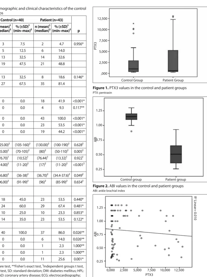
Figure 1. PTX3 values in the control and patient groups PTX: pentraxin Control Group PT X3 12,500 10,000 7,500 5,000 2,500 ,000 Patient Group
Figure 3. PTX3 and ABI correlation chart PTX: pentraxin; ABI: ankle brachial index
PTX3 0,000 2,500 5,000 7,500 10,000 12,500 ABI 1.25 1.00 0.75 0.50 0.25 R 2 Linear= 0.010
Figure 2. ABI values in the control and patient groups ABI: ankle brachial index
Control group ABI 1.25 1.00 0.75 0.50 0.25 Patient group
of the patient group. No significant correlation was determined be-tween the groups in terms of CAD, HT, HPL, or DM (p>0.05; Table 1). When participants’ electrocardiography (ECG) findings were com-pared, normal ECG results were determined in almost all in the con-trol group, while atrial fibrillation (AF) was determined in 14% of the patient group, atrial flutter (AFL) in 2.3%, non-ST changes in 2.3%, and ischemic ST elevation in 25.6% (Table 1).
Median PTX3 level in the control group was 1.027 ng/mL (25–75th
percentile: 0.395–2.902), and 0.585 ng/mL (25–75th percentile: 0.406–
5.467) in the patients with PAD(p=0.913; Figure 1). Median ABI in the control group was 1.15 (25–75th percentile: 1.10–1.20), significantly
higher than the median value of patients with PAD at 0.51 (25–75th
percentile: 0.36–0.66; p<0.001; Figure 2).
A weak and insignificant correlation was observed when we com-pared ABI values currently used as a diagnostic test with the new method PTX3 (r=0.016; p=0.886; Figure 3). PTX3 values investigated as a novel method also exhibited no significant correlation with any other measurement parameters (age, systolic BP, diastolic BP, heart rate, respiration rate, temperature, and SpO2) (p>0.05).
Examination of the relations between PTX3 and sociodemograph-ic variables, physsociodemograph-ical examination, and additional chronsociodemograph-ic diseases showed that none was a probable confounding factor. Accordingly, no correlation was determined between age, muscle atrophy, claudi-cation, rash, edema, CAD, HT, HPL, DM, or smoking status and PTX3 values (p>0.05).
Discussion
Mean PTX3 values were 1.027 (25–75th percentile: 0.395–2.902) in the
control group and 0.585 (25–75th percentile: 0.406–5.467) in patients
with PAD. The difference was not statistically significant (p=0.913). Comparison of ABI values used as an existing diagnostic test and PTX3 revealed a weak and non-significant correlation (r=0.016; p=0.886). No significant association was observed with factors shown in the literature with PTX3 values.
Studies have reported varying prevalence of PAD. The prevalence of PAD in the general population aged over 40 years in Spain was report-ed as 9.7% in women and 11.4% in men (4). A prevalence level of 5% for PAD (ABI <0.9) has been reported in individuals representing the gen-eral population aged ≥40 years in the USA (5). A study conducted in a care home in Turkey involving 507 individuals aged >60 years reported a prevalence of PAD of 5.9%, while a study that screened the general population aged >40 years reported a prevalence of 19.76% (6). Various studies have been performed for the early identification of PAD (7). However, the application of PTX3, an inflammatory marker, in the diagnosis of PAD has not been previously investigated. As described, PTX3 plays an important role in the primary inflammatory response. It is therefore included among the diagnostic tests for several diseases and cardiovascular diseases (CVDs) in particular, from ovarian torsion to pleural fluid effusion and pulmonary contusion (8, 9). Immunohis-tochemical studies have shown that plasma PTX3 levels increase in atherosclerotic lesions but not in non-atherosclerotic lesions and have
demonstrated that PTX3 is a marker of localized vascular inflammation. This has emphasized the importance of investigating the relation be-tween clinical atherosclerotic events and PTX3 levels. Additionally, the determination of higher PTX3 levels in subjects with CVD compared to those without CVD among patients with myocardial infarction with systolic BP elevation has led to PTX3 levels also being investigated in this group (10). Zhou et al. (11) reported a negative, highly significant correlation between PTX3 and ABI values. In our study, PTX3 and ABI did not correlate to age, systolic BP, diastolic BP, heart rate, respiration rate, body temperature, or SpO2. Although Tomandlova et al. (10) re-ferred to a significant correlation between age and PTX3, they report-ed no correlation between systolic or diastolic BP and PTX3, in agree-ment with our study. In a study investigating the relation between severity of PAD and endothelial progenitor cells (EPCs), Morishita et al. (12)reported that EPCs and PTX3 increased in a correlated manner in patients with PAD compared to those without PAD. In our study, and in contrast to Morishita et al. (12), PTX3 levels were lower in subjects with PAD, although the difference was not statistically significant. In addi-tion, Morishita et al. (12) reported a 33.3% prevalence of DM, 71.4% of HT, and 38.1% of CVD among subjects with PAD. In our study, the prev-alence values in patients with PAD were 53.5% for DM, 67.4% for HT, and 53.5% for CVD, indicating higher prevalence of DM and CVD but not HT. Additionally, mean systolic (137.38±26.20 mmHg) and diastol-ic (73.81±14.74 mmHg) BP values in the study by Morishita et al. (12) were similar to those of our study (systolic: 128.23±18.59 mmHg; dia-stolic: 77.58±12.55 mmHg). Inoue et al. (13) compared mean plasma PTX3 levels by collecting blood specimens from 252 patients undergo-ing angiography for CAD evaluation at a university hospital, with 162 patients under monitoring due to HT, HPL, DM, or CVD. Mean PTX-3 values of patients undergoing angiography with a preliminary diagno-sis of ischemic heart disease were significantly high. In the study of pa-tients with type 2 DM, Rashtchizadeh et al. (14) observed significantly higher mean PTX3 in patients with CAD compared to those with no CAD. However, no such relationship was observed in our study. Studies regarding PTX3 have revealed that the levels of PTX3 also in-crease in some inflammatory diseases. In a study of patients with ar-thritis, Ishihara et al. (15) observed higher serum PTX3 levels in periods when the disease was active compared to when it was not active and compared to a healthy control group. They reported that PTX3 is more specific than C-reactive protein (CRP) in showing arterial inflammation. Fazzini et al. (3) investigated PTX3 levels in 43 patients with vasculi-tis. They observed higher PTX3 levels in active vasculitis compared to during times of no activation. Moreover, PTX3 levels when the disease was not activated and in the healthy control group was similar. The study also observed high PTX3 levels in an untreated vasculitis group and low levels in subjects receiving immunosuppressive therapy. They concluded that PTX3 may be a reliable acute phase reactant in showing inflammation in active vasculitis (3). Studies investigating risk factors for PAD have identified DM, HL, HT, smoking, and obesity as such factors (16). More than 91% of patients with PAD were reported to have at least one risk factor for atherosclerosis (4). Similarly, in our study, 94% of patients had at least one such risk factor.
Several previously published studies have reported that coronary problems are encountered frequently in patients with PAD. In addi-tion, one necroscopy study involving cases with lower extremity isch-emia sufficiently severe to require amputation observed diffuse and
Eurasian J Emerg Med 2017 Katipoğlu et al.
severe coronary atherosclerotic and myocardial lesions in almost all these cases (17). CAD was diagnosed in 23 (53.5%) of the PAD cases in this study. A history of non-ST elevation was present in 1 (2.3%) patient and ischemic ST elevation in 11 (25.6%). In addition, medi-an value of ABI of 1.15 (25–75th percentiles: 1.10–1.20) in the control
group was significantly higher than median value of ABI 0.51 (25–75th
percentiles: 0.36–0.66) in the subjects with PAD (p<0.001), indicating that consistent results were obtained in the diagnosis of PAD.
Study limitations
The absence of any significant finding in this study despite PTX3 be-ing closely associated with atherosclerosis and the fact that the result did not change despite the grading of confounding factors, show the need for a larger and randomized sample.
Conclusion
We consider that PTX3 levels remained unchanged in this study be-cause despite its atherosclerotic foundation, PAD is a chronic process that does not develop acutely. Further studies with a larger patient group and with PAD classified according to subtypes are needed.
Ethics Committee Approval: Authors declared that the research was
con-ducted according to the principles of the World Medical Association Decla-ration of Helsinki “Ethical Principles for Medical Research Involving Human Subjects”, (amended in October 2013).
Informed Consent: Written informed consent was obtained from all
partici-pants who participated in this study.
Peer-review: Externally peer-reviewed.
Conflict of Interest: No conflict of interest was declared by the authors. Financial Disclosure: This study was supported by the Karadeniz Technical
University Scientific Research Foundation (Project Number: TTU-2015-5358), Trabzon, Turkey.
References
1. Cecilia A. Gutierrez. Peripheral Arterial Disease. In: Paulman PM, Paulman
AA, Harrison Taylor JD, editors.Taylor’s Manual of Family Medicine. Phila-delphia: Lippincott Williams &Wilkins; 2008. p.339-43.
2. Van der Wal AC, Becker AE, Das PK. Medial thinning and atherosclero-sis--evidence for involvement of a local inflammatory effect. Atheroscle-rosis 1993; 103: 55-64.
3. Fazzini F, Peri G, Doni A, Dell’Antonio G, Dal Cin E, Bozzolo E, et al. PTX3 in small-vessel vasculitides: an independent indicator of disease
activity produced at sites of inflammation. Arthritis Rheum 2001; 44: 2841-50.
4. Carbayo JA, Divisón JA, Escribano J, López-Abril J, de Coca EL, Artigao LM, et al. Using ankle-brachial index to detect peripheral arterial disease: prevalence and associated risk factors in a random population sample. Nutr Metab Cardiovasc Dis 2007; 17: 41-9.
5. Menke A, Muntner P, Wildman RP, Dreisbach AW, Raggi P. Relation of
bor-derline peripheral arterial disease to cardiovascular disease risk. Am J Cardiol 2006; 98: 1226-30.
6. Karabay O, Karacelik M, Yilik L, Tekin N, Iriz AB, Kumdereli S, et al. Ischemic
peripheral arterial disease: A screening survey. Turk J Thoracic Cardio-vasc Surg 2012; 20: 450-7.
7. Ambrosetti M. Timely diagnosis of lower extremity peripheral arterial disease: one of the many expected actions by the cardiologist. Int J Car-diol 2014; 175: 217.
8. Akman L, Erbas O, Terek MC, Aktug H, Taskiran D, Askar N. The long pen-traxin-3 is a useful marker for diagnosis of ovarian torsion: An experi-mental rat model. J Obstet Gynaecol 2016; 36: 399-402.
9. Tatlı O, Keha Kurt N, Karaca Y, Sahin A, Aygun A, Sahin E, et al. The diag-nostic value of serum pentraxin 3 levels in pulmonarycontusion. Am J Emerg Med 2017; 35: 425-8.
10. Tomandlova M, Jarkovsky J, Tomandl J, Kubkova L, Kala P, Littnerova S, et al. Prognostic value of pentraxin-3 level in patients with STEMI and its re-lationship with heart failure and markers of oxidative stress. Dis Markers 2015; 2015: 159051.
11. Zhou Y, Ni Z, Zhang J, Zhang W, Wu Q, Shen G, et al. Plasma pentraxin 3 may be a better marker of peripheral artery disease in hemodialysis patients than C-reactive protein. Vasc Med 2013; 18: 85-91.
12. Morishita T, Uzui H, Nakano A, Mitsuke Y, Geshi T, Ueda T, et al. Number of endothelial progenitor cells in peripheral artery disease as a marker of severity and association with pentraxin-3, malondialdehyde-modified low-density lipoprotein and membrane type-1 matrix metalloprotein-ase. J Atheroscler Thromb 2012; 19: 149-58.
13. Inoue K, Sugiyama A, Reid PC, Ito Y, Miyauchi K, Mukai S, et al. Estab-lishment of a high sensitivity plasma assay for human pentraxin3 as a marker for unstable angina pectoris. Arterioscler Thromb Vasc Biol 2007; 27: 161-7.
14. Rashtchizadeh N, Sede SA, Ghaffari MA, Mohammadzadeh G, Majidi S. Associations of pentraxin 3 with presence and severity of coronary ar-tery disease in type 2 diabetes patients. Turk J Biochemistry 2015; 40: 37-43.
15. Ishihara T, Haraguchi G, Kamiishi T, Tezuka D, Inagaki H, Isobe M. Sensitive assessment of activity of Takayasu’s arteritis by pentraxin3, a new bio-marker. J Am Coll Cardiol 2011; 57: 1712-3.
16. Ankle Brachial Index Collaboration. Ankle brachial index combined with Framingham risk score to predict cardiovascular events and mortality. JAMA 2008; 300: 197-208.
17. Mautner GC, Mautner SL, Roberts WC. Amounts of coronary arterial nar-rowing by atherosclerotic plaque at necropsy in patients with lower ex-tremity amputation. Am J Cardiol 1992; 70: 1147-51.
View publication stats View publication stats