A comparison of Chevron and Lindgren-Turan
osteotomy techniques in hallux valgus surgery:
a prospective randomized controlled study
Correspondence: Esat Uygur, MD. İstanbul Medeniyet Üniversitesi,
Göztepe Eğit. ve Araşt. Hast. Ortopedi ve Travmatoloji Kliniği, İstanbul, Turkey. Tel:+90 216 – 357 52 09 e-mail: [email protected]
Submitted: July 25, 2014 Accepted: January 16, 2016
©2016 Turkish Association of Orthopaedics and Traumatology
Available online at www.aott.org.tr doi: 10.3944/AOTT.2016.14.0272 QR (Quick Response) Code
doi: 10.3944/AOTT.2016.14.0272
Esat UYGUR1, Namık Kemal ÖZKAN2, Kaya AKAN1, Hakan ÇİFT3
1İstanbul Medeniyet University Göztepe Training and Research Hospital, Department of Orthopedics and Traumatology, İstanbul, Turkey 2Ersoy Hospital, Department of Orthopedics and Traumatology, İstanbul, Turkey
3Medipol University Faculty of Medicine, Department of Orthopedics and Traumatology, İstanbul, Turkey
Hallux valgus is a common deformity, with numerous techniques having been developed for its operative treat-ment. While various surgical approaches have been de-scribed, controversy remains regarding which method is most effective. In terms of the different stages of hallux valgus deformity, distal metatarsal osteotomy is
recom-mended for mild deformities, soft tissue correction with the addition of distal osteotomy is recommended for moderate deformities, and diaphyseal osteotomy, mul-tiple osteotomies, or proximal metatarsal osteotomy is recommended for advanced deformities.[1–3]
Chevron osteotomy was first described in 1976
Objective: The aim of this prospective randomized controlled single-blind study was to compare the
results of Chevron and Lindgren-Turan osteotomy techniques for treatment of moderate hallux valgus.
Methods: A total of 66 female patients (34 in Chevron group, 32 Lindgren-Turan group) were
recruit-ed in this study and followrecruit-ed up for an average of 26.08 months. Operative procrecruit-edures were performrecruit-ed by 2 surgeons, and patients were evaluated by an another researcher who was blinded to the surgical technique. The groups were compared for their radiological and clinical results.
Results: Both techniques was clinically and radiologically effective (p<0.01). However, no significant
differences were found between the 2 groups regarding American Orthopaedic Foot and Ankle Soci-ety’s clinical rating system, Painful Foot Evaluation scale of Maryland University scores, or radiologic evaluation (p>0.05). Compared to the Chevron group, the Lindgren-Turan group was found to have shorter surgical duration (p<0.05) and significantly more shortening at the first metatarsal (p<0.05).
Conclusion: In moderate hallux valgus deformity, both the Chevron and Lindgren-Turan osteotomy
techniques are clinically and radiologically safe, effective, and reliable alternatives. No superiority was detected in either technique. Although shortening at the first metatarsal in the Lindgren-Turan group was radiologically significant, the results were clinically tolerable.
Keywords: Chevron osteotomy; Hallux valgus; Lindgren-Turan osteotomy; operative treatment. Level of Evidence: Level I, Therapeutic Study.
and is still commonly used.[4,5] Subsequently, Lindgren-Turan osteotomy was described in 1983 as a modifica-tion of Wilson osteotomy.[6]
This study aims to compare Chevron osteotomy, a technique that is well known and accepted worldwide,[7,8] with the relatively less-known Lindgren-Turan tech-nique. Clinical and radiological results of distal Chevron and Lindgren-Turan osteotomy techniques in moderate hallux valgus deformity were compared.
Patients and methods
This study was designed as a prospective randomized single-blind study. After institutional review board ap-proval was obtained, 35 patients in each group with hal-lux valgus deformity who agreed to participate in the study between January 2010 and February 2012 were included.
Patients who had moderate deformity (hallux valgus angle [HVA]: 20°–40°; intermetatarsal angle [IMA]: 14°–20°),[9] persistent complaints despite at least 6 months of conservative treatment, and congruent meta-tarsophalangeal (MTP) joints were included in the study. Patients with incongruent MTP joints, degenera-tive arthritis, peripheral vasculopathy, diabetes mellitus, inflammatory arthritis, and fracture or history of foot surgery were excluded.
Preoperatively, clinical evaluation of the patients was performed by American Orthopaedic Foot and Ankle Society (AOFAS) forefoot score. Radiological evalua-tion was performed on weight-bearing lateral and dorso-plantar X-rays taken from a distance of 1 m and 15° of inclination.[10] Postoperative radiographs were taken in 3 days at the latest, after surgery.
Patients were operated randomly by 2 surgeons (the second and third authors). The Lindgren-Turan osteot-omy group was labeled group 1, and the distal Chevron osteotomy group was labeled group 2. In both groups, transosseous capsuloplasty, one of the soft tissue proce-dures in hallux valgus surgery, was performed as stan-dard procedure.[11] In all patients, osteotomies were per-formed by 1.10-mm surgical saw, after which a 3.5-mm cortical screw was inserted.
Thirty-five Lindgren-Turan osteotomies (35 feet) were performed by the second author in group 1, and 35 distal Chevron osteotomies (35 feet) were performed by the third author in group 2. In the postoperative pe-riod, patients were followed up by the first author, who was blinded as to which osteotomy had been performed. This investigator evaluated patients by AOFAS score as a single-blinded. Radiologic evaluation of all patients
was performed by the second and third authors sepa-rately, and the average values were calculated.
According to the anesthesia department’s decision, patients received general, spinal, or popliteal block anes-thesia by tourniquet (supramalleolar tourniquet in pop-liteal anesthesia patients and thigh tourniquet in other patients).
In group 1, a dorsomedial incision was made at the medial of the extensor hallucis longus (EHL), and the tendon was lateralized. Breaking from Lindgren-Turan’s original procedure, the joint capsule was opened longi-tudinally. Subsequently, returning to the original pro-cedure, the metatarsal head and bunion were revealed. After bunionectomy, osteotomy was performed 1.50 cm proximally from the metatarsal head at 30° oblique to the metatarsal joint surface.[6] Following maximal dis-tal segment lateralization, alignment was obtained and fixed by a screw.
In group 2, following skin incision at the medial of the MTP joint, the joint capsule was reached. After the joint capsule was opened longitudinally, the metatarsal head and bunion were revealed, and bunionectomy was performed. By inserting a Kirschner wire horizontally 1 cm proximally from the joint surface of the medial meta-tarsal head, the most distal edge of the osteotomy line was determined. A V-shaped Chevron osteotomy, which has 2 equal legs at the sagittal plane, was performed. Af-ter osteotomy was completed, the distal segment was lat-eralized maximally, and alignment was fixed by a screw.
In both groups, screws were angled from dorsomedi-al to plantolaterdorsomedi-al. By applying a countersink, the screw heads were embedded into the bone. The medial joint capsule was reinforced by tunneling a No. 2 poliglactin suture material (Vicryl, Philadelphia, PA, USA) into the proximal metatarsal.
After the surgery, the hallux was bandaged for 3 weeks to support neutral alignment.
In both groups, patients were encouraged to bear weight on special hallux valgus shoes as far as they could tolerate.
Mean follow-up time was 26.08 months (range: 19–34 months). Two patients were excluded from the study, 1 due to ankle fracture and 1 due to having a to-tal hip prosthesis. Two other patients were lost to follow up because they had moved to another city. In total, the study was conducted with 66 patients, with 32 patients in group 1 and 34 patients in group 2 completing follow-up.
Follow-up was conducted by the first author, who had no knowledge of the osteotomy type. In follow-up
visits, translated versions of the Maryland University Painful Foot Evaluation scale and AOFAS score were used. X-ray measurements were obtained, and statistical analyses were performed.
Radiologic evaluation was performed by measuring midlines of the metatarsals and phalanges, as first de-scribed by Miller.[12] While evaluating sesamoids, posi-tions of tibial sesamoids relative to the first metatarsal midline (0, 1, 2, 3) were taken into consideration.[10,13] While evaluating the alteration at length of the first metatarsal (MT1), MT1 length was proportioned to second metatarsal (MT2) length, which was not to be touched so length have not changed. Proportion was measured as percentage (%).
In statistical analyses, SPSS software (version 21.0, SPSS Inc., Chicago, IL, USA) was used. Normaliza-tion of the distribuNormaliza-tion was evaluated by Kolmogorov-Smirnov test. Normally distributed data were evaluated by t-test, and abnormally distributed data were evalu-ated by Mann-Whitney U and Wilcoxon signed-rank test; p values less than 0.05 were considered statistically significant.
Results
Average age of the study group was 46.3 years (range: 18–63 years). According to Kolmogorov-Smirnov test, preoperative age, HVA, IMA, and AOFAS values and sesamoid positions were abnormally distributed. The study groups were compared with an independent sam-ple Mann-Whitney U test. There was no significant dif-ference between the groups (Table 1).
Comparing preoperative and postoperative angular values (while dependent sample t-test was used in HVA evaluation, independent sample Wilcoxon-signed rank test was used in evaluation of IMA and sesamoid posi-tions) indicated that there was a significant difference in
postoperative values both in 2 groups (p<0.01).
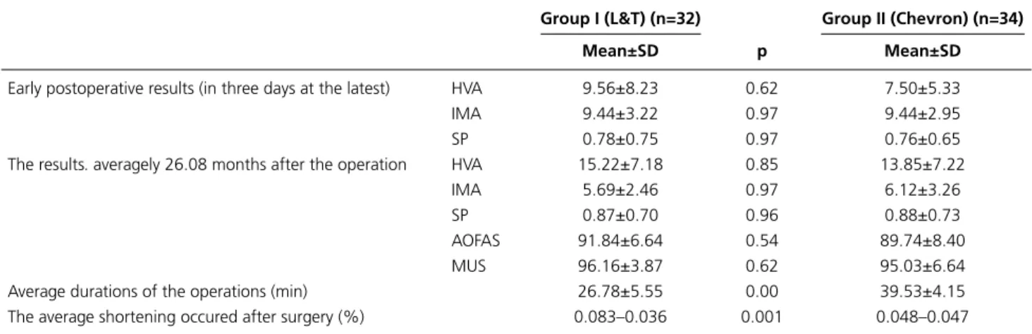
When HVA, IMA, and sesamoid positions on post-operative radiographies—which were performed in the first 3 days after the operation—were compared (inde-pendent sample Mann-Whitney U test was used), it was revealed that there was no significant difference between the 2 groups (p>0.05) (Table 2).
While HVA values measured at a mean of 26.08 months postoperatively were compared with indepen-dent t-test, IMA values and sesamoid positions were compared with independent sample Mann-Whitney U test. No significant differences were found between the groups (p>0.05) (Table 2).
Patient wellness was evaluated by AOFAS forefoot score and Maryland University Painful Foot Evaluation scale. When data from the 2 groups were compared by independent sample Mann-Whitney U test, no signifi-cant difference was detected (p>0.05) (Table 2).
While the average duration of the operation was 26.7 minutes in the Lindgren-Turan group, it was 39.5 min-utes in the Chevron group. These results were compared by Mann-Whitney U test, which found that duration of surgery in the Lindgren-Turan group was significantly shorter (p<0.01) (Table 2).
Shortening of the first metatarsal after surgery was noted as a ratio to the second metatarsal (MT1/MT2). Preoperatively, in the Lindgren-Turan group, average length of MT1 was 87.2% of MT2, and average short-ening was found to be 0.083%. However, this ratio was 88.9% in the Chevron group, and average shortening was found to be 0.048%. Postoperatively, alteration in MT1/MT2 ratio was found to be 81.6% in the Lind-gren-Turan group and 84.1% in the Chevron group. By using independent sample t-test, when comparing the 2 groups, more shortening was found in the Lindgren-Turan group (p<0.01) (Table 2).
Table 1. Preoperative values are compared between Lindgren-Turan and Chevron groups.
n Mean±SD p
Age Group I (Lindgren-Turan) 32 46.94±15.52 0.80
Group II (Chevron) 34 45.82±13.10
Hallux valgus angle Group I (Lindgren-Turan) 32 32.28±4.94 0.71
Group II (Chevron) 34 32.44±6.24
Intermetatarsal angle Group I (Lindgren-Turan) 32 13.91±2.44 0.90
Group II (Chevron) 34 14.15±1.96
Sesamoid position Group I (Lindgren-Turan) 32 2.03±0.69 0.60
Group II (Chevron) 34 1.94±0.69
American Ortopaedic Foot and Ankle Society score Group I (Lindgren-Turan) 32 24.06±5.99 0.43
Group II (Chevron) 34 24.68±4.98
Angular measurements on radiographs—which were initially performed within the first 3 days postoperative-ly and were continued for an average follow-up of 26.08 months—were compared. Statistical analysis showed that there was no significant difference between sesa-moid positions. However, in terms of HVA and IMA values, similar significant impairment was found in both groups (Tables 2 and 3).
One common problem was related with the 3.5-mm diameter fixation screw, which required extraction 4–6 months after surgery as an outpatient procedure. An-other common problem was that there was no appar-ent improvemappar-ent in patiappar-ents’ footwear habits: 58% of all patients reported that they were able to wear any shoes of their choosing following surgery. Moreover, most pa-tients reported that they were not wearing narrow or high-heel shoes, which may have caused discomfort.
Wound healing problems and skin necrosis were en-countered in 2 patients. Since lateral release was not per-formed in the present study and no comorbidities which impair healing were found in these 2 patients, skin ne-crosis was interpreted to be due to surgical procedure.
Skin necrosis healed under close follow-up and wound care.
Reflex sympathetic dystrophy was encountered in 2 patients. Both recovered with rehabilitation and encour-agement for weight bearing.
Discussion
Strengths of this study include its prospective random-ized design, that surgeries in both groups were per-formed by experienced surgeons, and that follow-ups were performed as a single-blinded procedure by a third investigator without knowledge of osteotomy type.
Chevron osteotomy is an accepted procedure in mild to moderate hallux valgus deformities.[4,5,7,8,14] Although the procedure is recommended in patients younger than 50 years by some authors,[7] Schneider et al. stated in their work[8] that there were no significant differences in patients under or over 50 years in a 10 year follow-up period. Sfollow-upporting this finding, Trnka et al. proved that the procedure is safe and not contraindicated for patients older than 50 years.[7] Therefore, there was no
Table 2. Peroperative and postoperative results are compared between two groups.
Group I (L&T) (n=32) Group II (Chevron) (n=34)
Mean±SD p Mean±SD
Early postoperative results (in three days at the latest) HVA 9.56±8.23 0.62 7.50±5.33
IMA 9.44±3.22 0.97 9.44±2.95
SP 0.78±0.75 0.97 0.76±0.65
The results. averagely 26.08 months after the operation HVA 15.22±7.18 0.85 13.85±7.22
IMA 5.69±2.46 0.97 6.12±3.26
SP 0.87±0.70 0.96 0.88±0.73
AOFAS 91.84±6.64 0.54 89.74±8.40
MUS 96.16±3.87 0.62 95.03±6.64
Average durations of the operations (min) 26.78±5.55 0.00 39.53±4.15
The average shortening occured after surgery (%) 0.083–0.036 0.001 0.048–0.047
L&T: Lindgren-Turan; n: Sample size. SS: Standard deviation; p: significance value; HVA: Hallux valgus angle; IMA: Intermetatarsal angle; SP: Sesamoid position; AOFAS: American Ortopaedic Foot and Ankle Society; MUS: Painful foot evaluation scale of Maryland University.
Table 3. Early and midterm radiological results are compared in two groups.
Group I (L&T) (n=32) Group II (Chevron) (n=34)
Mean±SD p Mean±SD p
Early postoperative Hallux valgus angle 9.56±8.23 0.01 7.50±5.33 <0.01
Midterm postoperative 15.22±7.18 13.85±7.22
Early postoperative Intermetatarsal angle 9.44±3.22 <0.01 9.44±2.95 <0.01
Midterm postoperative 5.69±2.46 6.11±3.26
Early postoperative Sesamoid position 0.78±0.75 0.44 0.76±0.65 0.37
Midterm postoperative 0.87±0.70 0.88±0.73
L&T: Lindgren-Turan; n: Sample size; SD: Standard deviation; p: significance value; Early postoperative: In three days at the latest after the operation; Midterm postoperative: averagely 26.08 months after the operation.
age-related division (under or above 50 years) in the population group of this study.
Trnka et al. published 2- and 5-year follow-up results of the Chevron procedure, finding that there was no dif-ference in clinical results.[7] Mean HVA was 18°±11° and IMA 9°±3° at 5-year follow-up in their study. In the present study, at 2-year follow-up, mean HVA was 13.8° (range: 2°–29°), and mean IMA was 6.1° (range: 3°–13°). For the Lindgren-Turan group, these values were 15.2° (range: 0°–28°) for HVA and 5.6° (range: 1°–11°) for IMA. These results are similar with those in the current literature. When early and late postopera-tive radiologic criteria were compared in this study, there were no significant diferences in sesamoid locations in both groups, but there was deterioration in HVA and IMA values. This disarrangement was clinically insig-nificant (Tables 2 and 3).
A study by Ertürer et al. demonstrated that good re-sults can be obtained in moderate hallux cases with the Lindgren-Turan procedure.[14] The authors evaluated not only HVA but also pain criteria and patient satisfac-tion in determining their findings. HVA was 27° preop-eratively and 20° postoppreop-eratively at 26-month follow-up. In our study, postoperative HVA values were lower than those of Ertürer et al. (Table 2). The better radiologic re-sults can be explained because our bone supported me-dial soft tissue reinforcement procedures. This can also increase the indications for distal osteotomies.[11]
Distal metatarsal osteotomies were offered for IMAs which were found to be lower than 15° in previous stud-ies. In our clinical experience, we accept an IMA of 15° as a guide, but this does not prohibit its usage in IMAs of higher degrees. The indications for distal metatarsal osteotomies can be broadened with our bone-supported soft tissue reconstruction technique. Deenik et al. sug-gested that IMA and other distal metatarsal joint mea-surements do not have a defining role in hallux valgus surgery, stating that HVA is the primary determinant.[15]
There are few comparative studies in the literature of the Lindgren-Turan and Chevron procedures. Saro et al. compared the 2 procedures in a study group of 100 patients and claimed that in the Lindgren-Turan procedure, the distal part was more shifted, resulting in more medialization of the tibial sesamoid bone. This re-sulted in a better correction of IMA and HVA than in the Chevron procedure.[4] Despite this, 4-year follow-up results showed similar clinical outcomes. In their study, Lindgren-Turan osteotomy was fixed with a cortical screw, but no fixation devices were used for the Chevron procedure, which may have affected the results. In the current study, both procedures were fixed with screws.
Translation of the distal part in distal metatarsal oste-otomies directly affects the surgical results.[7,16] Esemenli et al. concluded that Chevron osteotomy can produce only a 2- to 5-mm lateralization, which is insufficient.[13] However, Murawski and Beskin proved that they could translate approximately 90% of the distal bone part with this procedure, providing a good and reliable outcome. [17] Lateralization of the distal part was not measured in the present study, which can be accepted as a weakness of our study. In their study, Saro et al. showed that Lind-gren-Turan osteotomy is better in sesamoid correction than the Chevron procedure.[4] They again stated this was achieved with the help of distal part lateralization. In the present study, there was no difference between the 2 series in terms of sesamoid bone locations (Table 2).
In this study of moderate hallux cases, it has been demonstrated that postoperative AOFAS scores were significantly better than preoperative levels after a mean follow-up of 26 months in both series. Therefore, both procedures can be considered effective. Since there were no significant differences between the series in terms of both correction degrees and Maryland University Pain-ful Foot Evaluation scale scores in early and late follow-up evaluations, it can be concluded that neither of the 2 procedures is superior to the other (Table 2).
One difference found in this study was operation time. Duration of the Lindgren-Turan procedure was significantly shorter than that of the Chevron procedure (Table 2). This could be attributed to difference in sur-geons, but with the level of experience of the surgeons in the present study, it is likely that the procedures them-selves are the cause of the discrepancy. Nonetheless, this is a weakness of the study.
One difference between the 2 procedures was that metatarsal shortening was greater in the Lindgren-Turan than Chevron group (Table 2). This is the most criticized aspect of the Lindgren-Turan procedure.[4] Our study demonstrated that while the Lindgren-Turan procedure causes more shortening than the Chevron procedure, this was not reflected in clinical outcomes. Thus, although there is a statistically significant shorten-ing, it is tolerated at the clinical level.
Even if full radiologic correction in hallux valgus surgery is achieved, patients may not be satisfied with their results. In a study by Saro et al., quality of life was measured in hallux valgus patients pre- and postopera-tively,[18] finding that 86% of patients were satisfied with the procedure and 87% said that they would recommend the procedure to friends. In our study, all patients except 2 were satisfied with the procedure and recommended it to their friends.
The ability to wear shoes directly affects patient sat-isfaction, quality of life, and surgical results. Although Thordarson et al. could not find a relationship between surgery type and level of deformity with quality of life,[19] many articles state that the ability to wear shoes directly affects patient satisfaction.[4,18] Despite these facts, pa-tient satisfaction can vary widely between individuals, so a high satisfaction rate should not be expected. In an-other study, only 60% of patients reported that they were able to wear their shoes of choice.[20] This rate was 58% in the present study.
Osteonecrosis is more frequent in more distal oste-otomies such as the Chevron procedure. Lateral soft tis-sue release, which helps augmentation of the deformity correction, can also increase the risk of osteonecrosis by disturbing circulation. In a study by Jones et al., a safe zone was defined for the first metatarsal bone and Chev-ron osteotomy. In distal metatarsal osteotomy cases without lateral soft tissue release, lateralization of the surgical saw can cause arterial damage and is considered a surgical error.[2,21] Osteonecrosis was not encountered in our study population, a finding which could be related to surgical experience as well as the protection of the lat-eral soft tissues.
The major limitation of the study is that the op-erations were performed by 2 different surgeons in 2 groups. The other limitation is that the medial plication procedure which was applied is a relatively individual technique.
In conclusion, Chevron and Lindgren-Turan osteot-omies are both effective and safe procedures with similar midterm clinical results. Metatarsal shortening, which is the most criticized aspect of the Lindgren-Turan proce-dure, can be radiologically significant but is of no clinical importance.
Conflics of Interest: No conflicts declared. References
1. Easley ME, Trnka HJ. Current concepts review: hallux val-gus part 1: pathomechanics, clinical assessment, and non-operative management. Foot Ankle Int 2007;28:654–9. 2. Easley ME, Trnka HJ. Current concepts review:
hal-lux valgus part II: operative treatment. Foot Ankle Int 2007;28:748–58.
3. Thomas RL. The forefoot. In: Pinzur MS editör. Orho-paedic knowledge update. Foot Ankle Int Rosemont (IL) 2008.
4. Saro C, Andrén B, Wildemyr Z, Felländer-Tsai L. Out-come after distal metatarsal osteotomy for hallux valgus:
a prospective randomized controlled trial of two methods. Foot Ankle Int 2007;28:778–87.
5. Austin DW, Leventen EO. A new osteotomy for hallux valgus: a horizontally directed “V” displacement osteotomy of the metatarsal head for hallux valgus and primus varus. Clin Orthop Relat Res 1981;157:25–30.
6. Lindgren U, Turan I. A new operation for hallux valgus. Clin Orthop Relat Res 1983;175:179–83.
7. Trnka HJ, Zembsch A, Easley ME, Salzer M, Ritschl P, Myerson MS. The chevron osteotomy for correction of hal-lux valgus. Comparison of findings after two and five years of follow-up. J Bone Joint Surg Am 2000;82-A:1373–8. 8. Schneider W, Aigner N, Pinggera O, Knahr K. Chevron
osteotomy in hallux valgus. Ten-year results of 112 cases. J Bone Joint Surg Br 2004;86:106–20.
9. Doğan A, Üzümcügil O, Akman YE. Halluks valgus. TOTBİD Dergisi 2007;2:88–94.
10. Smith RW, Reynolds JC, Stewart MJ. Hallux valgus as-sessment: report of research committee of American Or-thopaedic Foot and Ankle Society. Foot Ankle 1984;5:92– 103.
11. Ozkan NK, Güven M, Akman B, Cakar M, Konal A, Turhan Y. Transosseous capsuloplasty improves the out-comes of Lindgren-Turan distal metatarsal osteotomy in moderate to severe hallux valgus deformity. Arch Orthop Trauma Surg 2010;130:1201–7.
12. Miller JW. Distal first metatarsal displacement osteotomy. Its place in the schema of bunion surgery. J Bone Joint Surg Am 1974;56:923–31.
13. Esemenli T, Yildirim Y, Bezer M. Lateral shifting of the first metatarsal head in hallux valgus surgery: effect on sesamoid reduction. Foot Ankle Int 2003;24:922–6. 14. Ertürer E, Aksoy B, Beki S, Toker S, Oztürk I.
Radio-graphic and functional results of the Lindgren-Turan oper-ation in the treatment of hallux valgus. [Article in Turkish] Acta Orthop Traumatol Turc 2004;38:125–9.
15. Deenik AR, de Visser E, Louwerens JW, de Waal Malefijt M, Draijer FF, de Bie RA. Hallux valgus angle as main pre-dictor for correction of hallux valgus. BMC Musculoskelet Disord 2008;9:70.
16. Coughlin MJ. Hallux Valgus. J Bone Joint Surg Am 1996;78:932–66.
17. Murawski DE, Beskin JL. Increased displacement maxi-mizes the utility of the distal chevron osteotomy for hallux valgus deformity correction. Foot Ankle Int 2008;29:155– 63.
18. Saro C, Jensen I, Lindgren U, Felländer-Tsai L. Quality-of-life outcome after hallux valgus surgery. Qual Life Res 2007;16:731–8.
Age-adjusted baseline data for women with hallux val-gus undergoing corrective surgery. J Bone Joint Surg Am 2005;87:66–75.
20. Mann RA, Pfeffinger L. Hallux valgus repair. DuVries modified McBride procedure. Clin Orthop Relat Res
1991;272:213–8.
21. Jones KJ, Feiwell LA, Freedman EL, Cracchiolo A 3rd. The effect of chevron osteotomy with lateral capsular release on the blood supply to the first metatarsal head. J Bone Joint Surg Am 1995;77:197–204.