i
PREPARATION AND CHARACTERIZATION OF ULTRA THIN FILMS CONTAINING SILVER-COPPER NANOALLOYS USING LAYER-BY-LAYER
DEPOSITON TECHNIQUE FOR ANTIBACTERIAL APPLICATIONS
A THESIS
SUBMITTED TO THE DEPARTMENT OF CHEMISTRY AND THE GRADUATE SCHOOL OF ENGINEERING AND SCIENCE
OF BILKENT UNIVERSITY
IN PARTIAL FULFILLMENT OF THE REQUIREMENTS FOR DEGREE OF
MASTER OF SCIENCE
By
MERVE TANER CAMCI JUNE 2012
ii
I certify that I have read this thesis and that in my opinion it is fully adequate, in scope and in quality, as a thesis for the degree of Master of Science
……….. Prof. Dr. Şefik Süzer (Principal Advisor)
I certify that I have read this thesis and that in my opinion it is fully adequate, in scope and in quality, as a thesis for the degree of Master of Science
……….. Prof. Dr. Ömer Dağ
I certify that I have read this thesis and that in my opinion it is fully adequate, in scope and in quality, as a thesis for the degree of Master of Science
……….. Assoc. Prof. Dr. DönüşTuncel
iii
I certify that I have read this thesis and that in my opinion it is fully adequate, in scope and in quality, as a thesis for the degree of Master of Science
……….. Assoc. Prof. Dr. Işık Yuluğ
I certify that I have read this thesis and that in my opinion it is fully adequate, in scope and in quality, as a thesis for the degree of Master of Science
……….. Assist. Prof. Dr. Gülay Ertaş
Approved for the Graduate School of Engineering and Science:
……….. Prof. Dr. Levent Onural
iv ABSTRACT
PREPARATION AND CHARACTERIZATION OF ULTRA THIN FILMS CONTAINING SILVER-COPPER NANOALLOYS USING LAYER-BY-LAYER
DEPOSITON TECHNIQUE FOR ANTIBACTERIAL APPLICATIONS
Merve TANER CAMCI M.S. in Chemistry
Advisor: Prof. Dr. Şefik SÜZER June, 2012
The main objective of this master thesis is to explore the preparation, characterization and antibacterial applications of Layer-by-Layer (LbL) assembled ultra thin films containing AgCu nanoalloys. Within this purpose, first part of the research mainly focused on the preparation of Ag nanoparticles and AgCu nanoalloys in order to prevent the oxidation of copper to copper oxide and the formation of polyelectrolyte-AgCu nanoalloy films by Layer-by-Layer assembly. Accordingly, Ag nanoparticles and AgCu nanoalloys were synthesized by the chemical reduction of silver and copper salts in aqueous solution with the help of strong reducing agents sodium borohydride or hydrazine hydrate in the presence of complexing agent and stabilizer, then ultra thin polyelectrolyte layers containing pre-prepared AgCu nanoalloys were constructed by Layer-by-Layer deposition technique in different combinations. Also the stability of these nano sized binary alloys in solution phase were
v
prolonged in the presence of third metal zinc as a sacrificial anode. In the second part of the study, characterization of LbL assembled ultra thin polyelectrolyte and metal nanoparticle thin films using Optical (UV-visible) and X-ray Photoelectron Spectroscopy (XPS) was studied. In order to get further information on the optical response of single and bimetallic nanoparticles, Ag nanoparticle and AgCu nanoalloy incorporated ultra thin polyelectrolyte films were investigated by optical spectroscopy. In addition the LbL films were analyzed by Static XPS to extract atomic level chemical information due to elemental and chemical state analysis. In order to get further understanding at the molecular level, samples were analyzed under external bias application by Dynamic XPS and it was shown that Ag and Cu respond in the same way to applying external electrical stimuli when both are in the same environment as a result of alloy formation, as reflected by the same shifts in Ag3d and Cu2p binding energy positions. Lastly, in the third part of the study detailed antibacterial analysis of synthesized monometallic and multimetallic nanoparticle solutions and the organized ultra thin polyelectrolyte layers containing Ag and AgCu nanoclusters against Escherichia coli strain was performed. These approaches enabled us to show the better antibacterial behavior of AgCu nanoalloys as a result of successful synthesis of AgCu nanoalloy without any copper oxide formation as the end product.
Keywords:Ultra-Thin Film Coatings, Layer-by-Layer Deposition, Ag Nanoparticles, AgCu Metal Nanoalloys, Antibacterial Applications, XPS
vi ÖZET
ANTİBAKTERİYEL UYGULAMALAR İÇİN GÜMÜŞ-BAKIR NANOALAŞIMLAR İÇEREN VE KATMAN-KATMAN-KAPLAMA YÖNTEMİ İLE HAZIRLANAN
ULTRA İNCE FİLMLERİN HAZIRLANMASI VE KARAKTERİZASYONU
Merve TANER CAMCI Kimya Yüksek Lisans Tezi Danışman: Prof. Dr. Şefik SÜZER
Haziran, 2012
Bu yüksek lisans tezinin ana amacı, AgCu nanoalaşımlar içeren ve katman-katman-kaplama (KKK) tekniği ile oluşturulmuş ultra ince filmlerin hazırlanması, karakterizasyonu ve antibakteriyel uygulamalarını incelemektir. Bu kapsamda araştırmanın ilk bölümü esas olarak Ag nanoparçacık ve bakır oksit içermeyen AgCu nanoalaşım sentezi, depolama süresince bakır oksit oluşumunun engellenmesi ve Katman-Katman Kaplama yöntemi ile polielektrolit filmlerin hazırlanması üzerine yoğunlaşmaktadır. Bu noktadan hareketle, Ag nanoparçacıkları ve AgCu nanoalaşımlar kompleks yapıcı ajan varlığında, çok güçlü indirgeyici ajanlar olan sodyum borohidrür ve hidrazin hidrat yardımı ile gümüş ve bakır tuzlarının sulu çözeltilerinin indirgenmesi ile sentezlenmiştir. Ayrıca, bu nano boyuttaki ikili alaşımların çözelti fazında kararlığı, üçüncü bir metal olan çinkonun ‘sacrificial anode’ olarak mevcudiyetinde uzatılabilmiştir. Daha sonra da, önceden sentezlenen Ag ve AgCu
nano-vii
parçacık/alaşım içeren ultra ince polielektrolit filmler Katman-Katman kaplama tekniği kullanılarak farklı kombinasyonlarda inşa edilmiştir. Çalışmanın ikinci kısmında katman-katman-kaplama tekniği kullanılarak hazırlanan ultra ince polielektrolit ve metal nanoparçacık içeren filmlerinin Optik (UV-visible) ve X-ray Fotoelektron Spektroskopi (XPS) teknikleri kullanılarak karakterizasyonu ele alınmıştır. Tek ve bimetalik nanoparçacıklar hakkında optik yanıt ve bilgi alabilmek için, Ag nanoparçacık ve AgCu nanoalaşım dahil edilmiş ultra ince polielektrolit filmleri de optik spektroskopi ile incelenmiştir. Bu noktadan sonra KKK filmleri, elemental ve kimyasal durum analizleri yapılarak atomik düzeyde bilgi elde etmek için ‘Statik XPS’ metodu ile incelenmiştir. Ayrıca, moleküler düzeyde daha fazla bilgi elde etmek amacıyla numuneler ‘Dinamik XPS’ yöntemi ile dışarıdan uygulanan elektriksel gerilim altında incelenmiştir. Gümüş ve bakırın nanoalaşım oluşturmasının sonucu olarak aynı yapısal çevrede bulunması halinde dışarıdan gelen elektriksel uyarana aynı şekilde tepki verdiği Ag3d ve Cu2p tepeciklerinin bağlanma enerjilerindeki kaymalarla kanıtlanmıştır. Son olarak, çalışmanın üçüncü bölümde sentezlenen mono-metalik ve multi-metalik nanoparçaçık çözeltilerinin ve Ag ya da AgCu nanopartiküller içeren organize ultra ince polielektrolit katmanlarının, Escherichia coli bakterisi oluşumuna karşı detaylı antibakteriyel özellikleri analiz edilmiştir. Bu yaklaşımlar AgCu nanoalaşımların çok daha iyi antibakteriyel özellik gösterdiğini ve bakır oksit içermeyen başarılı bir sentez yolunun antibakteriyel özellik üzerindeki etkisini açıkça ortaya koymaktadır.
Anahtar Kelimeler: Katman-Katman Kaplama, Ultra-İnce Filmler, Ag Nano-Parçacık, AgCu Metal Nano-Alaşımları, Anti-Bakteriyel Uygulamaları, XPS
viii
ACKNOWLEDGEMENTS
Before starting a new academic education and at the end of comprehensively scientific survey throughout last three years as impressive introduction to my academic carrier, I have always been proud of the privilege of work with my supervisor Prof. Dr. Şefik Süzer who taught me “how to swim in the sea”. I have my deepest gratitude and respect to him because of his great interaction, guidance and counseling in every stage of my life and scientific study. I would also like to show my appreciation with my sincere gratefulness to Assist. Prof. Dr. Gülay Ertaş, who supported me during each step of my life for the last six years. I express my thanks to Assoc. Prof. Işık Yuluğ from Molecular Biology and Genetics Department for her cooperation in this research study. I also thank to our past lab group members Hikmet Sezen, EdaYılmaz, Dr. Ivalina Abramova and Chemistry Department members Emine Yigit and Ethem Anber. I never forget my dear friends Müge Artar, Vusala İbrahimova, Gözde Barım, Özlem Ünal and Şeyma Ekiz and express my special thanks to Nilüfer Sayar for her contribution, friendship and support for this scientific study for three years.
I would like to express my thankfulness to my respectable doctor Assoc. Prof. Dr. Selahattin Özmen, who has provided me the medical treatment and enabled me to continue my life and reach these days, and also I thank his team in Gazi University, Faculty of Medicine profoundly.
My inclusive education and scientific research survey in Bilkent University has been most pleasant period of my life with my special family: a father whom I am always proud to be a daughter of him, a mother who devotes her life like an angel with her endless dearness, a sister who always shares each moment of her life and peerless friendship with me and a very special husband who makes every single day of my life special for me with his fabulous love and support.
ix
TABLE OF CONTENTS
1. INTRODUCTION………...1
1.1. METAL NANOPARTICLES………1
1.1.1. Synthesis of Metal Nanoparticles………1
1.1.1.1. Monometallic Nanoparticles………1
1.1.1.2. Bimetallic and Multimetallic Nanoparticles………...5
1.1.2. Antibacterial Applications of Metal Nanoparticles…………..……….7
1.2.
LAYER-BY-LAYER ASSEMBLED ULTRA THIN FILMS………101.2.1.Layer-By-Layer Assembly...…...10
1.2.2. Building Blocks of Layer-by-Layer Assembly………...12
1.2.3. Design of Antibacterial Ultra Thin Polyelectrolyte Layers Using Layer-by-Layer Assembly………...15
1.3. CHARACTERIZATION TECHNIQUES………...17
1.3.1. UV-visible Spectroscopy……….17
1.3.1.1 Optical Response of Metal Nanoparticles……….17
1.3.1.2. Optical Response of LbL Films Containing Metal Nanoparticles…...19
1.3.2. X-ray Photoelectron Spectroscopy………..21
1.3.2.1. Basic Principles of XPS………....21
1.3.2.2. Static and Dynamic XPS Measurements………..23
1.3.2.3. Application of XPS to Metal Nanoparticle Incorporated Ultra Thin Polyelectrolyte Multilayers………26
x 2. EXPERIMENTAL………..30 2.1. MATERIALS……….30 2.2. INSTRUMENTATIONS………...30 2.3. PROCEDURES………..32 2.3.1. Synthesis of Ag Nanoparticles………32
2.3.2. Synthesis of AgCu Nanoalloys………32
2.3.3. Synthesis of AgCu-Zn Nanoalloys………..………32
2.3.4. Preparation of Ultra Thin Polyelectrolyte Layers………33
2.3.5. Preparation of Metal Nanoparticle Inserted Ultra Thin Polyelectrolyte Layers………34
2.3.6. Antibacterial Activity Tests……….34
2.3.6.1. Antibacterial Investigation of Ag Nanoparticles – AgCu Nanoalloys Inserted Agar Plates………35
2.3.6.2. Batch Growth Profiles of Ag Nanoparticles – AgCu Nanoalloys for Antibacterial Efficiency………...35
2.3.6.3. Quantification of the Antimicrobial Properties of Ag Nanoparticles and AgCu Nanoalloys……….36
2.3.6.4. Antibacterial Investigation of Ag Nanoparticles and AgCu Nanoalloys Incorporated Ultra Thin Metal polyelectrolyte Films…….37
xi
3. RESULTS AND DISCUSSION……….39
3.1. PREPARATION OF Ag NANOPARTICLES………..39
3.2. PREPARATION OF AgCu NANOALLOYS………...39
3.2.1.Complexing Agent Effect on AgCu Nanoalloy Synthesis………...40
3.2.2. Reducing Agent Effect on AgCu Nanoalloy Synthesis………..42
3.2.3. Reaction Media Purging Effect on Ag-Cu Nanoalloy Synthesis………44
3.2.4. Stabilizer Effect on AgCu Nanoalloy Synthesis………..45
3.2.5. Effect of Zn-salt addition on AgCu Nanoalloy Synthesis………...48
3.3. CHARACTERIZATION OF NANOPARTICLE AND NANOALLOY SOLUTIONS.………52
3.4. CHARACTERIZATION OF LAYER-BY-LAYER DEPOSITED ULTRA THIN POLYELECTROLYTE-METAL NANOPARTICLE FILMS……….54
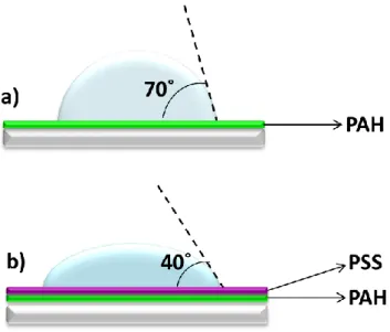
3.4.1. Water-Contact-Angle Measurements of Ultra Thin Films………..56
3.4.2. Characterization of Ag Nanoparticle Incorporated Ultra Thin Films….58 3.4.2.1. Optical Response of Ag Nanoparticle Ultra Thin Films…..…58
3.4.2.2. XPS Characterization of Ag Nanoparticle Ultra Thin Films…59 3.4.3. Characterization of AgCu Nanoalloy Incorporated Ultra ThinFilms.….61 3.4.3.1. Optical Response of AgCu Nanoalloy Ultra Thin Films……..61
3.4.3.2. XPS Characterization of AgCu Nanoalloys Ultra Thin Films via “Static XPS”………..62
3.4.3.3. XPS Characterization of AgCu Nanoalloys Ultra Thin Films via “Dynamic XPS”……….66
xii
3.5. ANTIBACTERIAL APPLICATIONS OF METAL NANOPARTICLES………71 3.5.1. Investigation of Antibacterial Effects of Ag Nanoparticles and AgCu Nanoalloys Dispersed in Solution…..…...………71
3.5.1.1. Antibacterial Test on Solid Agar Plates………71 3.5.1.2. Batch Growth Profiles of Ag Nanoparticles and AgCu Nanoalloys……….73 3.5.1.3. Quantification of Antibacterial Efficiency………...77 3.5.2. Antibacterial Investigation of Ag Nanoparticles and AgCu Nanoalloys Incorporated Ultra Thin Films………...83
4. CONCLUSIONS……….85
5. REFERENCES………87
6. Synthesis, Characterization and Antibacterial Investigation of Silver- Copper Nanoalloys………..…93
xiii
LIST OF FIGURES
Figure 1. Illustration of silver-only nanoparticles formation after reduction of silver ions (Ag+) to silver atoms (Ag0)………..2 Figure 2. Representation of reduction potentials of gold (Au3+), silver (Ag+) and
copper (Cu2+) ions………...4
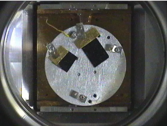
Figure 3. Representation of metal nanoparticle formation by reduction of metal ions and oxide contamination of synthesized nanoparticles………...5 Figure 4. Illustration of silver-copper nanoalloys formation after co-reduction of silver and copper ions (Ag+ and Cu2+)to silver and copper atoms (Ag0 and Cu0)………….6 Figure 5. Illustration ofantibacterial action of (a) larger (b) smaller nanoparticles…..9 Figure 6.Schematic representation of polyelectrolyte adsorption on negativelycharged substrate showing molecular structures for commonly used polyelectrolytes: Polyethyleneimine (PEI), Poly(styrene sulfonate) (PSS) and Poly(allylaminehydrochloride)(PAH)………..…….14 Figure 7.Schematic representation of metal-nanoalloy/polyelectrolyte ultrathin film constructionwith adsorption of negatively capped AgCu NAs on PEI/PSS/PAHpolyelectrolyte layers via LbL assembly……….………...16 Figure 8.Schematic representation of XPS measurements using Static XPS and Dynamic XPS techniques………..24 Figure 9.Cu2p region of XPS spectrum showing zero-valent Cu 0, Cu x+ peaks and copper-oxide satellites………...27 Figure 10. Representative image of rotatable sample holder used in Dynamic XPS measurements………31
xiv
Figure 11.Schematic representation of (a) AgCu NAs particle agglomeration in absence of complexing agents (b) Electrostatic protection of AgCu NAs via citrate ions against agglomeration with the help of attractive forces between negatively
charged COO- groups………41
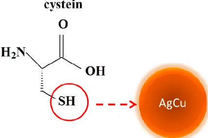
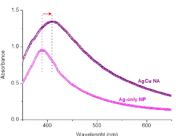
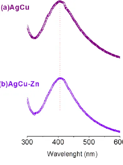
Figure 12.Cu 2p region of the XPS spectrum of AgCu nanoalloys which weresynthesized in the presence of NaBH4 as reducing agent………..43 Figure 13.Cu 2p region of the XPS spectrum of AgCu nanoalloys which weresynthesized in the presence of NH2NH2.H2O as reducing agent………...44 Figure 14.Schematic representation of affinity of thiol group at terminal position of cysteine toward AgCu nanoalloys……….47 Figure 15. Representation of sacrificial anode effect on metal nanoparticle formation by re-reduction of metal ions after oxide contamination………..48 Figure 16. Images of (a) fresh AgCu (b) 14weeks-old AgCu (c) fresh AgCu-Zn (d) 14weeks-old AgCu-Zn nanoalloys’ solutions………...49 Figure 17. Cu2p region of the XPS spectra of (a) AgCu binary and (b) AgCuZn ternary nanoalloys recorded in fresh, after 7 and 14 weeks………..51 Figure 18. UV-visible absorption spectrum of Ag-only and AgCu nanoparticle solution………..52 Figure 19. UV-visible absorption spectrum ofaqueous solutions of AgCu binary and AgCu-Zn ternary alloy nanoparticles………...53 Figure 20.Structures of ammonia, primary amine, secondary amine and tertiary amine groups………54 Figure 21.Schematic representation of Water Contact Angle measurements for
xv
Figure 22.UV-visible absorption spectrum of silver-only nanoparticle solution and layer-by-layer assembled PEI-PSS-PAH / Ag-only NPs ultra thin film………...58 Figure 23. XPS spectrum of PEI-PSS-PAH / Ag-only NPs ultra thin layers showing S2p, C1s and N1s regions……….59 Figure 24.Ag 3d region of the XPS spectrum of LbL assembled PEI-PSS-PAH / Ag-only NPs ultra thin film………...60 Figure 25. UV-visible absorption spectrum of layer-by-layer assembled PEI-PSS-PAH / AgCu NAs ultra thin film………...61 Figure 26.XPS survey of layer-by-layer assembled PEI-PSS-PAH / AgCu NAs ultra thin film……….62 Figure 27.XPS survey of AgCu-Zn ternary nanoalloys thin film……….…………...63 Figure 28.Cu 2p region of the XPS spectrum of LbL assembled PEI-PSS-PAH / AgCu NAs ultra thin film………...………...64 Figure 29. S2p region of the XPS spectrum of LbL assembled PEI-PSS-PAH / AgCu NAsultra thin film showing chemical structures of PSS and cysteine……….65 Figure 30. The XPS spectra of Cu2p and Ag3d regions of LbL assembled PEI-PSS-PAH / AgCu NAsultra thin film subjected to +10 V(red) and –10 V (blue) external DC stress under square wave excitation at 103 and 10-3Hz………..…...67 Figure 31.The XPS spectra of C1s, N1s and S2p regions of LbL assembled PEI-PSS-PAH / AgCu NAsultra thin film subjected to +10 V(red) and –10 V (blue) external DC stress under square wave excitation at 103 and 10-3Hz………...68 Figure 32. The XPS spectra of Zn2p, Cu2p and Ag3d regions of LbL assembled PEI-PSS-PAH / AgCu-Zn NAsultra thin film subjected to +10 V(red) and –10V (blue) external DC stress………..69
xvi
Figure 33.Representative images of agar plates containing an equal amount of Ag-only nanoparticles bacteria colony formation and AgCu nanoalloys without colonies with initial E. coli DH5α concentration at (a) 0.2OD (b) 0.1OD and (c)0.05OD……….………...72 Figure 34. Representative batch growth profiles in the presence of Ag-only nanoparticles and AgCu nanoalloys for initial concentrations of E.Coli DH5α: (a) 0.2 OD and (b) 0.1 OD and (c) 0.05 OD………...74 Figure 35.Representative batch grow profiles in the presence of AgCu and AgCuZn nanoalloys (a) fresh and (b) after 14 weeks………..76 Figure 36.Microscope images of (a) AgCu/PSS-PAH film right side (b) PEI-PSS-PAH film right side (c) AgCu/PEI-PEI-PSS-PAH film left side and PEI-PEI-PSS-PAH film left side………..84
xvii
LIST OF TABLES
Table 1.Water Contact Angle Measurements for given concentrations and time…………...57 Table2. MIC (μg ml−1) and MBC (μg ml−1) of silver nanoparticles and silver– copper nanoalloys for E. coli DH5α………..78 Table 3. Comparison of the Antibacterial Activity ( MIC and MBC values) of fresh, 7weeks-old and 14weeks-old AgCu NAs and AgCu-Zn ternary NAs towards E. coli DH5α……….79 Table 4.Effect of differentconcentrations of NP andNAs on dilutionseries of E. coli DH5α...…80 Table5:Effect of differentconcentrations ofNAs on dilutionseries of E. coli DH5α for fresh and 14 weeks old samples……….82
xviii
LIST OF ABBREVATIONS
CFA: Colony Forming Abilities CFU: Colony Forming Units DC: Direct Current
DF: Dilution Factor E.coli: Escherichia coli LbL: Layer - by -Layer LB: Langmuir – Blodgett LB: Luria Broth
MBC: Minimum Bactericidal Concentration MIC: Minimum Inhibitory Concentration NAs: Nanoalloys
NPs: Nanoparticles
PAH: Poly (allyamine hydrochloride) PE: Polyelectrolyte
PEI: Poly (ethyleneimine)
PSS: Poly (sodium 4- styrene- sulfonate) SAM: Self Assembled Monolayer SQW: Square wave
OD: Optical Density UNC: Uncountable
UV- vis: Ultraviolet- Visible
WCA: Water Contact Angle Measurements XPS: X-ray Photoelectron Spectroscopy
1 1. INTRODUCTION
1.1. METAL NANOPARTICLES
Metal nanoparticles have been heavily investigated because of their particle characteristics, size dependent properties, easy syntheses and chemical modifications. The metallic nanoparticles are also challenging for functionalization and surface modification. The increased surface-to-volume ratio imparts high reactivity to nano-sized clusters than their bulk counterparts. Accordingly smaller, smart and more efficient materials can be built up as nano-sized particles which behave different than their bulk and atomic forms. On account of the attractive and easily tunable properties of nanoparticles of the metals exhibiting increased photochemical activity, chemical and biological features because of their high surface to volume ratio, nanoparticles have been used in wide range of nano scale applications such as electronics, biology, catalysis, optics and chemical sensing.
1.1.1. Synthesis of Metal Nanoparticles
1.1.1.1. Monometallic Nanoparticles
Nanoparticles of metals can be synthesized from noble and transition metal precursors or even in the form of oxides of transition metals referring to smart materials in the 1nm – 100nm size range. In addition, bimetallic or multi-metallic nanoparticles being composed of combination of two or more metal atoms, have distinct and enhanced properties from their monometallic counterparts.
Preparation of metallic nano-clusters is executed by using a variety of methods such as galvanic exchange reactions, wet-chemical synthesis via assorted reducing agents, electrochemical, photo-reductive, microwave enhanced or biological techniques.1-9 Rather
2 than the sophisticated physical synthesis routes, metal nanoparticle synthesis can be regularly carried out via using solution chemistry. The nano-sized particles possess few meter square interface as in form of colloidal solutions, thus facilitate the surface modification in solution in terms of ease in controlling reaction parameters such as temperature, concentration, pH, reducing agent or solvent without the need of expensive or complex equipment. 4, 10-12
Monometallic nanostructures such as silver-only or copper-only nanoparticles can be synthesized by chemical reduction of silver and copper ions, which are produced by dissolving the salts of silver and copper in aqueous media, with the help of a reducing agent according to various wet-chemical reduction procedures. Reduction takes place with the formation of metal in zero-oxidation state on account of electron uptake of metal ions, then as a next step these reduced-metal atoms migrate and form nanoparticles.
Figure 1. Illustration of silver-only nanoparticles formation after reduction of silver ions (Ag+) to silver atoms (Ag0).
Ag+ Ag+ Ag+ Ag+ Ag+ Ag+ Ag+ Ag+ e- e- e- e- e- e- e- e - Ag+ e- Ag0 Ag0 Ag0 Ag0 Ag0 Ag0 Ag0 Ag0 Ag0
Ag
3 Because of the increased surface-to-volume ratio of the nano-sized materials, their high surface energy and reactivity impose limitations in their shell-life and use in different applications. Naked-nanoparticles are not stable in the absence of any protecting agents. For example naked nano-sized straightforwardly move towards each other causing aggregation and increase in particle size uncontrollably or react with other species in their medium yielding cumulative species with entirely different chemical and physical properties. Therefore, different kinds of stabilizer and / or surfactants are used to prevent particle aggregation, control over particle-size, impart stability to nanoparticles. In the presence of stabilizer and / or surfactants there exists a repulsive force between the capped nanoparticles preventing their attractive migration to each other. Henglein et al. demonstrated the capping of silver nanoparticles by citrate in terms of being an efficient protecting surfactant, furnishing long-term stability to the capped-nanoparticles. Furthermore the size and the structure of silver nanoparticles were controlled in the presence of citrate as capping agent via changing the concentration of this surfactant allowing the diameter dispersion.13 In order to impart further stability to the highly reactive nanoparticles and prevent nano-sized metal clusters at different oxidation states rather than zero, assortment of techniques are used in association with different kinds of surfactants by virtue of the steric effects, and by keeping nucleation sites of stabilizers and nanoparticles with the help of electrostatic interactions as far as possible, 13-15 chemical and biological protecting agents under different reaction conditions. 11, 16-20
Highly dispersed metal nanoparticles of uniform size, shape and purity can easily be synthesized in solution by reduction of metal ions that have higher standard reduction potentials whereas species having relatively low reduction potential have a tendency toward contamination with reactive species in the medium, forming non-zero oxidation states. Varghese et al. represented the advance stabilization of Ag nanoparticles using chemisorption
4 of very strong protecting agent which is an amino acid containing sulphhydryl group (-SH) 19 and found their stabilization with high affinity of metal atoms toward sulfur- containing reagents. 18, 21
In the presence of suitable complexing, reducing and protecting agents Ag and Au nanoparticles are stable under ambient conditions against oxidation. However for the synthesis of copper nanoparticles, the main handling problem is their high air sensivity, low stability and reactivity of the copper nanoparticles that cause the formation of copper oxide in the end product due its lower standard reduction potential (+0.34V) as compared to Ag (+0.80V) and Au (+1.50V). In the presence of appropriate reducing agents, reduction of Ag or Au is more favorable than Cu as a result of difference between their reduction potentials.
Figure 2. Representation of reduction potentials of gold (Au3+), silver (Ag+) and copper (Cu2+) ions.
Especially for the wet-chemical synthesis under ambient conditions oxidation of copper nanoparticles is nearly inevitable without presence of extremely strong reducing and protecting agents because of the highly reactive oxygen species in air or solution. To suppress the oxidation problem of copper, purging of the reaction media with inert gases, use of non-aqueous media or inert atmosphere, use of very strong reducing and, complexing agents, surfactants, stabilizers are used against copper oxide formation during preparation and/or storage periods.
5 Figure 3. Representation of metal nanoparticle formation by reduction of metal ions and oxide contamination of synthesized nanoparticles.
1.1.1.2. Bimetallic and Multimetallic Nanoparticles
Synthesis of the monometallic nano-sized clusters using different preparation routes have been heavily investigated for many years, whereas metal nanoalloy syntheses have not been so widespread due to handling problems and difficulties in preparation. Bimetallic or multi-metallic nanoclusters can be synthesized by the co-reduction of two or more kinds of metal ions forming nanoalloys sharing the same crystal structure. However, reductive deposition of one kind of metal ion over another reduced metal nucleus results in forming core-shell nanoparticles. 3
In favor of incorporating two different metals to form nanoalloys, suitable reducing agent which has the capability to reduce both types of the metal ions is used for the reduction of both metal ions to zero valent metals at the same time. Furthermore, as an alternative way to the monometallic counter parts of nano-sized structures, surfactants are utilized in bimetallic nanoclusters synthesis not only for stabilization of nanoclusters but also to bring the reduction potentials of two different metals closer in the direction of co-reduction of these metal ions together. 42
6 Figure 4. Illustration of silver-copper nanoalloys formation after co-reduction of silver and copper ions (Ag+ and Cu2+)to silver and copper atoms (Ag0 and Cu0).
Also, after the synthesis of monometallic or multimetallic nano-sized clusters with high dispersity and stability, these nanoclusters can be further functionalized by surfactants or capping agents in order to use in various applications. For instance, negatively capped Ag nanoparticles, which are produced by carboxylic group surrounding around zero-valent silver nano-sized metals, can be used to construct ultra thin multilayers as a result of electrostatic attachment of negatively charged nanoparticles in between the positively charged polyelectrolyte layers effectively.
7 1.1.2. Antibacterial Applications of Metal Nanoparticles
For many years metals such as zinc, gold, silver and copper containing materials have been used for antibacterial applications. While the exact mechanism responsible for antibacterial action is still unknown, these antibacterial metals have the ability to penetrate into bacteria causing cell death or else interact with microbial cell membranes and active proteins as a result of high affinity of transition metals toward sulphur and phosphorus containing proteins that cause enzyme denaturation and deactivation their bacterial activity as a result of interaction between metal and bacteria.17, 19, 22-29 These antibacterial species show strong and fatal toxic action against prokaryotes including all kinds of bacteria and microorganisms whereas much less toxicity can be omitted for eukaryotes including all other organisms. Thus, as compared to heavy metal ions, nano-sized metal particles show lower toxicity toward humans with remarkably higher antibacterial impact.
Antibacterial activity of these species robustly related with their size and decreases with increasing size, so nano-size metal particles exhibit higher antibacterial activity over their larger colloidal, ionic and bulk counterparts. High surface-to-volume ratio and small size of nanoparticles impart an advantage of close interaction with cell membranes or active sites of the bacteria cells causing deactivation of microbial action. Hence this nanometer-size advantage allows the penetration of nanoparticles through the cell membrane causing cell death. Bacteria cell membranes consist of many sulphur containing proteins, so these sulphur containing parts are privileged sites for transition metal nanoparticles to interact causing disruption of bacterial activity. The infiltrated metal nanoparticles lean towards the interior proteins containing sulphur or phosphorus such as DNA and react with these proteins causing cell death.
8 Aggregation of nanoparticles is also a crucial problem for antibacterial applications because when the nano-sized particles aggregate they deteriorate their antibacterial properties as a result of increasing size and alteration of chemical and physical properties. Immobilization of antibacterial metal nanoparticles on different kinds of host materials increase the effectiveness of antibacterial application in terms of longer release time and prevented particle aggregation via attachment on supported materials so imparting higher durability.
Subsequently, with recent technological developments and emerging demand for health promoting products, metal particles in nanometer size are suitable candidates for innovative biotechnological applications in industry and hospitals in terms of the desired antimicrobial modification of surfaces and materials to prevent bacterial activity of microorganisms. Advances in coating technology and ability to design intelligent nano-sized materials elicit innovation of antibacterial coatings for broad variety of applications such as in biomedical devices, packaging, textile and cosmetic industries.
Ruparelia and his co-workers 28 studied on bacteria growth profiles in the presence of 3 nm silver and 10 nm copper nanoparticles against various strains of Staphylococcus aureus, Escherichia coli and Bacillus subtilis bacteria and they showed the superior antibacterial characteristics of silver nanoparticles over oxide-contaminated copper nanoparticlesagainst E. coli and S. aureus where copper nanoparticles are more effective against B. subtilis. In addition, they stated that the combination of Ag and Cu nanoparticles, as physical mixtures, give rise to more effective antibacterial effect against mixed bacteria populations as in real life systems.
Sambhy et al.23 presented their work on antibacterial composites consisting of embedded AgBr nanoparticles in poly (4-vinyl-N-hexylpyridinium bromide) as cationic polymer matrix and the surfaces coated with these AgBr/polymer nano-composites forming
9 biofilms on glass microscope slides show antibacterial characteristics toward both Gram negative and Gram positive bacteria. Likewise, they demonstrated the tunable antibacterial properties of the AgBr/polymer nano-composite coating via changing the size of embedded AgBr nanoparticles impart higher antibacterial activity of 10 nm AgBr nanoparticles as compared to 71 nm AgBr nanoparticles to the higher surface-to-volume ratio of smaller nanoparticles.
10 1.1.2. LAYER-BY-LAYER ASSEMBLED ULTRA THIN FILMS
Layer-by-Layer assembled PEs multilayer incorporated with antibacterial nanoparticles are ideal materials for studying chemical and physical properties of desired surfaces and interface between these smart surfaces and bacteria cells. LbL deposition technique for this purpose is preferred over other deposition techniques because; it combines inorganic nanoparticles with organic polyelectrolyte layers using `very small amount of materials` and constructs surfaces without any chemical modification of coated polyelectrolytes, metal-nanoclusters or substrate as well as inducing stability during antibacterial applications.
LbL is also applicable to various kinds of materials and can be used to coat the common substrates like glass, with high efficiency. Besides the control of ultra thin layers in nanometer scale is easy because of the tunable parameters such as pH of the medium, temperature, supporting PEs’ concentration present during the adsorption process. Ability to control the chemical and physical properties of layer components allows the design of ultra thin films as surface properties of the multilayers can be controlled and modified. In addition, this highly efficient and cheap technique can be carried out at room temperature without requiring vacuum equipment or any kind of special instrumentation for chemical and biological applications.
1.2.1. Layer-By-Layer Assembly
In 1960s, with the help of Langmuir- Blodget (LB) technique30, which uses the principle of monolayer formation on water-air interface firstly and transfer of the monolayers onto a solid support, Kuhn and co-workers showed that functional units can be formed exhibiting different properties for individual layers in nanoscale.31 In 1966 a new method was
11 developed in order to provide alternative technique to limitations of LB technique and Iler had proposed a method that constructs multilayers by using inorganic colloids.30 Afterward this application was used as basis of multilayer formation process, and in 1990s Decher proposed a method for fabrication of multi composite films depending on electrostatic attraction between oppositely charged polyelectrolyte species. This method is now called the Layer-by-Layer (LbL) assembly that opens a new route potentially leading other new developments.
For the biological and material applications, thin solid films consisting up many different polyelectrolyte layers using the LbL deposition technique provides advanced coatings and smart surfaces with the creation of functional and controllable properties using polyelectrolytes for modification of surfaces. This LbL process assembles the organic polymers with the inorganic nanoparticles combining the unique responses of macro molecules and micro molecules. Also LbL technique imparts unique properties to nano-materials in opposition to the expensive instrumentation, long fabrication period for film preparation, instability of prepared films resulting from weak interactions under ambient conditions and limited number of substrates in LB technique and Self Assembled Multilayers (SAMs).32 Owing to the fact that, particles are most useful in the form of thin films LbL technique allows the formation of tunable and homogenous films that contain metal nanoparticles rather than the mechanical mixing of nanoparticles and building blocks which may result non-homogenous thin films and unsuccessful coverage of materials with unusual properties of geometry.33
1.2.2. Building Blocks of Layer-by-Layer Assembly
The building blocks of the LbL technique are polyelectrolytes (PEs), and it was developed for polyelectrolyte-polyelectrolyte systems because oppositely charged polyelectrolytes have also the complexation ability in multilayer structures.30 Polyelectrolytes
12 have been used in analytical and colloidal chemistry which are defined as macromolecular species dissociating into charged polymeric molecules in solution. The multilayers constructed from PEs show indistinguishable properties than each other in terms of multilayer structures due to their high penetration ability in molecular level. 34
LbL deposition technique is used for consecutive adsorption of oppositely charged polyelectrolytes for constructing multilayer structures and the type of polyelectrolyte which is used for deposition process to form the first layer is an important factor on the stability of the ultra thin multilayer, because the attaching of the whole film to the different substrates is highly dependent of the first layer. When the adsorption of first layer on the substrate is completed, surface charge is inverted by the adsorption of oppositely charged second layer using enough concentration of opposite charges. With the purpose of stabilization of each PE layer, washing process is used after adsorption of monolayer to remove either unreacted or weakly bonded PEs.
In LbL process generally poly (allylaminehydrochloride) (PAH), poly(diallyldimethylammoniumchloride) (PDDA), polyethyleneimine (PEI) are used as the positively charged polyelectrolytes and poly (acrylic acid) (PAA), poly (styrene sulfonate) (PSS), poly (vinyl sulfate) are used as the negatively charged polyelectrolytes, where all polyelectrolytes are broadly available and have enough density of positive or negative charges on their polymer chains for the preparation of multilayers easier.30-32, 35-37
Manipulation for improvement of LbL ultra thin layers is related not only with the choice of oppositely charged polymer partner but also parameters such as ionic strength to control the availability and density of ionic charges on polymer chains. In an ionizing solvent, successful deposition of these PEs requires maintenance of electro-neutrality for the neutralization of charge on repeating units of PEs with the help of smaller counter ions which are oppositely charged since macromolecular species such as PEs likely to be in their most
13 expanded form with higher availability of charges on polymer sides, due to intermolecular repulsions of each charged monomeric unit when ionic strength of solution.34, 38, 39 Therefore the charges on the monomeric units of PEs are balanced either by oppositely charged PEs or counter ions.
Using LbL approach, to reach the desired multilayer combination after the deposition of charged polyelectrolyte layer, oppositely charged polyelectrolyte layer is deposited. This sequential cyclic process is continued until the desired multilayer configuration is achieved and single monomeric layers can also be easily obtained by adsorption of the positively charged polycation or negatively charged polyanion on charged substrate directly.
LbL deposition technique is available not only for PEs but also any type of common type of charged species. In order to incorporate three dimensional inorganic metallic nanoclusters with various functionalities in the ultra thin PE layers, after the deposition of the charged polyelectrolyte layers, the charged metallic nanoclusters are deposited imparting a net negative charge to the nanoclusters requires oppositely charged polyelectrolyte partner for LbL assembly constructing nano scale functional ultra thin films consisting of organic and inorganic layers.40-44 Also by the use of colloidal nanoclusters three dimensional construction of these ultra thin films basically uses the irreversible strong electrostatic forces between charged PE layers and the required – oppositely charged nanoclusters partners as driving force for stable film assembly but also multiple intermolecular interactions such as hydrogen bonding 37, 41, 45, hydrophobic interaction 46 and charge transfer 47 cooperatively contributes to the formation so as the stability of the films.
14 Figure 6. Schematic representation of polyelectrolyte adsorption on negatively charged substrate showing molecular structures for commonly used polyelectrolytes: Polyethyleneimine (PEI), Poly(styrene sulfonate) (PSS) and Poly(allylaminehydrochloride) (PAH).
The thickness of polymer layer is related to both the amount of polyelectrolyte and the nature of polyelectrolyte and the films can be constructed by LbL deposition at the molecular level. Also, the nanoscale control over thickness of adsorbed thin layers related to the dipping time of the substrate in PEs’ aqueous solution on account of improvement in the amount of adsorbed charged polyelectrolyte chains.48 As very important parameter for nano scale precision thickness of single bilayer consisting of one positively charged and one negatively charged layer is in the range of 1nm- 5nm allowing the nanometer scale control for thickness of these layers.32, 49
15 1.2.3. Design of Antibacterial Ultra Thin Polyelectrolyte Layers Using Layer-by-Layer Assembly
Surface modification has been advocated for better controlling of the interactions between bacteria and substrate inducing biochemical death of bacteria in consequence of interaction with antibacterial functional groups 50-52 and propagation of antibacterial compounds over time from material surface to bacteria with respect to releasing ability of antibacterial agents from polymer multilayers.17, 32, 53-55
Both Gram negative bacteria, having single peptidoglycan layer between lipopolysaccharide rich layers, and Gram positive bacteria, having multilayered peptidoglycan cell at outermost layer, are susceptible for the antibacterial activity control of nanoparticle loaded multilayer thin films.54, 55 Additionally, ultra thin antibacterial layers can be studied in terms of their effectiveness by different kinds of methods as well as counting the number of bacteria after a certain incubation time, antibacterial drop test, determination of colony forming units, following the optical density at 600nm referring to the bacteria number in aqueous medium after definite incubation time or disk-diffusion (Kirby- Bauer) test.26, 28, 54-59
Polyelectrolyte multilayers provide numerous opportunities to design intelligent surfaces with nanostructures to explore antibacterial characteristics combined with chemical properties for biotechnological applications. Incorporation of antimicrobial metal nanoparticles with charged PEs via immobilization on to the various types of materials for antibacterial applications in this intelligent ultra thin surface construction is effectively used for antibacterial purposes.26, 42, 55, 60-62 With the use nanoparticles with charged polymers these ultra thin layers achieve high reactivity as a result of large interface area between nanoparticles and PEs. 17, 22 These immobilized antibacterial nanostructures reveal long term stability and are much more environment friendly. The surface density and arrangement of the resulting antibacterial PE thin layers are controlled by different number of antibacterial
16 reagent-PEs dipping cycles and time. As a function of nanoparticle containing thin layer thickness and nanoparticle density, the effectiveness of the layers can be controlled for antibacterial purposes.55, 60, 63
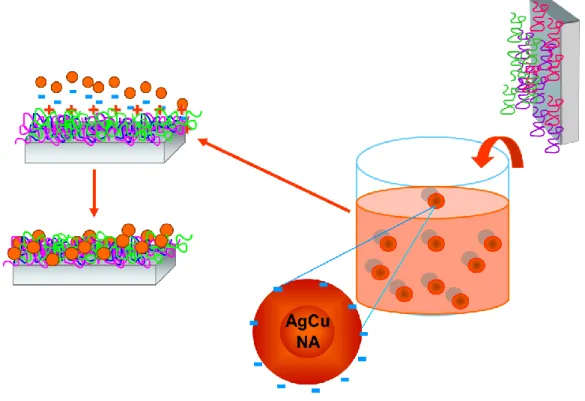
Figure 7. Schematic representation of metal-nanoalloy/polyelectrolyte ultra thin film construction with adsorption of negatively capped AgCu NAs on PEI/PSS/PAH polyelectrolyte layers via LbL assembly.
17 1.3. CHARACTERIZATION TECHNIQUES
1.3.1. UV-visible Spectroscopy
For the characterization of the nanoparticles NMR, IR, Raman, UV-Visible and X-ray Photoelectron Spectroscopy, AFM, SEM, TEM, and XRD diffraction are the commonly used techniques. Unfortunately, the most common techniques used by chemist like Infrared or Nuclear Magnetic Resonance cannot be used for the characterization of ultra thin layers containing metal nanoclusters because of their lack of sensivity.
UV-visible spectroscopy, that gives important informations for samples, is the most commonly used optical technique because it is easy in application and cheaper than other techniques. UV-visible spectroscopy records the absorption spectrum in ultraviolet and visible region in electromagnetic radiation containing electronic transition in molecular orbitals from ground state to excited state in terms of absorbed light at different wavelengths. This technique is also used for determination of different properties such as average particle size, shape, concentration and composition of metal nanoparticles.
In this work, UV-visible spectroscopy is used to characterize both the nanoparticles which are prepared by chemical reduction via solution chemistry, and the nanoalloys embedded into the ultra thin multi-layers which are prepared by LbL assembly.
1.3.1.1 Optical Response of Metal Nanoparticles
Metal nanoparticles exhibit strong plasmon resonance (SPR) extinction band in the visible spectrum as a consequence of interaction between conduction electrons of metal nanoparticles and incident electromagnetic radiations.4 SPR band is the outcome of resonance frequency as a result of restoring force to compensate polarization that is generated by
18 electron motion in phase for the particles having much smaller size than wavelengths of incident light. 4
Particles which have smaller diameter than 3 nm do not exhibit significant surface plasmon absorption covering visible region unless the particle size becomes higher than 5 nm surface plasmon bands broaden where the dimensions of the nanoparticles are comparable with the wavelength of interacting light.4, 64 The metal particles of silver and gold show distinct and well-defined plasmon absorption in the visible region exhibiting drastic color effects due to the surface plasmon absorption in the metal nanoparticles, as a result of the collective oscillations of the free conduction electrons.65 For the bimetallic nanoclusters, in case of nanoalloys where the particles of two metals distributed homogenously, position of surface plasmon band lies between two distinct absorption bands of single metal nanoparticles.64, 66 Alternatively for the core-shell bimetallic nanoclusters, surface plasmon absorption is mainly determined by absorption of the metal at the shell as a result of alternating oscillation mode of surface conduction electrons.67 In addition, since resonance wavelength depends on the orientation of the electrical fields and the oscillation modes of the shape of the nanoclusters, optical properties also change as shape of the nanoclusters change.7 Silver nanoparticle existence can easily be detected with their optical spectra with a characteristic peak around 390-400nm.68 However, characterization of copper as nanoparticle or in nanoalloy cannot be directly observed with their UV-visible spectra, because the surface plasmon band for copper nanoparticles is not in the UV-visible region and the oxidative property of copper makes plasmon frequency determination in this region harder. In order to prove the existence of copper nanoparticles by UV-visible optical spectra, indirect characterization techniques are used such as controlling the decrease in the broad absorption band of the cupric ions in the solutions with the addition of reducing agent to the reaction media.69, 70 What is more, absence of copper plasmon peak in UV-visible region depends on
19 the small cluster size with high dispersity of nanoparticles whereas the silver nanoparticle surface plasmon peak is much more intense as it can be detectable as compared to copper nanoparticle counterparts. 16, 71, 72
For the optical characterization of co-reduced silver and copper nanoparticles from the metal salts of silver and copper in aqueous media, silver in silver-copper alloy nanoparticles can be detected by the broadening of the peak in absorption spectrum towards 410 nm-450 nm, which has a different position than the characteristic Ag nanoparticle peak, as a result of the Cu and Ag interaction and alloy formation. Thus the red shift in the Ag-Cu nanoalloy optical spectrum to the higher wavelengths and the broadening in the absorption spectrum is a proof of the existence of the silver and copper together in the nanoalloy.
1.3.1.2. Optical Response of LbL Films Containing Metal Nanoparticles
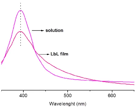
Layer-by-layer assembly allows the nano-scale organization of nanoparticles and the thickness via alternating adsorption of the optically active nanoclusters and polymeric electrolytes resulting ultra thin film formation.33, 37 Tunable properties of metal nanoparticle-polyelectrolyte films at the molecular level give opportunity for tunable optical properties. Red-shift is expected in the absorption spectrum of these particles since introduction of the nanoclusters into an organic matrix with different dielectric constant affect the dipolar-dipolar interactions resulting surface plasmon resonance shifts to different position in their spectrum.49, 67
Nanoclusters are not only subjected to the dipolar interaction of the nanoclusters with their matrix and adjacent layer but also the interparticle distance between nanoclusters and embedding matrix have effect on the optical properties of ultra-thin polyelectrolyte layers.
20 In view of the fact that measured absorbance is directly related with the concentration of the nanoparticles absorbing species due to Beert-Lambert law, which defines absorption at specific wavelength (A) as multiplication of extinction coefficient (ε), path length (b) and the concentration of absorbing species (c), there exists a significant decrease at the intensity of absorbance in the optical spectrum of LbL deposited metal nanoclusters films and this can be clarified by the fact that LbL assembly uses very small amounts of materials to construct ultra thin films. Besides as the number of nanoclusters-polyelectrolyte layers increases, absorbance in the optical spectrum also increases with respect to the deposited bilayer on the substrate indicating the uniform assembly and distribution of the nanoclusters in each bilayer.37
21 1.3.2. X-ray Photoelectron Spectroscopy
X-ray Photoelectron Spectroscopy (XPS) which is used for surface analysis of materials is one of the most common techniques for characterization of nano-particles which are obtained by layer-by-layer deposition on polyelectrolyte multilayers. This non-destructive technique gives valuable quantitative and qualitative information about the surfaces with high precision and sensivity. As being a powerful technique for surface analysis, XPS has ability to give chemical information and high surface sensitivity for thin films, solids and nano clusters.73, 74
1.3.2.1. Basic Principles of XPS
XPS is based on the kinetic energy determination of emitted photoelectrons of the sample under radiation with highly energetic X-rays. X-rays of the photon source are directed to the analysis sample and photoelectrons are emitted from the sample. Generally conventional XPS instruments work with photon energies of 1486.6 eV for Al Kα and 1253.6 eV for Mg Kα. Electron analyzer collects the emitted photoelectrons and analyze their kinetic energies. Ultra high vacuum conditions are necessary to increase the probability of emitted photoelectron to reach the electron energy analyzer where only the escaped photoelectrons can arrive at the detector.75 Binding energies of emitted photoelectrons are determined by the theory of conservation of energy 73-77 using following Einstein relation:
BE = hν – KE – Φ
where BE is the binding energy of emitted photoelectrons, hν is the incident photon energy of X-rays, KE is the measured kinetic energy of the analyzed photoelectrons and Φ is the work function of spectrophotometer in terms of minimum energy required to eject one electron
22 from the Fermi level of the solid matrix to the vacuum level. Each spectrometer has its own characteristic work function and electronic behavior and this work function correction in binding energy eliminates the errors coming from spectrometer. After kinetic energy determination of the emitted photoelectrons by analyzer, spectrum of the photoelectron peaks in terms of their corresponding binding energies is obtained.
Ejection of electrons by X-rays during the measurements generates a positive charge on the surface of the sample as photoelectron flow toward electron analyzer. In case of conductive materials, generated positive charge is compensated by the electron flow from the ground, whereas non-conductive, poor conductive or not grounded materials cannot compensate this generated charge on the surface of the sample and cause shifts and drifts in the binding energy peak positions. Therefore, flood gun which supplies flow of low energy electrons to the sample surface is used to eliminate these generated positive charges and the unwanted shifts in the binding energies.
From the binding energies of the photoelectron peaks, XPS spectrum gives characteristic and element specific information about the sample. Binding energies are robustly specific to each atom and give distinctive information about corresponding atoms and elements except for hydrogen and helium. Chemically specific XPS not only allows the determination of type of atoms on the surface but also give information about chemical states and type of atomic orbitals where photoelectron is ejected such as s, p, d or f with respect to binding energies. In addition to qualitative analysis via XPS, quantitative analysis can be performed by calculating relative atomic ratios of the surface with the help of intensity of peaks and the monitored area of corresponding photoelectron peaks in the XPS spectrum.78, 79 This property enables the determination of the stoichiometry between elements of the sample.
23 The chemical and physical information of the sample can be collected from about few outermost atomic layers of the sample’s surface by XPS. X-rays have the ability to penetrate micron-depth through the sample but information can only be collected from 10-20 nm probe length. Emitted photoelectrons cannot travel micron-depth distances through the sample without any loss in their kinetic energy; accordingly due to the energy loss as a result of inelastic collisions between emitted photoelectron and other atoms in traveled-solid ejected photoelectron can only result from the outermost 10-20nm depth. This short probe length is material specific and is determined by the inelastic mean-free path (λ), which is defined as the distance traveled by ejected photoelectron without loosing its energy when intensity reduced to value 1/e factor of the its initial intensity, also enables the high surface sensitive investigations for nano-sized structures and thin films.74, 76
1.3.2.2. Static and Dynamic XPS Measurements
Flow of X-rays generated photoelectrons from sample to the vacuum generates a current during XPS measurements. This current for conductive and grounded samples, is the result of the withdrawn electrons from the ground. Also, in the presence of extra electrons from flood gun current is altered. XPS studies which are performed with a grounded sample system under no additional or external electron sources to control surface charging other than the flood gun are called “Static XPS Measurements”. In another application, the degree and the sign of the surface charging is controlled by an external bias applied to the sample, the charging of the sample during photoemission process is controlled, which is an essential tool to obtain additional information about the sample. The XPS studies regarding charging as a tool under use of a time varying external bias are called as “Dynamic XPS Measurements”. 80-85
24 Figure 8. Schematic representation of XPS measurements using Static XPS and Dynamic XPS techniques.
Applying external DC voltage stress to the sample is one of the ways to use surface charging as a tool at various voltages. For conductive materials positions of the photoelectron peaks are displaced by application of either positive or negative external bias potential as much as the bias applied in a linear fashion. For non-conductive or poorly-conductive materials this behavior is non-linear. For example, if +10V is applied to the conductive sample, photoelectron peaks will shift towards higher binding energy positions by exactly 10 eV. On the other hand, application of same +10V to non-conductive material results shifts in binding energies smaller than 10 eV because of the created positive potential on the non-conductive material surface. If -10V is applied to the non-conductive material, negative surface charge accumulates on the surface of sample so electrons are repelled from the surface and the peak positions shift toward lower binding energies. Again for the case of non-conductive materials the overall shift as a result of negative voltage application is smaller than 10 eV
25 because of a shift toward higher binding energies due to positive charge accumulation on the surface.
In order to extract additional information, application of external square wave (SQW) pulses to the sample under analysis is an alternative way to utilize surface charging as a tool. SQW pulses can be described as alternating potentials at a given frequency and potential, so application of SQW pulses enables the use of response time via alternating potentials with different time intervals. Amplitude of the pulses is generally +10V / -10V while the frequency of pulses ranges in 105 to 10-3 Hz for the sample under investigation. Application of positive and negative voltages leads the splitting of the binding energy peak into two peaks at a given frequency toward higher and lower binding energies respectively. Similar to the DC voltage stress results, if the sample is non-charging exactly 20 eV difference is observed between two splitted peaks’ binding energies but with the increasing charging phenomena the binding energy difference decreases. As well as external DC voltage application facilitates the extracting information on extent of surface charging, SQW pulse application allows getting additional information about the degree of the response of sample to the potential changes in the applied frequency range, giving enough time for the sample to build up surface charging. In short either external DC stress or SQW pulse application are new tool to control surface charging and extract additional information from charging of the samples, DC stress furnishes information on resistance of the sample whereas SQW pulse furnishes information on both resistance and capacitance.
26 1.3.2.3. Application of XPS to Metal Nanoparticle Incorporated Ultra Thin Polyelectrolyte Multilayers
XPS that give valuable information in elemental identification, composition and chemical state depending on the characteristic binding energies is an essential tool for the investigation of metal nanoparticles and metal nanoparticle embedded ultra thin polyelectrolyte multilayers. For metal nanoparticles, besides the elemental analysis successful synthesis of the nano-structures is followed by the specific binding energies of the elements in their zero-oxidation state. Additionally, the successful nanoalloy synthesis can be analyzed by XPS without the need for electron microscopy. If the reduced nano-sized metal particles of different metals are in the same chemical structure forming nanoalloys, the binding energy shifts in the XPS spectra of different regions such as Ag3d and Cu2p for AgCu nanoalloys under external DC bias or SQW pulses are expected to be equivalent as a result of the response coming from the same domains where non-equivalent binding energy shifts are expected for the non-uniform or core-shell structured nano-sized particles.
Also the reduction level to determine the ratio of reduced / non-reduced species can be followed by the characteristic binding energies of the elements at different oxidation states and peak shape analysis in their XPS spectra.86-88 Especially, in the case of copper nanoparticles shake up satellites and the peak-shape analysis in the spectrum of Cu2p region, precisely determine the oxidation state of copper whether at zero-one or two and the formation of different kinds of copper-oxide species such as CuO or Cu2O.89, 90
27 Figure 9. Cu2p region of XPS spectrum showing zero-valent Cu 0, Cu x+ peaks and copper-oxide satellites.
Construction of ultra thin multilayers containing metal nanoparticles can be monitored directly by XPS. Since adsorption of each polyelectrolyte layers can be screened by the elemental analysis of XPS spectra of constructed films corresponding specific elements of the construction materials. Conger et al. represented their XPS analysis on polyelectrolyte multilayers on SiO2 / Si substrate and proved the adsorption of positively charged polyelectrolyte PAH (Poly (ally amine hydrochloride)) with the presence of N1s peak and the adsorption of negatively charged PSS (Poly (styrene sulfonate)) with the presence of S2p peak in XPS spectrum of layer-by-layer assembled bilayer system.82 Furthermore, incorporation of the metal nanoparticles in ultra thin polyelectrolyte layers can be attested by the characteristic spin orbit splittings of metals such as Au4f or Ag3d corresponding to the presence of gold and silver nanoparticles on LbL multilayers respectively. The thickness of each adsorbed
28 polyelectrolyte layers can also be determined by using the proper relationships of the electron attenuation of XPS signals or analysis of collected spectra at grazing angles.82, 91, 92
More to the point of XPS studies on nanoparticles, Chen et al. presented the size-controlled synthesis of copper nanoparticles and comparison of Cu nanoparticles, Cu fibers and polycrystalline Cu in terms of size effect using core-level XPS spectra of Cu2p region.93 Formation of nanoparticles is confirmed by XPS and by comparing binding energies in the core-level spectra of nanoparticles, fibers and polycrystalline they demonstrate the size effect with the light of information of that as the particle size reduces to nano scale, the core-level binding energy shifts to larger values as compared with that of bulk one.
Above and beyond of qualitative analysis, quantitative analysis can be performed by XPS investigations by means of combining monitored peak areas of corresponding XPS peaks with cross-sections of the interested orbitals and determined kinetic energies. In the light of recorded peak intensities so as the peak areas, stoichiometry between functional groups, surface atomic concentration of polyelectrolyte multilayers and the atomic ratios of elements or species are determined and constructed thin films can be developed for further applications.79, 94-96
29 1.4. AIM OF THE STUDY
This thesis focuses on three main objectives: (i) synthesis of silver nanoparticles and silver-copper nanoalloys, and preparation of ultra thin polyelectrolyte layers via incorporation of synthesized nanoclusters into the layer-by-layer assembled polyelectrolyte multilayers, (ii) characterization of metal nanoclusters and the metal nanoclusters/polyelectrolyte ultra thin films using different spectroscopic techniques, and (iii) antibacterial investigation of synthesized nanoclusters solutions and nanoclusters containing ultra thin polyelectrolyte multilayers.
In the first part of the study, preparation of ultra thin polyelectrolyte multilayers containing silver and silver-copper nanoclusters will be discussed. This part includes the wet-chemical synthesis of silver-only nanoparticles and silver-copper nanoalloys using different chemical procedures with different reagents for stable nanoclusters formation in details.
Second part focuses on primarily the characterization of synthesized nanoparticle and nanoalloys solution using UV-visible absorption spectroscopy and X-ray photoelectron spectroscopy and then the characterization of metal nanoclusters incorporated ultra thin polyelectrolyte layers by both optical and X-ray photoelectron spectroscopy. Also, investigation of response of ultra thin multilayers under external stimulus during XPS measurements will be discussed.
The last part of the study discusses the antibacterial properties of Ag-only and AgCu nanoalloys solutions following colony forming abilities and optical density changes against Gram negative bacteria Escherichia coli. Additionally, antibacterial effects of layer-by-layer assembled ultra thin films containing these metal nanoclusters will be discussed.