Received : 30.08.2016 Accepted : 26.10.2016
Correspondence: İlknur ÇetİnAslAn tÜRKMen
İstanbul Medipol Üniversitesi, Tıp Fakültesi, Patoloji Anabilim Dalı, İsTAnbul, Turkey
(Turk Patoloji Derg 2017, 33:134-143)
ABstRACt
Objective: There is no other screening program close to the success rate of PAP test. Cervical cytology constitutes a large workload so that quality
control in cervical cytology is important for the quality assurance of pathology laboratories.
Material and Method: In this study, we collected the cervical cytology results from all over Turkey and discussed the parameters influencing the
quality of the PAP test. The study was conducted with Turkish gynaecopathology working group and 38 centers (totally 45 hospitals) agreed to contribute from 24 different cities. The study was designed to cover the cervical cytology results during 2013. The results were evaluated from the data based on an online questionnaire.
Results: The total number of epithelial Cell Abnormality was 18,020 and the global epithelial Cell Abnormality rate was 5.08% in the total 354,725
smears and ranging between 0.3% to 16.64% among centers. The Atypical squamous cells /squamous intraepithelial lesion ratios changed within the range of 0.21-13.94 with an average of 2.61. When the centers were asked whether they performed quality assurance studies, only 14 out of 28 centers, which shared the information, had such a control study and some quality parameters were better in these centers.
Conclusion: There is an increase in the global epithelial Cell Abnormality rate and there are great differences among centers. Quality control
studies including the Atypical squamous cells/squamous intraepithelial lesion ratio are important. Corrective and preventive action according to quality control parameters is a must. A cervical cytology subspecialist in every center can be utopic but a dedicated pathologist in the center is certainly needed.
Key Words: Cervical smear, epithelial cell abnormality, Turkey
A part of this study was presented as an oral presentation at 24th National Pathology Congress, 2014, Trabzon, Turkey
The study was submitted to 28th Congress of ESP, 2016, Cologne, and accepted as poster presentation.
What does the Data of 354,725 Patients from Turkey Tell
Us About Cervical Smear Epithelial Cell Abnormalities?
- The Epithelial Cell Abnormality Rate is Increasing
- Quality Control Studies and Corrective Activity are Musts
İlknur ÇETİnASlAn TÜRKMEn1, Alp USUBÜTÜn2, Aslı ÇAKIR1, Özlem AyDIn3, Filiz AKA BOlAT4,
Metin AKBUlUT5, Serdar AlTInAy6, Sema ARICI7, Figen ASlAn8, Müzeyyen ASTARCI9,
Emine KIlIÇ BAĞIR10, yılmaz BAŞ11, nuray BAŞSÜllÜ12, Betül ÇElİK13, Handan ÇETİnER14,
Bengü ÇOBAnOĞlU15, Abdullah AyDIn15, Hale DEMİR16, Hayriye TATlI DOĞAn17, Kamile Gülçin EKEn18,
Selma ŞEnGİz ERHAn19, Gülgün ERDOĞAn20, Kıvılcım EREn ERDOĞAn21, Funda EREn22, Şafak ERSÖz23,
Pınar FIRAT24, İclal GÜRSES25, nihan HABERAl26, Dudu SOlAKOĞlU KAHRAMAn27,
Gülçin HARMAn KAMAlI19, yasemin yUyUCU KARABUlUT28, Mehmet KEFElİ29, Meral KOyUnCUOĞlU30,
R. Doğan KÖSEOĞlU31, Bahar MÜEzzİnOĞlU32, Binnur OnAl33, Sevgen ÖnDER2, zuhal ÖzCAn34,
Elife KIMIlOĞlU34, Hatice ÖzER35, F. Cavide SÖnMEz7, Sevinç ŞAHİn36, nurhan ŞAHİn37, Tülin yAlTA38
Department of Pathology, 1Istanbul Medipol University Faculty of Medicine, İstanbUl, 2Hacettepe University Faculty of Medicine, ankara, 3acibadem University
Faculty of Medicine, İstanbUl, 4başkent University ErH, aDana, 5Pamukkale University Faculty of Medicine, DEnİZlİ, 6bagcilar ErH, IstanbUl, 7bezmialem Vakif
University Faculty of Medicine, IstanbUl, 8Dumlupinar University Evliya Celebi ErH, kÜtaHYa, 9abant Izzet baysal University Faculty of Medicine, bOlU, 10Cukurova University Faculty of Medicine, aDana, 11Hitit University Çorum ErH, ÇOrUM, 12bilim University Faculty of Medicine, IstanbUl, 13antalya ErH, antalYa, 14Zeynep kamil ErH, İstanbUl, 15Medeniyet University Goztepe ErH, İstanbUl, 16Istanbul University Cerrahpaşa Medical Faculty, İstanbUl, 17Yildirim beyazit University
ankara ataturk ErH, ankara, 18sisli Hamidiye Etfal ErH, İstanbUl, 19Okmeydani ErH, İstanbUl, 20akdeniz University Faculty of Medicine, antalYa, 21Mersin state
Hospital, MErsİn, 22Marmara University Faculty of Medicine, İstanbUl, 23karadeniz technical University Faculty of Medicine, trabZOn, 24Istanbul University Faculty of
Medicine, İstanbUl, 25Mersin University Faculty of Medicine, MErsİn, 26baskent University Faculty of Medicine, ankara, 27tepecik ErH, İZMİr, 28Cankiri state Hospital,
ÇankIrI, 29Ondokuz Mayis University Faculty of Medicine, saMsUn, 30Dokuz Eylul University Faculty of Medicine, İZMİr, 31Gaziosmanpasa University Faculty of
Medicine, tOkat, 32kocaeli University Faculty of Medicine, kOCaElİ, 33Diskapi ErH, ankara, 34taksim ErH, İstanbUl, 35Cumhuriyet University Faculty of Medicine, sİVas, 36bozok University Faculty of Medicine, YOZGat, 37Inonu University Faculty of Medicine, MalatYa, 38trakya University Faculty of Medicine, EDİrnE, tUrkEY
IntRODUCtIOn
Cervical cancer has shown a decreasing incidence since the use of the Papanicolaou (PAP) test starting at the 1950’s. According to the GlObOCAn 2012 database, cervical cancer is the 7th most common cancer in the world and
4th among women (1). According to the 2012 data of the
Cancer Department of the Health Ministry of Turkey, it is the 10th most common cancer among women with a 2.4%
incidence rate (2).
since the use of the PAP test, there has been no other screening program with a similar success. There is a population-based screening program carried out by the Ministry of Health in Turkey, but hospital-based (opportunistic) screening still constitutes a great volume in cervical cancer screening. There are several articles representing the hospital-based results of cervical cytology from different centers (3-13) and one multi-center study representing data from 33 centers comprising 140,334 patients in Turkey (14).
Cytology slides are usually evaluated by pathologists and only few centers have cytotechnologists in Turkey. Cervical cytology constitutes a large volume in the workload within the pathology routine so that quality control in cervical cytology is important for the quality assurance (QA) of the pathology laboratories. There are several proposed quality control parameters for cervical cytology such as cytology histology correlation (CHC), retrospective review of negative smears that have a following positive biopsy, monitoring diagnostic rates, multi-head review of difficult cases, and high risk Human Papilloma Virus (hrHPV) positivity of atypical squamous cells of undetermined significance (AsCus) cases (15). Two reliable and widely used criteria are the Atypical squamous cells (AsC)/ squamous intraepithelial lesion (sIl) ratio and CHC. AsC is expressed as the total of AsCus and AsC – cannot exclude High grade squamous Intraepithelial lesion (HsIl) (AsC-H) (16-18). The recommended AsCus/sIl ratio is 2 to 3 in different publications (16,19,20) and between 0.87 and 4.49 in others (16,20,21). The reported hospital based ratios from Turkey vary between 3.28 and 12.6 (3-13) and 2.83 in the multi-center study of the Turkish Cervical Cancer and Cervical Cytology research Group (14). The CHC is another important parameter to be followed in terms of QA. In 90.8% of the patients with a positive cervical cytology, sIl was determined on biopsy and the biopsy was found to be normal in 62.8% of the cases where no epithelial cell abnormality (eCA) was seen on cytology (22). The CHC increases in direct proportion with the increase of atypia degree on cytology and can be as high as 100% for HsIl and decreases to 54% for AsCus and to 20% for atypical glandular cells (AGC) (4,6,7,9,23,24).
The workload is also an important determinant affecting the quality parameters in pathology. The workload limit can vary; it is reported to be assessed by daily or hourly slide number, time spent nonstop on screening or daily total time of screening (25). The daily workload of cervical cytology is important, but the more correlated parameter is reported to be the eCA-adjusted workload of 7 slides/day, which means 70 slides/day with a 10% eCA (26).
Cervical cancer screening is very important and several methods can be used. In Turkey, cervical cancer screening is mostly by opportunistic hospital-based screening. There are studies for public based screening. The first studies with this aim were based on the cervical PAP smear test. However, the current method in Turkey is HPV screening, and the cervical smear is evaluated if the patient is HPV positive (27).
In this study, we aimed to collect the cervical cytology results from all over Turkey to represent the whole country data as close as possible and discuss the parameters influencing the quality of the PAP smear reporting.
MAteRIAl and MetHODs
The study proposal was shared with Turkish gynaecopathology working group and all the laboratories were asked to contribute to the study. Thirty-eight centers, one of which having 8 different hospitals (in total 45 centers), from 24 different cities agreed to contribute to the study. The study was designed to cover the cervical cytology results within the period of 1 January-31 December 2013. An online questionnaire was conducted. laboratories were asked to state their annual biopsy/cytology/cervical cytology counts, method of cervical cytology, pathologist number responsible for PAP smear and number of total pathologists, annual diagnoses of eCA with subgroups of AsCus, AsC-H, low grade squamous intraepithelial lesion (lsIl), HsIl, AGC and carcinoma (CA). Additionally, the biopsy or follow-up PAP smear results of the patients for whom the eCA diagnosis was available were collected. The eCA and AsC/sIl ratio were calculated from participant-reported data. The eCA rate was defined by the ratio of eCA to the total cervical cytology number. AsC/sIl defined by the ratio of the sum of AsCus and AsC-H to the sum of lsIl, HsIl, and CA cases.
ResUlts
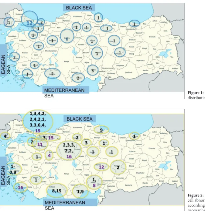
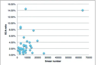
The study group was composed of 38 pathology laboratories and 45 different hospitals from 24 cities. The participant distribution is shown in Figure 1. Among the participant centers, 12 (26%) were state hospitals while 8 (18%) were private hospitals and 25 (56%) were university hospitals. The annual total number of smears was 354,725 and cervical
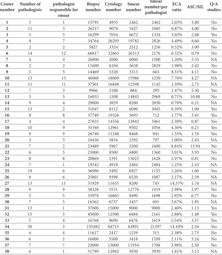
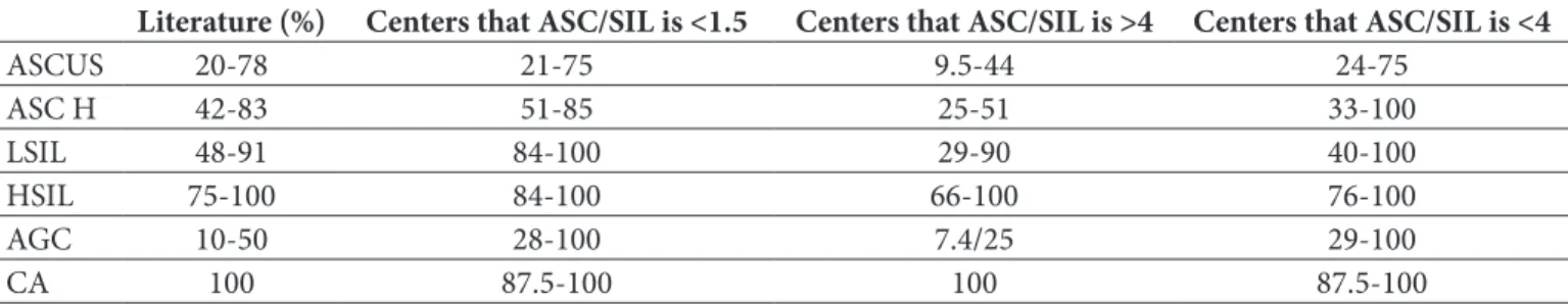
The annual total number of eCA was 18,020 in the study group and the global eCA ratio of the study group was 5.08% in the total 354,725 smears. Among the centers, the lowest reported eCA rate was 0.3% and the highest eCA was 16.64%. The eCA rates of the laboratories are shown in Table I. The distribution of eCA rates varied among the geographic regions (Figure 2). The relation between the eCA rates and the total smear number is shown in the graph (Figure 3). When analyzed based on the type of the centers, eCA rate varied between 0.52 and 7.76% (mean: 2.81%, median: 2.18%) in state hospitals while it was 3.71-16.64% (mean: 12.32%, median: 14% ) in private hospitals and 0.39-14.57% (mean: 3.16%, median: 2.38%) in university hospitals.
cytology numbers of the laboratories ranged between 674-49,483 (Table I) while total annual biopsy numbers were in the range of 5827 to 155,497. The cervical smear constituted 28% of the total biopsy volume in average. The type of method used in cervical cytology was conventional smears in 18 and liquid based cytology (lbs) in 20 laboratories while 9 centers were using automated screening program. The number of pathologists working in the centers ranged between 1 and 30 and the number of pathologist responsible for cervical cytology were in the range of 1-13. The annual number of smears per pathologist varied from 294 to 21,297. Working cytotechnologists were present in 2 centers. Cervical cytology constituted 28% of the volume on average with a range of 8.87%-92.46%.
Figure 1: The participant distribution of the study.
Figure 2: The epithelial cell abnormality rates according to the geographic distribution.
table I: The laboratory characteristics of the participants
Center
number pathologistsnumber of
number of pathologist responsible for
smear
Biopsy
number Cytology number numbersmear
smear number/per
pathologist
eCA
ratio AsC/sIl studiesQ/A
1 5 1 15791 4935 2462 2462 2.03% 3.80 no 2 11 5 26217 9078 5427 1085 0.87% 4.00 no 3 5 5 16299 7034 6672 1334 3.03% 2.88 yes 4 8 7 54764 26293 19782 2826 4.49% 8.60 yes 5 2 2 5827 3324 2512 1256 0.52% 3.00 no 6 14 12 66817 32863 26113 2176 0.52% 0.79 no 7 4 4 26000 2000 6000 1500 1.20% 5.55 nA 8 2 2 11690 6104 5658 2829 1.98% 2.42 no 9 5 5 14469 5328 3315 663 8.51% 4.11 no 10 13 13 46060 18060 15986 1230 7.76% 4.27 nA 11 11 11 37501 16848 12598 1145 1.59% 2.75 nA 12 7 3 9966 2180 884 295 1.47% 5.50 yes 13 5 5 16055 1508 14845 2969 0.71% 10.88 no 14 9 4 29000 3859 8200 2050 0.78% 0.21 nA 15 15 2 31047 8112 6090 3045 0.39% 1.00 yes 16 8 8 37749 19326 5693 712 1.77% 3.41 yes 17 4 4 27655 14556 13843 3461 2.38% 0.87 yes 18 10 9 35348 12961 9502 1056 4.36% 6.21 yes 19 9 9 26740 11248 8468 941 1.55% 1.76 yes 20 3 3 10430 3834 2392 797 1.00% 2.43 nA 21 7 2 23489 5967 3200 1600 8.84% 13.94 no 22 6 5 25000 8500 6800 1360 3.01% 3.93 no 23 8 8 20869 2391 13023 1628 2.97% 0.81 no 24 7 1 19342 4918 2484 2484 1.25% 2.43 nA 25 19 6 36996 3492 6927 1155 1.26% 1.00 yes 26 6 6 25801 9398 6520 1087 3.17% 2.59 nA 27 13 11 31929 11635 8200 745 14.57% 1.74 nA 28 9 9 38328 3531 12770 1419 2.98% 2.97 no 29 5 5 33978 16865 8490 1698 2.92% 6.77 no 30 7 5 16562 6737 3457 691 3.67% 1.95 nA 31 13 1 37000 15000 9000 9000 2.40% 1.13 yes 32 15 3 45000 12500 6484 2161 2.88% 1.48 yes 33 5 4 16768 9690 6476 1619 5.54% 3.37 yes 34 30 3 155492 84715 63891 21297 14.10% 2.54 yes 35 4 4 11617 2417 1259 315 2.38% 2.75 no 36 6 2 16000 5500 3418 1709 2.11% 3.24 no 37 7 7 22000 13000 11954 1708 3.98% 2.30 yes 38 6 1 31799 12943 3930 3930 1.81% 3.12 nA
Figure 4: The AsC/sIl ratios distribution of the centers.
AsC: Atypical squamous cell, sIl: squamous intraepithelial lesion).
Figure 5: The AsC/sIl ratio in relation to eCA rates of the centers.
AsC: Atypical squamous cell, sIl: squamous intraepithelial lesion, eCA: epithelial cell abnormality.
Figure 6: The AsC/sIl ratio in relation to annual smear number.
AsC: Atypical squamous cell, sIl: squamous intraepithelial lesion. Among 18020 eCA, the most common lesion was AsCus
(11557/18020, 64.2%) followed by lsIl (4106/18020, 22.8%), AsCH (1029/18020, 5.8%), HsIl (700/18020, 4%), AGC (548/18020, 3%) and CA (18/18020, 0.2%), in descending order.
The AsC/sIl ratios ranged between 0.21 and 13.94 with an average of 2.61 (±2.95) and median of 2.87. eight centers had an AsC/sIl ratio lower than 1.5 and 13 centers had a value between 1.5 and 3, while 7 centers had an AsC/ sIl ratio of 3-4 and 10 of them had a value higher than or equal to 4 (Figure 4). When analyzed according to the type of the centers, the AsC/sIl ratio varied between 0.79 and 10.87 (mean: 4.23, median: 3.05) in state hospitals while it was in the range of 2.07-5.25 (mean: 2.08, median: 3.74) in private hospitals and 0.21-13.94 (mean: 3.17, median: 2.75) in university hospitals.
The graphical distribution of eCA percentages in relation to AsC/sIl ratios is shown in Figure 5.
The AsC/sIl ratios in relation to annual cervical cytology per pathologist in the centers are shown in Figure 6. When the centers were asked for whether they perform QA studies, 28 centers shared the information and 14 of them had such a control study. eight centers were following the CHC results, another 5 were following both CHC and AsC/ sIl ratio and in 1 center the annual malignancy rate, AsC/ sIl ratio, and CHC rates were followed and 10% of negative reported cases were re-evaluated. In the QA-performing group, the AsC/sIl ratio range was 0.87-8.6 with an average of 3. In the QA-non performing group AsC/sIl ratio range was 0.79-13.94 with an average of 4.5.
Figure 3: eCA rate distribution according to the total smear number of the center.
of cases with negative biopsy was 138. The biopsy results of 1461 patients whose smear diagnosis had been lsIl were documented and sIl was verified in 1005 (726 lsIl, 276 HsIl and 3 CA) (68.8%). The sIl verification rate by biopsy in patients diagnosed with HsIl was 87% (55 lsIl, 259 HsIl and 48 CA). The positive predictive value (PPV) of AGC was 47.8% (14 lsIl, 9 HsIl and 35 CA and glandular pathology in 39 cases) and 97.4% (12 lsIl, 1 HsIl and 24 CA) for carcinoma diagnosis. The biopsy follow-up results are shown in Table II.
The PPV of the each center according to the smear diagnosis showed variations. The lowest PPVs for AsCus, AsC-H, lsIl, HsIl, CA, and AGC were 9.5; 25; 25; 66.6; 7.4, and 87.5, respectively. When these results were evaluated according to the AsC/sIl ratio, the PPVs for AsCus, AsC-H, lsIl, HsIl, CA, and AGC were 21-75.51-85, 84-100, 84-100, 87.5 -100, and 28-100 respectively for the centers having an AsC/sIl ratio lower than 1.5. For the centers having an AsC/sIl ratio higher than 4, the PPVs for AsCus, AsC-H, lsIl, HsIl, CA and AGC were 9.5-44; 25-51; 29-90; 66.6-100; 7.4-25 and 100, respectively (Table III). When we analyzed the centers according to AsC/sIl higher
than 3 and QA study performance, 35.7% (5 of 14) of the centers in the QA-performing group had an AsC/sIl value higher than 3 but this was as high as 57.1% (8 of 13) in the QA-non performing group.
There were lower AsC/sIl ratios in private hospitals and no difference between university/non-university hospitals. Private hospitals had a common feature of having quality control studies in 5 out of 6 centers. Despite such QA studies, AsC/sIl was higher than 3 (3.37-3.41-5.5-6.21-8.6) in 5 out of these 14 centers. Among the 8 centers with AsC/ sIl ratios higher than 4, we found that 4 had no QA studies, 3 were performing QA studies and no data was obtained from 1 of them.
Follow-up data of the patients was reported from 41 centers. In the study group, 4173 patients in total had at least one biopsy. Out of the 1951 biopsy-verified AsCus cases, 722 were sIl and CA (559 lsIl, 152 HsIl, 11 CA) and the biopsy was negative in 1229 patients. Follow-up biopsy was performed in 344 patients reported as AsC-H and the results were 93 lsIl, 98 HsIl, and 15 CA while the number
table II: The biopsy results of the patients with follow-up
smear Diagnosis
Biopsy Diagnosis
neG lsIl HsIl CA Glandular pathology (dysplasia, hyperplasia, polyp, etc.) total Mean PPV AsCus 1229 (62.9%) 559 (28.7%) 152 (7.8%) 11 (0.6%) - 1951 37.1 AsC-H 138 (40%) 93 (27%) 98 (28.5%) 15 (4.5%) - 344 60 lsIl 456 (31.2% 726 (49.7%) 276 (18.9%) 3 (0.2%) - 1461 68.8 HsIl 55 (13%) 55 (13%) 259 (62.5%) 48 (11.5%) - 417 87 AGC 106 (52.2%) 14 (6.7%) 9 (4.5%) 35 (17.3%) 39 (19.3%) 203 47.8 CA 1 (2.6%) 12 (31.6%) 1 (2.6%) 24 (63.2%) - 38 97.4
AsCUs: Atypical squamous cell of undetermined significance, AsC-H: Atypical squamous cell-HsIl can not excluded, lsIl: low grade squamous
intraepithelial lesion, HsIl: High grade squamous intraepithelial lesion, AGC: Atypical glandular cell, CA: Carcinoma, neG: negative, PPV: Positive predictive value
table III: The PPV of centers according to the diagnosis
literature (%) Centers that AsC/sIl is <1.5 Centers that AsC/sIl is >4 Centers that AsC/sIl is <4
AsCus 20-78 21-75 9.5-44 24-75 AsC H 42-83 51-85 25-51 33-100 lsIl 48-91 84-100 29-90 40-100 HsIl 75-100 84-100 66-100 76-100 AGC 10-50 28-100 7.4/25 29-100 CA 100 87.5-100 100 87.5-100
PPV: Positive predictive value, AsC: Atypical squamous cell, sIl: squamous intraepithelial lesion, AsCUs: Atypical squamous cell of undetermined
significance, AsC-H: Atypical squamous cell-HsIl can not excluded, lsIl: low grade squamous intraepithelial lesion, HsIl: High grade squamous intraepithelial lesion, AGC: Atypical glandular cell, CA: Carcinoma, neG: negative
unfortunately, we do not have data from the eastern part of the country, and there is a gap in that area. There are studies representing data from this region (32) and the eCA rate was found to be 1.81% by those studies. Although we cannot discuss the regional data, there is a lower eCA rate in this region of Turkey in comparison to the averages of the whole study group.
The differences in eCA rates among different cities show an increase in some cities on the coast. This can be attributed to socio-cultural differences. However, this cannot be the only explanation as there are low rates from the some cities such as Mersin, Adana, Istanbul and low rates reported from Izmir –another city on the coast. One striking result of the eCA rates is that a laboratory having 8 different hospitals reported the highest rates (shown in purple in Figure 2) among other hospitals in the same cities. In terms of QA, this laboratory has a highly acceptable AsC/sIl ratio smaller than 3, so the eCA rates can be considered reliable. This center is a private laboratory and most of the hospitals are A+ hospitals where the patient group consisted mostly upper socio-economic group of high-risk patients. This may reflect the difference in socio-economical difference among other centers. The data from this center is similar to the data of high income countries (28-30).
The ranges of eCA rates showed no great differences when evaluated according to hospital type as public-private or university-nonuniversity hospitals.
The AsC/sIl is reported to be less variable when compared to eCA and creates mathematical data recommended for use in QA (16,19,21). This ratio is recommended to be 1.73 - 2.05 for cytopathologists and 0.87- 4.5 for cytotechnologists in several articles (16,21) and under 2-3 in others (16,19,20). The AsC/sIl ratio in hospital-based studies from Turkey varies between 2.25 and 12.6 (3-13). This value was 2.83 in the multi-center study from Turkey (14). The AsC/sIl ratios from this study showed a great range among centers. The values changed from 0.21 to 13.94 with a mean of 2.83. When grouped, 8 centers had a ratio lower than 1.5 (21%) and 13 centers reported a ratio of 1.5-3 (34%). The number of centers having a ratio higher than 3 was 17 (45%) and 9 of them had a ratio higher than 4. These values are not totally correlated to eCA percentages, as shown in the graph in Figure 5. It is seen that there are very low AsC/ sIl values in some high eCA reporting centers as well as high AsC/sIl values in some low eCA reporting centers, although the majority are grouped in the left part of the graph with acceptable values in terms of QA. It is stated that the AsC/sIl ratio may be lower in high-risk populations DIsCUssIOn
The time-honored PAP test is currently the most effective cancer screening method. The cervical smear data from Turkey is documented in hospital-based reports (3-13) and only one study (14) represents a multi-center data reporting results from 22 cities, 34 hospitals and a total of 140,334 smears. There is an ongoing public-based national cervical carcinoma-screening program searching hrHPV presence since 2014, and the Turkish Ministry of Health runs it, but their data is not published yet. Hence our study is the largest existing study showing the results based on 354,725 patients.
Objective data is required in order to detect the efficiency of cervical smears. Various data such as the eCA rate and AsC/sIl ratio can be used for QA. When the eCA rate was evaluated, it varied between 1.5% and 7.3% in countries with high-income (28-30). reported rates from Turkey range from 1.2% to 12.6% (3-13) and were 1.8% in the multi-center study (14). The average eCA rate of 5.08% in our study is higher than the unreported current data of the Ministry of Health national screening program (31). This difference can be considered to be partly related to the patient population as our study used hospital-based rather than public-based data. To a smaller extent, possible patient duplications can be a reason, but our data was collected as patient-based, not test-based, and this cannot be the explanation for this high rate. Our study method is similar to the multi-center hospital-based study reported in 2009 in Turkey (14) and our current results are also higher than this study. The difference may be attributed to the difference in hospital types, as the previous study did not include private hospitals, and the number of the patients involved in the studies. Our study includes 2.5 times more patients than the other study so we believe that our results are more reliable. As our study also includes private hospitals, we can also argue that our data represents a more homogeneous distribution among the Turkish population and hence it is more generalizable since the previous study did not represent the private hospitals that play a major role in the health care system. It is found that there is an increase in eCA rate in the 6-year period from 2007 (the data of the previous study shows the 2007 data) to 2013 (the year of data collection of the present study). The eCA rates of the private hospitals are seen to be higher than state hospitals, and this may be attributed to the upper socio-economic group of high-risk patients. This may reflect the difference in socio-economical difference among other centers, similar to the data of countries with high income (28-30).
Centers with an AsC/sIl ratio higher than 4 have another common property that cervical cytology was evaluated by all of the pathologists working in the laboratory and there was no dedicated pathologist in most of these centers. In 89% of the centers that have a ratio higher than 4, cervical cytology is evaluated by all of the pathologists while only 46% of the centers reporting low AsC/sIl values have such a working principle. based on this result, one can discuss whether cervical cytology should be reported by all pathologists or whether it should be a subspecialty or at least reported by some dedicated pathologist at the centers. In the pathology routine, cervical cytology is easily shared and with the current health performance system, every pathologist wants to evaluate it in order to “increase performance”. This is also discussed in other countries and it is stated that when health care professionals are paid for each service they provide, the health care neither becomes more efficient and well coordinated nor has high quality (33).
Cervical cytology constitutes a large work volume of pathology routine and it made up an average of 28% of the patients in our study group. In this context, the adequacy of cytology training during pathology training should be discussed. At a survey of pathologists training in cytopathology in european countries (responded from 26 countries), pathologists without specific training in cytopathology signed out cytology reports in 54.7%, more often in centers where training was 3-6 months or less in duration. However, 92.2% of respondents thought that cytology should not be reported by pathologists without experience in cytopathology (cervical cytology workloads of survey respondents varied from 500 to 200,000 requests per year; those defined as “large” processed a range of 13,000-200,000 with an average of 38,000 per year) (34). Another important point to note is that cervical smears are accepted as the most efficient screening test and but are now becoming a somewhat “diagnostic” test due to the changing screening methods with the use of HPV typing as the first step. The curriculum of pathology residents should also be reviewed according to this changing profile.
In terms of QA of cervical cytology, the best parameter that can be used is eCA confirmed by cervical biopsy. The specificity and sensitivity rates of cervical cytology are reported to be highly variable (29,30,35). specificity is reported as 14 to 97% with a mean of 69% and sensitivity as 11 to 99% with a mean of 58% in the literature (23,30,35,36). The CHC increases in direct proportion with the increase of degree of atypia on cytology to as high as 100% for HsIl, and decreases to 54% for AsCus and 20% for AGC and higher in low-risk populations (28). However, this
statement cannot explain the difference in our study group, as there are centers with high AsC/sIl ratios although they have low eCA rates, as well as some low AsC/sIl ratios with high eCA rates.
The workload is an important determinant, affecting the quality parameters in pathology. The workload limit can be variable, it can be assessed by daily or hourly slide number or time spent nonstop on screening or daily total time of screening (25). As well as the daily workload, a more highly correlated parameter is reported to be the “eCA-adjusted workload” as 7 slides/day, which means 70 slides/day with a 10% eCA (26). However, similar to eCA rates, the great range of AsC/sIl among centers in this study cannot be ascribed solely to workload as there are high values with low workload and very accurate values with very high workload. Figure 6 shows that there are very low AsC/sIl values in 2 high workload centers as well as AsC/sIl values higher than 4 in 7 centers that of which have a low workload of cervical cytology per pathologist although most of the centers are grouped in left part of the graph. The laboratories having the two highest values are different than the others, as they have cytotechnologists working with pathologists. Therefore, they can be evaluated separately. Other than these two highest volume centers, the annual PAP smear load is under 3000 cases in the 16 laboratories with AsC/ sIl higher than 3 when AsC/sIl ratio is evaluated in terms of the workload (defined by PAP smear per pathologist). so the AsC/sIl ratio cannot be said to depended on the workload, based on our study.
The QA is important in pathology laboratories as in all other fields. The Hawthorne effect describes the better performance of observed subjects than unobserved subjects and it is stated that pathologists in small laboratories working as solo screeners have the highest failure rate (15). Quality control studies were performed in 14 centers. There was no difference in parameters among hospital type, other than the low AsC/sIl ratios in private hospitals. It is seen that this group has a common feature to have quality control studies in 5 out of 6 centers. It is obvious that this cannot be the only explanation but the effect of quality control studies cannot be denied. However, it is observed that AsC/sIl was higher than 3 (3.37-5.5-6.21-8.6) in 4 out of the 11 centers with quality control studies. In this context, it is noted that not only does the presence of quality control studies ensure quality but also the presence of corrective and preventive action is a must. Most of the centers declared they have QA studies documenting the CHC data required by the Ministry of Health.
7. boztosun A, Mutlu AM, Özer H, Aker H, yanık A: servikovajinal smearde epitelyal hücre anomalisi saptanan hastaların kolposkopik biyopsi sonuçlarının değerlendirilmesi. Türk Jinekolojik Onkoloji Dergisi 2012, 1:13-19.
8. Atilgan r, Celik A, boztosun A, Ilter e, yalta T, Ozercan r: evaluation of cervical cytological abnormalities in Turkish population. Indian J Pathol Microbiol 2012, 55:52-55.
9. kır G, karateke A, Aker F: AsCus, lsIl ve HsIl tanılarının sito-histolojik korelasyonu, bethesda 2001 AsCus alt gruplarının irdelenmesi. Turk Patoloji Derg 2003, 19:14-16.
10. Coşkun A, köstü b, kıran G, Arıkan DC, Analan A: Pap smear screening results in kahramanmaraş. Gynecol Obstet reprod Med 2008,14:182-185.
11. ersöz Ş, reis A, baki n: Trabzon ilinde servikal Tarama Programı. Türk Jinekolojik Onkoloji Dergisi 2010, 7:35-39.
12. Isaoglu u, yilmaz M, Delibas Ib, bilici Ae, kabalar Me. evaluation of 37,438 consecutive cervical smear results in the Turkish population. Arch Med sci. 2015 Apr 25;11(2):402-5. 13. nayir T, Okyay rA, nazlican e, yesilyurt H, Akbaba M, Ilhan b,
kemik A. Cervical Cancer screening in an early Diagnosis and screening Center in Mersin, Turkey. Asian Pac J Cancer Prev. 2015;16(16):6909-12.
14. Turkish Cervical Cancer And Cervical Cytology research Group: Prevalence of cervical cytological abnormalities in Turkey. Int J Gynaecol Obstet 2009, 106:206-209.
15. Tworek J, nayar r, savaloja l, Tabbara s, Thomas n, Winkler b, Howell lP. General quality practices in gynecologic cytopathology: findings from the College of American Pathologists Gynecologic Cytopathology Quality Consensus Conference working group 3. Arch Pathol lab Med. 2013 Feb;137(2):190-8.
16. Alessandra F. nascimento, edmund s. Cibas. The AsC/sIl ratio for Cytopathologists as a Quality Control Measure. A Follow-up study Am J Clin Pathol 2007;128:653-656.
17. renshaw AA, Deschênes M, Auger M. AsC/sIl ratio for Cytotechnologists: A surrogate marker of screening sensitivity. Am J Clin Pathol. 2009 Jun;131(6):776-81.
18. Crothers bA1, Jones bA, Cahill lA, Moriarty AT, Mody Dr, Tench WD, souers rJ. Quality improvement opportunities in gynecologic cytologic-histologic correlations: findings from the College of American Pathologists Gynecologic Cytopathology Quality Consensus Conference working group 4. Arch Pathol lab Med. 2013 Feb;137(2):199-213.
19. solomon D, nayar r: The bethesda system for reporting Cervical Cytology: Definitions, Criteria, and explanatory notes. 2nd ed. new york, ny, springer; 2004: v-vii.
20. renshaw AA, Deschênes M, Auger M: AsC/sIl ratio for Cytotechnologists: A surrogate marker of screening sensitivity. Am J Clin Pathol 2009, 131:776-781.
21. renshaw AA, brimo F, Auger M: surrogate indicators of sensitivity in gynecologic cytology: can they be used to improve the measurement of sensitivity in the laboratory? Cytojournal 2009, 6:19 P
22. Anschau F, Guimarães Gonçalves MA: Discordance between cytology and biopsy histology of the cervix: What to consider and what to do. Acta Cytol 2011, 55:158-16.
(4,6,7,9,23,24). When PPVs are compared with the literature values, the mean rates of the study group are in accordance with the literature. low PPV values are noteworthy in the centers that had AsC/sIl > 4. Quality control studies and follow-up of the AsC/sIl ratio are important. However, the number of patients with biopsy follow-up varied. some centers that had very low follow-up numbers may therefore have appeared to have very high or very low PPVs.
In conclusion, this study shows that there is an increase in eCA rates in Turkey. There are great differences among centers. The most important result of this study is that QA studies including AsC/sIl are vital. However, QA activity does not mean to record the data to be sent to the Ministry of Health and taking corrective and preventive action according to quality control parameters is a must. A cervical cytology subspecialist in each center can be a dream at this stage but assigning dedicated pathologists for cervical cytology should be discussed and attempted. The pathology community should overview cervical cytology training during pathology residency and should be ready to the changing role of the pathologist in the future with cervical screening by hrHPV testing.
COnFlICt of InteRest
The authors have no conflicts of interest to disclose. ACKnOWleDGeMent
We want to thank leyla Durmuş, nevin kaya, Zeki Günlüoğlu, emel Filiz Özbay and Gökhan Türkmen for their support in the preparation of the article.
ReFeRenCes
1. GlObOCAn 2012, Available at: http://globocan.iarc.fr/Pages/ fact_sheets_cancer.aspx
2. Türkiye kanser istatistikleri, 2012, Available at: http://kanser.gov. tr/Dosya/2015haberler1/AnA_rapor_2012v02.pdf
3. Türkmen IÇ, başsüllü n, korkmaz P, Günenç b, baykal CM, Güdücü n, Işçı H, Dünder I, Doğusoy Gb. Patients with epithelial cell abnormality in PAP smears: Correlation of results with follow-up smears and cervical biopsies. Turk Patoloji Derg. 2013;29:179-84.
4. kaygusuz eI, Cetiner H, sahin D. lsIl/AsC-H (lsIl-H) in cervicovaginal smear: Histopathological outcomes and clinical significance. Turk Patoloji Derg 2011;27:46-50.
5. Mehmetoglu HC, sadikoglu G, Ozcakir A, bilgel n: Pap smear screening in the primary health care setting: A study from Turkey. n Am J Med sci 2010;2:467-72.
6. Abali r, bacanakgıl bH, Celik s, Aras O, koca P, boran b, Dursun n. Histopathological correlation of squamous cell abnormalities detected on cervical cytology. Turk Patoloji Derg. 2011;27:144-8.
30. sigurdsson k, sigvaldason H: longitudinal trends in cervical cytological lesions and the effect of risk factors. A 30-year overview. Acta Obstet Gynecol scand 2006, 85:350-358.
31. Işın Pak. ulusal HPV tarama projesi, 25. ulusal Patoloji kongresi, 6. ulusal sitopatoloji kongresi, Available at: http://www. patoloji2015.org/izinli_pdf/isinpak.pdf.
32. Özgül n. in: Tuncer M (ed), Cancer Control in Turkey, Ankara, Onur Press, Health Ministry Publication, 2008;74:347-56. 33. The external environment Influencing Diagnosis: reporting,
Medical liability, and Payment. In: balogh eP, Miller bT, ball Jr. Improving diagnosis in health care. 1st ed. national Academies Press; 2016. pp326.
34. Anshu, Herbert A, Cochand-Priollet b, Cross P, Desai M, Dina r, Duskova J, evered A, Farnsworth A, Gray W, Gupta s.s, kapila k, kardum-skelin I, kloboves-Prevodnik V, kobayashi T.k, koutselini H, Olszewski W, Önal b, Pitman M.b, Marinšek Ž. survey of medical training in Cytopathology carried out by the journal Cytopathology. Cytopathology, 2010,Vol. 21 Issue 3, p147-156.
35. yesil C, Onder s, boynukalin k, ergul Ö, Fırat P, kuzey GM, usubutun A: Correlation between cytological and histopathological diagnosis in premalignant lesions of the cervix. Turk Patoloji Derg 2010, 26:38-43.
36. bofin AM, nygård JF, skare Gb, Dybdahl bM, Westerhagen u, sauer T: Papanicolaou smear history in women with low- grade cytology before cervical cancer diagnosis. Cancer 2007 25, 111:210-216.
23. Mete Ö, yavuz e, Tuzlalı s, İlhan r, Özlük y, Topuz s, İyibozkurt C, İplikçi A: kolposkopik biyopsi yapılan 112 hastanın retrospektif incelemesi: sitolojik bulguların histoloji ile karşılaştırılması. Turk Patoloji Derg 2007, 23:33-37.
24. keskin Hl, seçen eİ, Taş ee, kaya s, Avşar AF: servikal smear sitolojisi ile kolposkopi eşliğinde biyopsi servikal biyopsi korrelasyonu. Türk Jinekolojik Onkoloji Dergisi 2011, 3:71-75. 25. Wiener HG, klinkhamer P, schenck u, Arbyn M, bulten J,
bergeron C, Herbert A. european guidelines for quality assurance in cervical cancer screening: recommendations for cytology laboratories. Cytopathology. 2007 Apr;18(2):67-78. erratum in: Cytopathology. 2007 Jun;18(3):207.
26. renshaw AA, elsheikh TM. Predicting screening sensitivity from workload in gynecologic cytology: a review. Diagn Cytopathol. 2011 nov;39(11):832-6.
27. serviks kanseri tarama programı ulusal standartları. Available at: http://kanser.gov.tr/Dosya/tarama/serviks.pdf .
28. eversole GM, Moriarty AT, schwartz Mr, Clayton AC, souers r, Fatheree lA, Chmara bA, Tench WD, Henry Mr, Wilbur DC: Practices of participants in the College of American Pathologists interlaboratory comparison program in cervicovaginal cytology, 2006. Arch Pathol lab Med 2010, 134:331-335.
29. llewellyn H: relationship between the cytologic reporting rate for high-grade squamous intraepithelial lesion or worse and Papanicolaou smear sensitivity: A simple mathematic proof with practical implications. Cancer 2007, 111:137-40; discussion 141-142.