http://journals.tubitak.gov.tr/medical/ © TÜBİTAK
doi:10.3906/sag-1401-4
Endothelial dysfunction and insulin resistance in young women with
polycystic ovarian syndrome
Mine YAVUZ TAŞLIPINAR1,*, Nedret KILIÇ2, Nilüfer BAYRAKTAR3, İsmail GÜLER4, Yasemin GÜLCAN KURT5, Tayfun GÖKTAŞ6, Mehmet Zeki TANER4, Mehmet Özdemir HİMMETOĞLU4, Halil YAMAN5, Abdullah TAŞLIPINAR7
1Department of Biochemistry, Dışkapı Yıldırım Beyazıt Training and Research Hospital, Ankara, Turkey 2Department of Biochemistry, Faculty of Medicine, Gazi University, Ankara, Turkey
3Department of Biochemistry, Faculty of Medicine, Başkent University, Ankara, Turkey 4Department of Obstetrics and Gynecology, Faculty of Medicine, Gazi University, Ankara, Turkey
5Department of Biochemistry, Gülhane Military Medicine Faculty, Ankara, Turkey 6Department of Physiology, Faculty of Medicine, Gazi University, Ankara, Turkey 7Department of Endocrinology, Gülhane Military Medicine Faculty, Ankara, Turkey
1. Introduction
Polycystic ovarian syndrome (PCOS) is a common endocrine and metabolic disorder in women of reproductive age, which is characterized by ovulatory dysfunction, hyperandrogenism, and polycystic ovaries (1). PCOS is known to be related to increased insulin resistance, obesity, type II diabetes, and cardiovascular diseases (2). Different theories have been proposed for the pathogenesis of PCOS; one of the major hypotheses is the insulin theory. According to this theory, insulin resistance and its resultant compensatory hyperinsulinemia are thought be etiologic factors of this syndrome and its related complications (3–6). Insulin resistance and hyperinsulinemia are known to disturb nitric oxide-mediated vasodilatation and endothelial functions, which
results in vascular damage and leads to increased risk for long-term metabolic disorders.
The homeostasis model assessment of insulin resistance (HOMA-IR) is an alternative tool to the glucose clamp for the evaluation of insulin resistance by using the fasting glucose and insulin levels of patients. It is commonly used, especially for investigations including a large number of subjects.
N-Dimethyl-L-arginine is an inhibitor of nitric oxide synthase and is also known as asymmetric dimethylarginine (ADMA) (7). It is synthesized by the action of the protein arginine methyltransferases and is eliminated by renal excretion or by the enzymatic action of dimethylarginine dimethylaminohydrolase (DDAH). High ADMA values inhibit nitric oxide (NO) production Background/aim: To evaluate whether there is a correlation between insulin resistance and nitric oxide-related endothelial dysfunction
in patients with polycystic ovarian syndrome (PCOS).
Materials and methods: The study was conducted with 25 young women with PCOS and 25 young healthy women, between 18 and 35
years of age. Plasma asymmetric dimethylarginine (ADMA) levels, serum nitric oxide (NO) levels, and homeostatic model assessment of insulin resistance (HOMA-IR) rates were measured in both the patient and control groups.
Results: Plasma ADMA levels were significantly higher in PCOS patients than in the controls (P = 0.001). Serum NO levels were
significantly lower in patients than in the controls (P = 0.008). The HOMA-IR rates, accepted as an insulin resistance parameter, were significantly higher in patients than in the controls (P = 0.001).
Conclusion: Results of the present study indicate that, independent of age, body mass index, and blood lipid profile, there is significant
insulin resistance in PCOS patients. However, no correlation was found between HOMA-IR as an insulin resistance determinant and altered ADMA and NO levels. This finding may indicate that there are additional mechanisms of cardiovascular risks in PCOS patients other than insulin resistance.
Key words: Polycystic ovarian syndrome, endothelial dysfunction, insulin resistance, asymmetric dimethylarginine, nitric oxide Received: 02.01.2014 Accepted: 13.01.2014 Published Online: 15.08.2014 Printed: 12.09.2014
and generate superoxides. However, NO is needed to promote vasodilation. ADMA is considered to be a marker of endothelial dysfunction because of the relation between ADMA and NO metabolism. High levels of ADMA in human plasma are associated with cardiovascular and metabolic diseases (8). Therefore, the strong association between PCOS and insulin resistance, diabetes, obesity, and cardiovascular disease, which are almost always found in conjunction with endothelial dysfunction, makes the relationship of PCOS and ADMA important.
The aim of this study was to find out whether there is any correlation between insulin resistance and NO-related endothelial dysfunction in patients with PCOS.
2. Materials and methods
Following local ethics board approval (19.12.2005/346), a total of 25 patients with PCOS and 25 healthy female volunteers were included in the study. The diagnosis of PCOS was done according to the 2003 Rotterdam Criteria (9). Patients who had received steroid or sex hormones in the previous 6 months or any medication related to hirsutism; patients with hyperprolactinemia, congenital adrenal hyperplasia (or with high 17α-hydroxyprogesterone), Cushing’s syndrome, thyroid diseases, functional tumors that may cause hyperandrogenemia, hypertension, glucose intolerance, or diabetes; and smokers were all excluded from the study.
The control group was composed of volunteers with normal ovarian ultrasound findings and with no hormonal, metabolic, and cardiovascular diseases or menstrual irregularities. Smokers were also excluded from the control group. The anthropometric measurements of both groups were recorded.
The blood samples of subjects of both groups were obtained on days 3–5 of their menstrual cycles. The sampling was performed around 0900 hours after 8–10 h of overnight fasting. These fasting venous blood samples were used for the analyses of glucose, insulin, free testosterone, dehydroepiandrostenedione (DHEA), follicle-stimulating hormone (FSH), luteinizing hormone (LH), estradiol, prolactin (PRL), 17-hydroxyprogesterone, total cholesterol, triglycerides, high-density lipoprotein cholesterol (HDL-C), ADMA, and NO.
The oral glucose tolerance test was performed by giving 75 g of glucose. Blood samples were taken at time intervals of 0 and 120 min. HOMA-IR was calculated as [(fasting glucose × fasting insulin) / 405]. Serum low-density lipoprotein (LDL) cholesterol levels were calculated by using the Friedewald formula [LDL-C = total cholesterol – (HDL-C + triglyceride / 5)].
Glucose levels were measured by autoanalyzer (Abbott Aeroset, Toshiba, Japan) with the adapted hexokinase method. Serum cholesterol measurements
were performed with an autoanalyzer (Abbott Aeroset, Toshiba) adapted for cholesterol esterase. Serum HDL levels were determined using the photometric elimination method (Abbott Aeroset, Toshiba). Serum triglyceride levels were measured by autoanalyzer (Abbott Aeroset, Toshiba) and an adapted photometric method. Serum LH, FSH, PRL, insulin, and DHEA levels were measured with commercially available chemiluminescence kits (Abbott Architect, Toshiba). Serum estradiol, free testosterone, and 17-hydroxyprogesterone levels were measured by radioimmunoassay (RIA) method (DSL RIA Kits, Webster, TX, USA). Serum NO levels of both the control and study groups were measured using the diazotization method, which is based on the Griess reaction (10). NO values are expressed as µmol/L. Plasma ADMA concentrations were measured by high-performance liquid chromatography method, which was described by Chen et al. (11). ADMA values are expressed as µmol/L.
2.1. Statistical analyses
All data were recorded as mean ± SD and median (minimum–maximum). Statistical analyses were performed with SPSS 10.01 for Windows. Using the Kolmogorov–Smirnov test, each variable was evaluated for relevance to normal distribution. For the variables relevant to normal distribution, a t-test was used for comparison of the study and control groups. For variables not relevant to normal distribution, the Mann–Whitney U test was used for comparison of variables. The Shapiro–Wilk test was also used. Correlation coefficients were calculated with Pearson and Spearman correlation tests. P < 0.05 was accepted to be statistically significant.
3. Results
The mean age and mean body mass index (BMI) of the study group were 24.64 ± 4.93 (range: 19–36) years and 26.35 ± 7.05 (range: 16.48–39.55) kg/m2, respectively. The
mean age of the control group was 26.32 ± 3.89 (range: 20–34) years and the mean BMI of the control group was 22.46 ± 3.43 (17.10–29.06) kg/m2.
Our results showed that the mean HOMA-IR rates of patients with PCOS were significantly higher than those of the control group (P = 0.001). We also found statistically higher ADMA levels in the PCOS group (P = 0.001). However, the NO concentrations were significantly lower in the PCOS patients (P = 0.008). In terms of the other serum parameters measured in the present study, no statistically significant alterations were detected between the study and control groups. The data obtained from the study are summarized in Tables 1 and 2.
Measurements of correlations among HOMA-IR, ADMA, and NO showed no significant correlations between the variables.
4. Discussion
The relationships between PCOS and obesity, insulin resistance, endothelial dysfunction, and endothelial dysfunction-related cardiovascular risks have complex features. Endothelial dysfunction is not only an early and reversible indicator of vascular anomalies, but it is also a diagnostic indicator of late cardiovascular morbidity (12).
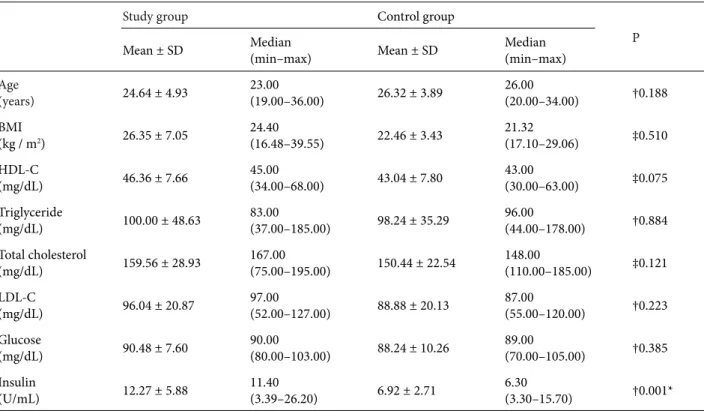
Insulin resistance in PCOS is both a reason and a result, which affects nearly 50%–70% of the PCOS patients. Pamuk et al. found higher HOMA-IR levels but no significant differences in ADMA levels in the PCOS group compared to the control group (13). A cardiovascular study done in Quebec showed that hyperinsulinemia is an independent risk factor of cardiovascular diseases Table 1. Comparison of routine data of the groups.
Study group Control group
P
Mean ± SD Median(min–max) Mean ± SD Median(min–max)
Age (years) 24.64 ± 4.93 23.00(19.00–36.00) 26.32 ± 3.89 26.00(20.00–34.00) †0.188 BMI (kg / m2) 26.35 ± 7.05 24.40(16.48–39.55) 22.46 ± 3.43 21.32(17.10–29.06) ‡0.510 HDL-C (mg/dL) 46.36 ± 7.66 45.00(34.00–68.00) 43.04 ± 7.80 43.00(30.00–63.00) ‡0.075 Triglyceride (mg/dL) 100.00 ± 48.63 83.00(37.00–185.00) 98.24 ± 35.29 96.00(44.00–178.00) †0.884 Total cholesterol (mg/dL) 159.56 ± 28.93 167.00(75.00–195.00) 150.44 ± 22.54 148.00(110.00–185.00) ‡0.121 LDL-C (mg/dL) 96.04 ± 20.87 97.00(52.00–127.00) 88.88 ± 20.13 87.00(55.00–120.00) †0.223 Glucose (mg/dL) 90.48 ± 7.60 90.00(80.00–103.00) 88.24 ± 10.26 89.00(70.00–105.00) †0.385 Insulin (U/mL) 12.27 ± 5.88 11.40(3.39–26.20) 6.92 ± 2.71 6.30(3.30–15.70) †0.001*
‡: Mann–Whitney U test, †: t-test, * P < 0.05.
BMI: Body mass index, HDL-C: high-density-lipoprotein cholesterol, LDL-C: low-density lipoprotein cholesterol.
Table 2. Comparison of HOMA-IR, plasma ADMA, and serum NO levels of the groups.
Study group Control Group
P
Mean ± SD Median(min–max) Mean ± SD Median(min–max)
HOMA-IR 2.76 ± 1.37 2.65(0.79–6.10) 1.52 ± 0.66 1.42(0.64–3.30) ‡0.001* ADMA (µmol/L) 1.20 ± 0.47 1.07(0.43–2.51) 0.75 ± 0.45 0.73(0.28–2.27) ‡0.001* NO (µmol/L) 10.66 ± 9.55 5.99(0.49–35.74) 25.85 ± 24.96 16.23(2.74–105.73) ‡0.008* ‡: Mann–Whitney U test, * P < 0.05.
(14). HOMA-IR levels of the PCOS group measured in the present study were significantly higher than those of the control group. Furthermore, we could not find any correlation between HOMA-IR levels and BMI or the age of the PCOS patients. Obesity is a risk factor for insulin resistance. However, results of the present study indicate that increased insulin resistance in PCOS patients may be due to the additional risk factors. Further studies are needed to find these putative risk factors.
Endothelium regulates the vascular tonus by secreting vasodilators, such as NO, and vasoconstrictors, such as endothelin (15). In the presence of insulin resistance, the relation between insulin and NO production is disturbed (16). Mather et al. showed normal endothelial functions in PCOS patients (17). However, Paradisi et al. reported endothelial dysfunction and insulin resistance in women with PCOS (18). Orio et al. evaluated young PCOS patients with no metabolic and cardiovascular disease and reported disturbances in endothelial functions in the early period of the disease (19). Sorensen et al. showed that endothelial dysfunction in PCOS patients is independent from age, BMI, and total cholesterol levels (12). In our study, serum NO levels of women with PCOS were significantly lower than those of healthy women. Low levels of serum NO may show an endothelial dysfunction. Thus, our results are in accordance with the findings of previous researchers.
There are several studies showing a strong correlation between increased ADMA levels and cardiovascular mortality and morbidity (7,20,21). These findings suggest that ADMA is not only an indicator of cardiovascular disease but also that it plays a role in the etiopathogenesis of the disease. In a recent metaanalysis, Toulis et al. showed increased cardiovascular disease risk markers in women with PCOS (22). They also demonstrated that ADMA levels were significantly elevated in the PCOS group (22). Moran et al. found increased ADMA levels in women with PCOS, independent of age and adiposity (23). Choi et al. suggested that nonobese women with
PCOS are at an increased risk for cardiovascular diseases. They also showed increased ADMA levels in the PCOS group (24). In our study, PCOS patients were found to have significantly higher plasma ADMA levels. These high levels of ADMA may be explained by endothelial dysfunction caused by insulin resistance. The incidence of hypercholesterolemia, hyperglycemia, and hyperhomocysteinemia is higher in PCOS patients than in normal controls. These pathologic conditions may inhibit the enzyme DDAH, which metabolizes ADMA, and cause an increase in ADMA levels. Ngo et al. showed increased ADMA levels with low NO responsiveness (25). However, in our study, we found no correlation between ADMA and NO levels. This difference may be due to the low number of the PCOS patients enrolled in this study.
Studies showed that a high-fat diet also causes an increase in plasma ADMA levels (26). It was shown that, in hypercholesterolemic rabbits, DDAH activity in both vascular and nonvascular tissues is decreased and plasma ADMA levels are increased (27). Chan et al. showed that mononuclear cell adhesion in hypercholesterolemic patients has a high correlation with plasma ADMA concentrations (28). In cell cultures, the adhesion of these cells is increased by the addition of ADMA-stimulated vascular cells to the culture. In our study, there was no dyslipidemia in the PCOS patients, and there was no difference between the study and control groups in terms of blood lipid parameters. Thus, we were able to exclude the effect of high lipid levels on ADMA concentrations.
In conclusion, we found that there is significant insulin resistance in PCOS patients, independently of age, BMI, and blood lipid profile. Altered ADMA and NO levels in PCOS patients indicate the presence of endothelial dysfunction. However, no correlation was detected between HOMA-IR as an insulin resistance determinant and altered ADMA and NO levels, which may show that there are additional mechanisms of cardiovascular risks in PCOS other than insulin resistance.
References
1. Ahles BL. Toward a new approach: primary and preventive care of the women with polycystic ovarian syndrome. Prim Care Update Ob Gyns 2000; 7: 275–278.
2. Lewandowski KC, Szosland K, Callaghan CO, Tan BK, Randeva HS, Lewinski A. Adiponectin and resistin serum levels in women with polycystic ovary syndrome during oral glucose tolerance test: a significant reciprocal correlation between adiponectin and resistin independent of insulin resistance indices. Mol Genet Metab 2005; 85: 61–69.
3. Buyalos RP, Geffner ME, Bersch N, Judd HL, Watanabe RM, Bergman RN, Golde DW. Insulin and insulin-like growth factor-1 responsiveness in polycystic ovarian syndrome. Fertil Steril 1992; 57: 796–803.
4. Dunaif A, Segal K, Futterweit W, Dobrjansky A. Profound peripheral insulin resistance independent of obesity in polycystic ovary syndrome. Diabetes 1989; 38: 1165–1174.
5. Kielstein J, Boger R, Bode-Boger S, Schaoffer J, Barbey M, Koch K, Frolich J. Asymmetric dimethylarginine plasma concentrations differ in patients with end-stage renal disease: relationship to treatment method and atherosclerotic disease. J Am Soc Nephrol 1999; 10: 594–600.
6. Kazer RR, Kessel B, Yen SSC. Circulating luteinizing hormone pulse frequency in woman with polycystic ovary syndrome. J Clin Enocrinol Metab 1989; 65: 233–236.
7. Cooke JP. Does ADMA cause endothelial dysfunction? Arterioscler Thromb Vasc Biol 2000; 20: 2032–2037.
8. Böger RH. The emerging role of asymmetric dimethylarginine as a novel cardiovascular risk factor. Cardiovasc Res 2003; 59: 824–833.
9. Rotterdam ESHRE/ASRM-Sponsored PCOS Consensus Workshop Group. Revised 2003 consensus on diagnostic criteria and long term health risks related to polycystic ovary syndrome. Fertil Steril 2003; 8: 1–25.
10. Miranda KM, Espey MG, Wink DA. A rapid, simple spectrophotometric method for simultaneous detection of nitrate and nitrite. Nitric Oxide 2001; 5: 62–71.
11. Chen BM, Xia LW, Zhao RQ. Determination of NC,NG-dimethylarginine in human plasma by high-performance liquid chromatography. J Chromatogr B Biomed Sci Appl 1997; 692: 467–471.
12. Sorensen MB, Franks S, Robertson C, Pennell DJ, Collins P. Severe endothelial dysfunction in young women with polycystic ovary syndrome is only partially explained by known cardiovascular risk factors. Clin Endocrinol 2006; 65: 655–659.
13. Pamuk BO, Torun AN, Kulaksizoglu M, Ertugrul D, Ciftci O, Kulaksizoglu S, Yildirim E, Demirag NG. Asymmetric dimethylarginine levels and carotid intima-media thickness in obese patients with polycystic ovary syndrome and their relationship to metabolic parameters. Fertil Steril 2010; 93: 1227–1233.
14. Depres JP, Lamarche M, Mauriege P, Cantin B, Dagenais GR, Moorjani S, Lupien PJ. Hyperinsulinemia as an independent risk factor for ischemic heart disease. N Engl J Med 1996; 334: 952–957.
15. Kravariti M, Naka KK, Kalantaridou SN, Kazakos N, Katsouras CS, Makrigiannakis A, Paraskevaidis AE, Chrousos GP, Tsatsoulis A, Michalis LK. Predictors of endothelial dysfunction in young women with polycystic ovary syndrome. J Clin Endocrinol Metab 2005; 90: 5088–5095.
16. Caballero AE. Endothelial dysfunction in obesity and ınsulin resistance: a road to diabetes and heart disease. Obes Res 2003; 11: 1278–1289.
17. Mather KJ, Verma S, Corenblum B, Anderson TJ. Normal endothelial function despite insulin resistance in healthy women with the polycystic ovary syndrome. J Clin Endocrinol Metab 2000; 85: 1851–1856.
18. Paradisi G, Steinberg HO, Hempfling A, Cronin J, Hook G, Shepard MK, Baron A. Polycystic ovary syndrome is associated with endothelial dysfunction. Circulation 2001; 103: 1410– 1415.
19. Orio F, Palomba S, Spinelli L, Cascella T, De Simone B, Di Biase S, Russo T, Labella D, Zullo F, Lombardi G et al. Early impairment of endothelial structure and function in young normal-weight women with polycystic ovary syndrome. J Clin Endocrinol Metab 2004; 89: 4588–4593.
20. Boger RH, Bode-Boger SM, Thiele W, Junker W, Alexander K, Frolich JC. Biochemical evidence for impaired nitric oxide synthesis in patients with peripheral arterial occlusive disease. Circulation 1997; 95: 2068–2074.
21. Miyazaki H, Matsuoka H, Cooke JP, Usui M, Ueda S, Okuda S, Imaizumi T. Endogenous nitric oxide synthase inhibitor: a novel marker of atherosclerosis. Circulation 1999; 99: 1141– 1146.
22. Toulis KA, Goulis DG, Mintziori G, Kintiraki E, Eukarpidis E, Mouratoglou SA, Pavlaki A, Stergianos S, Poulasouchidou M, Tzellos TG et al. Meta-analysis of cardiovascular disease risk markers in women with polycystic ovary syndrome. Hum Reprod Update 2011; 17: 741–760.
23. Moran LJ, Cameron JD, Strauss BJ, Teede HJ. Vascular function in the diagnostic categories of polycystic ovary syndrome. Hum Reprod 2011; 26: 2192–2199.
24. Choi YS, Yang HI, Cho S, Jung JA, Jeon YE, Kim HY, Seo SK, Lee BS. Serum asymmetric dimethylarginine, apelin, and tumor necrosis factor-α levels in non-obese women with polycystic ovary syndrome. Steroids 2012; 77: 1352–1358. 25. Ngo DT, Chan WP, Rajendran S, Heresztyn T, Amarasekera
A, Sverdlov AL, O’Loughlin PD, Morris HA, Chirkov YY, Norman RJ et al. Determinants of insulin responsiveness in young women: impact of polycystic ovarian syndrome, nitric oxide, and vitamin D. Nitric Oxide 2011; 25: 326–330. 26. Fard A, Tuck C, Donis J, Sciacca R, DiTullio M, Wu H,
Bryant T, Chen N, Torres Tamayo M, Ramasamy R et al. Acute elevations of plasma asymmetric dimethylarginine and impaired endothelial function in response to a high-fat meal in patients with type 2 diabetes. Arterioscler Thromb Vasc Biol 2000; 20: 2039–2044.
27. Ito A, Tsao PS, Adimoolam S, Kimoto M, Ogawa T, Cooke JP. Novel mechanism for endothelial dysfunction: dysregulation of dimethylarginine dimethylaminohydrolase. Circulation 1999; 99: 3092–3095.
28. Chan JR, Boger RH, Bode-Boger SM, Tanqphao O, Tsao PS, Blaschke TF, Cooke JP. Asymmetric dimethylarginine increases mononuclear cell adhesiveness in hypercholesterolemic humans. Arterioscler Thromb Vasc Biol 2000; 20: 1040–1046.