Corresponding author: Mustafa GOK E-mail: [email protected]
Original Investigation
Published Online: 09.02.2017Mustafa GOK
1, Elcin AYDIN
2, Serkan GUNEYLI
3, Ali AKAY
4, Celal CINAR
5, Ismail ORAN
51Adnan Menderes University, Department of Radiology, Aydın, Turkey 2Baskent University, Department of Radiology, Izmir, Turkey
3Bulent Ecevit University, Department of Radiology, Zonguldak, Turkey 4Ege University, Department of Neurosurgery, Izmir, Turkey
5Ege University, Department of Radiology, Izmir, Turkey
Iatrogenic Vascular Injuries Due to Spinal Surgeries:
Endovascular Perspective
ABSTRACT
are therefore crucial. As the early surgical management of those iatrogenic injuries is not easy, endovascular management of such injuries is a good option with acceptable morbidity and mortality rates.
We aimed to define the role of endovascular treatment in patients with iatrogenic vascular injuries in spinal surgery. The study focused on clinical presentations, imaging findings, management and outcomes.
█
INTRODUCTION
I
atrogenic vascular injuries due to spinal surgeries are rare but serious complications with high mortality and morbidity rates. Iatrogenic vascular injuries have been categorized according to the type of procedure responsible for the injury, like anterior-posterior cervical spine surgery, scoliosis surgery or tumor surgery (11). Vascular injuries during spinal surgery can be life-threatening, and early diagnosis and managementAIM: Iatrogenic vascular injuries due to spinal surgeries are rare but serious complications. Surgical management of these injuries is challenging with high morbidity rates. In this study we aim to present the results of endovascular management of iatrogenic vascular injuries due to spinal surgeries.
MATERIAL and METHODS: We retrospectively reviewed 11 patients (5 male, 6 female) who had vascular injuries due to cervical and lumbar spinal surgeries. Clinical findings were bleeding (n=5), leg edema (n=6) and right heart failure with severe dyspnea (n=1). The age range of the patients were between 42-67 (mean: 57.1). Six patients were reviewed with imaging before the procedures and the rest of the patients (n=5) were directly referred to the angiography unit for diagnosis and possible endovascular treatment. RESULTS: The types of surgeries were; cervical surgery (n=5) and lumbar disc operation (n=6). The type of vascular injuries were; vertebral artery stenosis (n=1), vertebral artery pseudoaneurysm (n=3), vertebral artery occlusion (n=1) and iliac arteriovenous fistula (n=6). The type of endovascular treatments were; parent artery occlusion (PAO) (n=2), covered stent graft implantation (n=6) and intrasaccular coil embolization of pseudoaneurysm (n=1). The remaining 2 patients were managed conservatively. No major complications or mortality occured during endovascular interventions. No bleeding or ischemia occured in the follow-up period. CONCLUSION: Iatrogenic vascular injuries due to spinal surgeries are rare but serious complications. Endovascular interventions are safe and effective in the diagnosis and treatment of such vascular injuries.
█
MATERIAL and METHODS
A retrospective review was performed for all patients who underwent endovascular management for iatrogenic spinal vascular complications during the last 15-years period. A total of 11 patients (five men, six women; between 42-67 years of age) with vascular injury after spinal surgery were included in this study. The patients were referred for diagnosis and possible endovascular treatment on the same day if intraoperative vascular injury was present. The remaining patients were scheduled for endovascular management. Angiographic findings were labeled as vertebral artery (VA) occlusion, VA stenosis, pseudoaneurysm (PA), and iliac arteriovenous fistula (AVF).
Patients’ demographic information, their presenting signs and symptoms, as well as conventional angiographic findings, treatment, follow-up period and outcomes were collected from patients’ files, images and reports.
█
RESULTS
Patients’ information, their presenting signs and symptoms, conventional angiographic findings, type of treatment and outcomes are summarized in Table I. A total of 11 patients (five men, six women; between ages: 42-67 years and mean age: 57.1 years) with vascular injury after spinal surgery were included in this study.
All 5 patients having vascular injuries in the cervical region were suspected intraoperatively; therefore these patients were managed urgently with imaging and endovascular treatment (if any). However vascular injuries in lumbar region were not suspected initially (during operation) in all 6 patients. These patients presented later on with local and systemic signs and symptoms related to high flow AVF in injured iliac vessel side. Therefore these 6 patients with iliac AVF were treated later, some time (1 week to 6 months) after the primary operation. Patients who suffered from vascular injury in the cervical region presented with intraoperative bleeding (n=5). The location for vascular injury in the cervical region was vertebral artery (n=5). With VA, the types of injuries were occlusion (patient #2), stenosis (patient #8) and PA (patient #1, 9 and 10). For occlusion and stenosis only diagnostic conventional angiography was performed while parent artery occlusion with coils was performed for the PAs.
Six patients had vascular injury due to lumbar disc operation. The type of injury was AVF in all six patients with lumbar disc operation. The location of AVF in all six patients was the iliac artery and vein. The time interval between the vascular injury and endovascular treatment was 1 week to 6 months. All patients who suffered vascular injury due to lumbar disc operation complained of leg edema and one patient (patient #4) complained of heart failure in addition to leg edema. All patients with AVF due to lumbar disc operation were treated with a covered stent. All patients’ symptoms disappeared after treatment. All patient outcomes were good (GOS “Glasgow Outcome Scale” score=0-2). No further symptoms due to iatrogenic vascular injury occurred. The
follow-up period varied from 4 years to 15 years. All patient were assessed with at least one radiological examination during this period, including magnetic resonance angiography (MRA) or computed tomography angiography (CTA).
█
DISCUSSION
Vascular injury is an uncommon though serious complication in spinal surgery. This study reviewed iatrogenic vascular injuries after spinal surgeries and their endovascular management and clinical outcomes. The initial results were promising with high technical and clinical success rates. During the follow-up period, no complications were seen. In literature, there are case series and case reports individually for vascular injuries for lumbar spinal surgery and for cervical spinal surgery. To the best of our knowledge, we report the largest series of iatrogenic vascular injuries after lumbar and cervical spinal surgeries together and their endovascular management in the literature (2,4-9,11-23).
Injuries following lumbar disc operations are common in the literature. The incidence of vascular injury in all cases of lumbar disc surgery is 1-5/10,000 disc operations (15). Risk factors for vascular injury during lumbar disc surgery include history of adhesions between the retroperitoneal vessels and vertebral bodies due to previous disc operations or other abdominal operations, chronic disc pathology, peridiscal fibrosis, improper position of the patient, vertebral anomalies and the retroperitoneal wrong approach to the operated disc due to the pillows placed under the abdomen in the prone position (4,15). The types of vascular injuries in lumbar surgery are vascular laceration, AVF and PA (15). The clinical signs of the vascular injury are hypotension with/without a dropping hematocrit, abdominal distention (suggests intraoperative bleeding - retroperitoneal hematoma), high output cardiac failure, leg edema, dyspnea, intermittent claudication, abdominal thrill (suggests AVF) (9,15,16,19,22). Proximal lumbar (L3-4) level is associated with injuries predominantly to the aorta and the inferior vena cava, while for distal lumbar (L4-5 and L5-S1) levels vascular injury side is predominantly to the iliac vessels (15,19). In lumbar region the only structure that separates the iliac vessels from intervertebral discs is anterior spinal ligament. The damage to the anterior spinal ligament can cause iliac vessel injury. The laceration or the PA formation is detected during the operation, however AVF can be detected after years. In the literature, the mortality rate of patients with AVF is 10% (15). Although color Doppler ultrasonography, CTA and MRA play an important role in the diagnosis of lower extremity AVF, in most cases conventional angiography is still required for accurate lesion localization for the treatment (3,5). This is why conventional angiographies and endovascular treatments for patients with suspicion of a vascular injury can be life saving. Most common surgical techniques in the management of lower extremity AVF include; 1) closure of the fistula through an arteriotomy and repair of the artery, 2) resection of the fistula with primary closure of the vein and reconstruction of the artery with graft, 3) simple division of the fistula and primary closure of the fistula from the inside and re-anastomosis of the artery, 4) simple ligation
of the AVF, 5) multiple ligation and 6) AVF exclusion procedure (15). Because of dilated venous structures and high risk of nerve damage, a surgical approach in the treatment of delayed diagnosed AVF cases is more difficult (3,5). In AVF, the venous structures of lower extremity are dilated and have a relatively thin wall with high flow. Any kind of vascular damage to these venous structures during the surgery causes massive hemorrhage and surgical management of these patients therefore has high mortality and morbidity rates (5). With new technical improvements, endovascular management has become an effective alternative to surgical management (5). With the latest improvements in stent technology, using covered stent grafts for traumatic AVF and PA is preferred endovascular technique (1-3,10,21). However, the subacute occlusion rate is relatively higher with covered stents. Therefore, an antiaggregating medication should be used for at least 3 months after procedure (5). The one-year patency rate of covered stent grafts in lower extremity arteries is reported to be around 83%-88% (5,10,21). Because of these findings, a covered stent graft implantation was the choice of
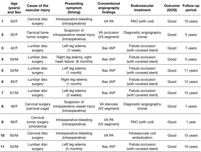
endovascular technique for all of our iatrogenic vascular injury patients with AVF due to a lumbar disc operation (Figure 1A, B).
For the cervical region, anterior cervical discectomy and spine fusion (ACDF) is the most common method for most cervical herniated discs (as in our patients #1, #8 and #10). Vertebral artery injury due to anterior cervical spine surgery is an uncommon complication with an incidence of 0.1-0.5% (14). Vertebral artery is 1-2 mm lateral to the joints of Luschka and fulsome lateral bone removal by high-speed drill may cause VA injury. Surgeons should be careful not to expose the dural root sleeve more than 5 mm when removing joints of Luschka. During lateral extension of central decompression procedures in the cephalad vertebra, the VA injury risk is also higher due to the increased inter-Luschka distance from C3 to C7. Because of the 3% incidence of anomalous tortuous VA, preoperative imaging is required in cases that need resection of Luschka joints (14,20). When intraoperative VA injury occurs, temporary hemostasis can be achieved by compression and
Table I: Summary of Patient Information Age (years) and Sex Cause of the vascular injury Presenting symptom (timing) Conventional angiography findings Endovascular
treatment Outcome(GOS) Follow-up period 1 63/F Cervical disc surgery Intraoperative bleeding (intraoperative) VA PA PAO (with coil) Good 10 years
2 62/F Cervical bone tumor surgery intraoperative vessel injury Suspicion of (intraoperative)
VA occlusion
(V3 segment) Diagnostic angiography (none) Good 5 years
3 42/F Lumbar disc surgery Left leg edema (1 week) Iliac AVF (with covered stent)Fistula occlusion Good 7 years
4 50/M Lumbar disc surgery heart failure (6 months)Right leg edema, right Iliac AVF (with covered stent)Fistula occlusion Good 5 years
5 50/M Lumbar disc surgery Left leg edema (1 month) Iliac AVF (with covered stent)Fistula occlusion Good 11 years
6 65/F Lumbar disc surgery Right leg edema (1 month) Iliac AVF (with covered stent)Fistula occlusion Good 12 years
7 67/M Lumbar disc surgery Left leg edema (2 weeks) İliac AVF (with covered stent)Fistula occlusion Good 15 years
8 46/F Cervical surgery (cervical cage) intraoperative vessel injury Suspicion of (intraoperative)
VA stenosis
(V2 segment) Diagnostic angiography (none) Good 7 years
9 66/F tumor surgery Cervical (chordoma)
Intraoperative bleeding
(intraoperative) (V2 segment)VA PA PAO (with coil) Good 1 year
10 65/M Cervical disc surgery Intraoperative bleeding (intraoperative) VA PA Intrasaccular coil embolization Good 15 years
11 52/M Lumbar disc surgery Left leg edema (2 months) Iliac AVF (with covered stent)Fistula occlusion Good 10 years
angiography was performed in two patients who suffered from stenosis/occlusion of VAs. These patients also did not have any complications during the follow-up period.
In our patients, the other type of surgery in the cervical region was chordoma surgery (patient #2 and #9). Chordoma, which is a low-grade malignant neoplasm, is a rare cancer that occurs in the bones of the skull and spine. It is thought to arise from remnants of the embryonic notochord, a rod-shaped, cartilage-like structure that serves as a scaffold for the formation of the spinal column. The primary treatment of this neoplasm is still surgery. This locally invasive neoplasm has a high rate of recurrence after treatment. Because of the high rate of recurrence, surgical treatment has become more aggressive in recent years, evolving from intralesional or partial excision to en-bloc resection. Because of the aggressiveness packing with cottonoids, bone wax or fibrillated collagen.
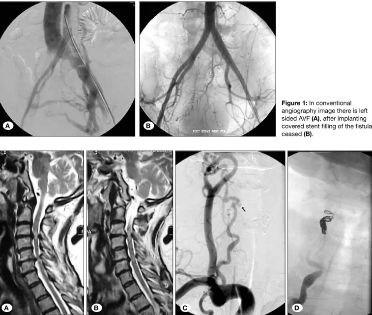
Direct exposure of the injured VA and suturing of the laceration can be achieved but this is not technically feasible. However, the use of endovascular techniques has been favored in the recent literature (11,20). Small pseudoaneurysms can be occluded with intraaneursymal coil implantation or with covered stents. Complicated lesions with those not having the possibility of saving the parent artery, after reviewing other VA and Wills Polygon, aneurysm and VA can be occluded (Parent Artery Occlusion “PAO”). The detachable coils are used as embolization devices in PAO. Three of our patients with VA PA had intraoperative bleeding (patient #1, #9 and #10). Two of those patients (patient #1 and #9) were treated with PAO with coil (Figure 2A-D) and the other patient (patient #10) was treated with intrasaccular coil embolization. During the follow-up period no patient re-bled. Only diagnostic conventional
Figure 1: In conventional angiography image there is left sided AVF (A), after implanting covered stent filling of the fistula ceased (B).
Figure 2: In sagittal T2-weighted MRIs in the cervical spinal region there are multiple level discal protrusions (A,B), in the conventional angiography image there is a PA in the right VA after the operation (C, arrow), parent artery occlusion with coil (D).
A B
of the tumor and the surgery, the complication rates are high. Major complications are VA injury, cerebrospinal fluid (CSF) leak, nerve palsies, and paralysis (12).
In the cervical region, most of the vascular injuries occur during the surgery and intraoperative bleeding would be mostly seen. If case of such injury, one has to urgently verify the type of vascular injury with imaging. Because of 24-hour availability, CTA would be the choice of imaging. In the lumbar region, due to the mechanism of vascular injury and anatomic reasons, intraoperative bleeding will not be detected. Surgeons must be alert in terms of peroperative unexpected hemodynamic changes to suspect possible vascular injury. If this vascular injury involves both the iliac artery and veins, then iatrogenic AVF may occur. This type of vascular injury usually presents late, after the patient is discharged. One should always consider the possibility of vascular injury after spinal operation as soon as there is a suspicion of vascular injury. After verification, immediate endovascular management would be a good option for patients who experience vascular injury during spinal surgery.
The limitation of our study was the limited patient population with spinal iatrogenic vascular injury. Future studies on spinal iatrogenic vascular injuries managed endovascularly with a larger patient series will help identify the effectiveness of endovascular treatment.
█
CONCLUSION
Iatrogenic vascular injuries due to spinal surgeries are rare but serious complications. Early diagnosis and management are crucial in such cases. Endovascular treatment is a good option with low mortality and morbidity rates.
█
REFERENCES
1. Aydin E, Gok M, Esenkaya A, Cinar C, Oran I: Endovascular management of iatrogenic vascular injury in the craniocervical region. Turk Neurosurg 28(1):72-78,2018
2. Baltacıoglu F, Cimsit NC, Cil BE, Cekirge S, Ispir S: Endovascular stent-graft applications in iatrogenic vascular injuries. Cardiovasc Intervent Radiol 26: 434-439, 2003 3. Beregi JP, Prat A, Willoteaux S, Vasseur MA, Boularand V,
Desmoucelle F: Covered stents in the treatment of peripheral arterial aneurysms: Procedural results and midterm follow-up. Cardiovasc Intervent Radiol 22: 13-19, 1999
4. Brewster DC, May ARL, Darling RC, Abbott WM, Moncure AC: Variable manifestations of vascular injury during lumbar disc surgery. Arch Surg 114: 1026-1030, 1979
5. Cil BE, Akmangit I, Peynircioglu B, Karcaaltincaba M, Cekirge S: Iatrogenic femoral arteriovenous fistula: Endovascular treatment with covered stent implantation and 4-year follow-up. Diagn Interv Radiol 12: 50-52, 2006
6. Fantini GA, Pappou IP, Girardi FP, Sandhu HS, Cammisa FP Jr: Major vascular injury during anterior lumbar spinal surgery: Incidence, risk factors, and management. Spine (Phila Pa 1976) 32(24): 2751-2758, 2007
7. Flouzat-Lachaniette CH, Delblond W, Poignard A, Allain J: Analysis of intraoperative difficulties and management of
operative complications in revision anterior exposure of the lumbar spine: A report of 25 consecutive cases. Eur Spine J 22(4): 766-774, 2013
8. Golfinos JG, Dickman CA, Zabramski JM, Sonntag VK, Spetzler RF: Repair of vertebral artery injury during anterior cervical decompression. Spine 19: 2552-2556, 1994
9. Goodkin R, Laska LL: Vascular and visceral injuries associated with lumbar disc surgery: Medicolegal implications. Surg Neurol 49: 358-372, 1998
10. Henry M, Amor M, Henry I, Klonaris C, Tzvetanov K, Buniet JM, Amicabile C, Drawin T: Percutaneous endovascular treatment of peripheral aneurysms. J Cardiovasc Surg (Torino) 41: 871-883, 2000
11. Inamasu J, Guiot BH: Vascular injury and complication in neurosurgical spine surgery. Acta Neurochir (Wien) 28: 239-247, 2006
12. Jiang L, Liu ZJ, Liu XG, Ma QJ, Wei F, Lv Y, Dang GT: Upper cervical spine chordoma of C2-C3. Eur Spine J 18: 293-300, 2009
13. Natali J: Forensic medical implications of vascular injuries in orthopedic surgery. J Mal Vasc 21(4): 206-215, 1996
14. Neo M, Fujibayashi S, Miyata M, Takamoto M, Nakamura T: Vertebral artery injury during cervical spine surgery: A survey of more than 5600 operations. Spine 33: 779-785, 2008 15. Papadoulas S, Konstantinou D, Kourea HP, Kritikos N,
Haftouras N, Tsolakis JA: Vascular injury complicating lumbar disc surgery. A systematic review. Eur J Vasc Endovasc Surg 24: 189-195, 2002
16. Parker SL, Amin AG, Santiago-Dieppa D, Liauw JA, Bydon A, Sciubba DM, Wolinsky JP, Gokaslan ZL, Witham TF: Incidence and clinical significance of vascular encroachment resulting from freehand placement of pedicle screws in the thoracic and lumbar spine: Analysis of 6816 consecutive screws. Spine (Phila Pa 1976) 39(8): 683-687, 2014
17. Parodi JC, Schonholz C, Ferreira LM, Bergan J: Endovascular stent-graft treatment of traumatic arterial lesions. Ann Vasc Surg 13: 121-129, 1999
18. Smith DW, Lawerence BD: Vascular complications of lumbar decompression, laminectomy and foraminotomy. Spine 16: 387-390, 1991
19. Smith MD, Emery SE, Dudley A, Murray KJ, Leventhal M: Vertebral artery injury during anterior decompression of the cervical spine. A retrospective review of ten patients. J Bone Joint Surg Br 75: 410–415, 1993
20. Suguawara T: Anterior cervical spine surgery for degenerative disease: A Review. Neurol Med Chir (Tokyo) 55: 540-546, 2015 21. Thalhammer C, Kirchherr AS, Uhlich F, Waigand J, Gross CM.
Postcatheterization pseudoaneurysms and arteriovenous fistulas: repair with percutaneous implantation of endovascular covered stents. Radiology 214: 127-131, 2000
22. Tonnessen BH: Iatrogenic injury from vascular access and endovascular procedures. Perspect Vasc Surg Endovasc Ther 23(2): 128-135, 2011
23. Veras LM, Pedraza-Gutierrez S, Castellanos J, Capellades J, Casamitjana J, Rovira-Canellas A: Vertebral artery occlusion after acute cervical spine trauma. Spine 25 (9): 1171-1177, 2000