ѵД' ^ 'Ч А *. . * - k Л і
А * ■* *■ ****
< ? 2
200
N Fl MUTATION ANALYSIS IN A CHILD HOMOZYGOUS
YO RM LH l MUTATION
A THESIS SUBMITTED TO
THE DEPARTMENT OF MOLECULAR BIOLOGY AND GENETICS
AND THE INSTITUTE OF ENGINEERING AND SCIENCE OF
BILKENT UNIVERSITY
IN PARTIAL FULFILLMENT OF THE REQUIREMENTS
FOR THE DEGREE OF MASTER OF SCIENCE
BY
HANI S. M. AL-OTAIBI
JULY 1999
I certify that I read this thesis and that in my opinion it is fully adequate, in
scope and in quality, as thesis for the degree of Master of Science.
Prof. Dr. Ash Tolun
I certify that I read this thesis and that in my opinion it is fully adequate, in
scope and in quality, as thesis for the degree of Master of Science.
___
Assist. Prof. Marie Ricciardone
I certify that I read this thesis and that in my opinion it is fully adequate, in
scope and in quality, as thesis for the degree of Master of Science.
Approved for Institute of Engineering and Science.
Prof.Dr. Mehmet Baray,
Director of Institute of Engineering and Science
ABSTRACT
NFl MUTATION ANALYSIS IN A CHILD HOMOZYGOUS
FOR MLHl MUTATION
Hani S. M. A]-Otaibi
M.S. in Molecular Biology and Genetics Supervisor: Assist. Prof. Marie D. Ricciardone
July 1999, 93 Pages
Hereditary nonpolyposis colorectal cancer (HNPCC) is a common autosomal dominant disease characterized by an inherited predisposition to early onset colorectal cancer and an increased risk o f certain other cancers. Cancer susceptibility is due to a heterozygous germ line mutation in one o f five mismatch repair (MMR) genes, with the majority o f mutations found in
M LHl
andMSH2.
This study focused on a child with a homozygous germ line mutation inMLHl
[C676T—>Arg226Stop] inherited from consanguineous parents (Ricciardoneef a i,
1999). This child presented with neurofibromatosis type 1 and hematological malignancy, instead o f the colorectal cancer phenotype usually seen in HNPCC individuals. This severe tumorigenic syndrome most likely resulted from a downstream mutation in theNFl
gene that could not be repaired due to complete absence o f DNA MMR activity. To confiiTn this hypothesis and possibly clarify the genetic mechanism involved in this phenotype, exons in the functional domaino(N F l
that contained mononucleotide or dinucleotide repeat sequences were analyzed by single strand conformation polymorphism (SSCP) and DNA sequence analysis for the presence o f mutations.SSCP analysis o f the child’s DNA identified altered mobility o f
NF'l
exon 22. Subsequent DNA sequence analysis revealed a heterozygous C3721T transition mutation.Taql
restriction digestion o f the exon fragment confirmed loss o f the restriction site, thus, verifying the sequencing results. The C3721T mutation results in substitution o f a stop codon for an arginine codon at position 1241. The resultingN Fl
gene product is truncated at the beginning o f the functional GTPase activation domain. A putative somatic mutation in the wild-type allele caused complete loss o f neurofibromin function. The resulting impaired regulation o f Ras*GTP signaling probably contributed to tumor development -- neurofibromatosis and hematological malignancy. ThisN Fl
mutation was not present in the mother, father or sibling, indicating that this was ade novo
mutation that occurred in early embryogenesis, mostÖZET
MLHİ MUTASYONU İÇİN HOMOZİGOT OLAN BİR ÇOCUKTA
NFI MUTASYON ANALİZİ
Hani S. M. Al-Otaibi
Moleküler Biyoloji ve Genetik Yüksek Lisans Tez Yöneticisi; Yardımcı Doçent Dr. Marie D. Ricciardone
Temmuz 1999, 93 Sayfa
Kalıtsal polipoz olmayan kolorektal kanser, erken yaşta ortaya çıkan, diğer kanser türlerinin görülme insidansını artıran ve otozomal dominant kalıtım özelliği gösteren bir hastalıktır. Kansere yatkınlık, çoğunluğu
MLHİ
veMSH2
genlerinde olmak üzere, beş yanlış eşleşme DNA tamir genlerinden herhangi birinde olanheterozigot eşey hücresi mutasyonundan kaynaklanmaktadır. Bu çalışma, bir akraba evliliğinden doğan ve homozigot eşey hücresinde
MLHİ
geni mutasyonu[C 676T ^A rg226Stop] taşıyan bir çocuk üzerine yoğunlaşmıştır. Bu çocukta, genellikle HNPCC bireylerinde görülen kolorektal kanser fenotipi yerine,
nörofibromatoz tip 1 ve hematolojik malignité görülmektedir. Bu ciddi tümorijenik sendrom, büyük olasılıkla, DN A MMR aktivitesi olmadığı için
NFI
geninde bulunan bir mutasyonun tamir edilememesi sonucu gelişmiştir. Bu hipotezi doğrulamak ve bu fenotipin oluşmasını etkileyen genetik mekanizmaya açıklık getirmek amacıyla, mononükleotid ya da dinükleotid tekrar dizileri içerenNL'V'm
fonksiyonel bölgesinde bulunan eksonlar tek iplikçikli yapısal çeşitlilik (SSCP) analizi ve DNA dizi analizi kullanılarak mutasyon varlığı açısından incelenmiştir.DNA'nın SSCP analizi sonucunda,
NLL
ekson 22'de farklılık tanımlanmış ve DNA dizi analizi sonucunda, bir heterozigot C3721T geçiş mutasyonu ortaya çıkarılmıştır. Ayrıca,Taq\
restriksiyon enzim analizi ile, bu eksondaki restriksiyon bölgesinin kaybolduğu saptanarak sekans sonuçları doğrulanmıştır. C3721Tmutasyonu, bir arjinin kodonunun bir stop kodonuna dönüşümüne yol açmakta ve elde edilen
NLL
gen ürünü, fonksiyonel GTPase aktivasyon bölgesini taşımamaktadır. Yabanıl aleldeki olası bir somatik mutasyon, nörofıbromin fonksiyonunun tamamen kaybolmasına sebep olmuştur. Böylece bozulan Ras*GTP sinyal regülasyonu, büyük bir olasılıkla tümör oluşumuna — nörofıbromatoza ve hematolojik maligniteye neden olmuştur.NFI
mutasyonunun anne, baba ve kardeşte olmayışı, bunun, erkenACKNOWLEDGEMENTS
First o f all, I would like to express my gratitude to my supervisor. Assist. P ro f Marie D. Ricciardone, for her kindness, advice, encouragement and all she gave me during my work. She taught me much, and I am thankfiil for knowing her.
Many thanks to P ro f Mehmet Öztürk for his valuable advice and
encouragement. I would like also to thank all my teachers at Bilkent: Tayfun, Uğur, Işık, Rengül, Günay, David, Can, Cengiz and Ergün.
For the one who stayed beside me, who cheered me up through my bad times, special thanks to Neslihani. Her support was o f a different kind and I am grateful to be with her.
^ Special thanks to Birsen, Hilal, Cemaliye and Gökçe for their assistance in the lab. They were there whenever I asked for help.
Many thanks to all my friends in the department: Gürol, Arzu, Tuba, Tolga Çağatay, Berna, Emre, Burcu, Esra, Aslı, Bilada, Gülayşe, Tolga Emre, Nuri,
Abdullah, Lutfıye, Sevim, Füsun and Tülay for providing me a good atmosphere and for making me feel like home.
TABLE OF CONTENTS
Page SIGNATURE P A G E ... ii A B STR A C T... iii Ö Z E T ... iv ACKNOW LEDGEMENTS... ... viTABLE OF CO N TEN TS... vii
LIST OF TABLES...x
LIST OF FIG U R E S... xi
1. IN TRO D U CTIO N ...1
1.1. Description o f TF3 K indred... 1
1.2. Colorectal C ancer...2
1.2.1. Sporadic Colorectal Cancer...3
1.2.2. Familial / Hereditary Colorectal C ancer... 4
1.3. Hereditary Non-Polyposis Colorectal Cancer (H N PC C )... 6
1.3.1. Incidence o f H N P C C ... 6
1.3.2. Microsatellite instability in H N PC C ... 7
1.3.3. Molecular genetics o f H N PC C ... 8
1.3.4. DNA Mismatch Repair... 10
1.3.4.1. DNA Mismatch Repair in Prokaryotes...11
1.3.5. M ouse Models for H N PC C ... 15
1.3.6. Mechanism o f Tumorigenesis in H N P C C ...16
1.4. Neurofibromatosis Type 1... 17
1.4.1. Incidence o f Neurofibromatosis Type 1 ... 18
1.4.2.
N Fl
Gene Structure...191.4.3.
N Fl
Gene Expression...201.4.3.1. Alternative Splicing...20
1.4.3.2. mRNA E diting... 22
1.4.4.
N Fl
Gene Product; N eurofibrom in...231.4.4.1. Neurofibromin S tructure... 24
1.4.4.2. Neurofibromin Functions... 26
1.4.4.2.1. Tumor Suppressor... 26
1.4.4.2.2. GTPase Activating Protein (G A P )...27
1.4.4.2.3. Other Functions... 29 1.4.5. M '7 Mutation R ate ... 29 1.4.5.1. Location o f Mutations in
N F l
...30 1.4.5.2. Types o f Mutations inN F l
... 31 1.5. Mutation Screening... 32 1.5.1. Protein Truncation T e s t... 32 1.5.2. R T -P C R ... 33 1.5.3. Heteroduplex Analysis (H A )... 331.5.4. Single Strand Conformation Polymorphism (S S C P )... 33
1.5.5. DNA Sequence Analysis... 35
1.5.6. Restriction Enzyme Analysis... 36
1.6. Hypothesis... 37
2. MATERIALS AND M ETHODS... 39
2.1. Patient Sam ples... 39
2.1.1. DNA Isolation... 40
2.1.2. Spectrophotometric Determination o f DNA Concentration...42
2.2. Polymerase Chain Reaction (P C R )...43
2.2.1. Primer Selection...43
2.2.2. PCR Conditions...43
2.3. Restriction Enzyme Cleavage o f PCR Products...45
2.4. Single Strand Conformation Polymorphism (S S C P )... 46
2.5. DNA Sequence Analysis... 49
2.6.
Taq\
Restriction Enzy^me Digestion...512.7. Allele Specific Amplification o f
M L H l
... 513. RESU LTS...53
3.1. Prioritization o f
NFI
Exons for Mutation D etection... 533.2. DNA Isolation... 55
3.3. Polymerase Chain Reaction Optimization... 57
3.4. Restriction Digestion
o iN F l
Exons 16 and 2 3 a ... 593.5. Single Strand Conformation Polymorphism (SSCP) A nalysis... 60
3.5.1. Conventional SSCP Protocols...61
3.5.2. Low pH SSCP Protocol... 64
3.6. DNA Sequence Analysis... 65
3.7. Mutation Verification by
Taql
Digestion...673.8. Allele-specific Amplification
o f M LH J
... 684. DISCUSSION...71
Perspectives... 76
LIST O F TABLES
Page
Table 1.1. Expression o f Alternative Transcripts... 21
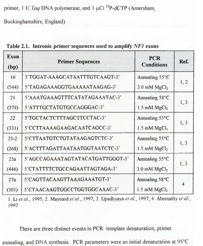
Table 2.1. Intronic primer sequences used to amplify
NFI
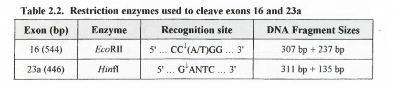
e x o n s... 44Table 2.2. Restriction enzymes used to cleave exons 16 and 2 3a... 45
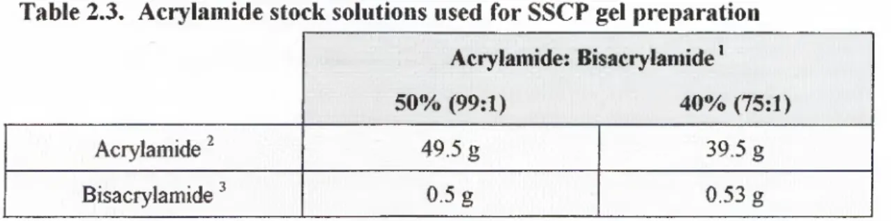
Table 2.3. Acrylamide stock solutions used for SSCP gel preparation... 46
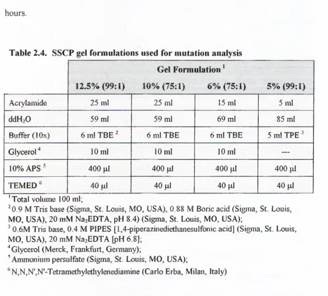
Table 2.4. SSCP gel formulations used for mutation analysis... 47
Table 2.5. SSCP protocols used for mutation detection... 48
Table 3.1.
NFl
exons containing microsatellite repeats...54Table 3.2.
NF I
exon priorities for mutation detection...55Table 3.3. Spectrophotometric analysis o f genomic DNA sam ples... 56
Table 3 .4. PCR parameters for priority-one e x o n s...58
LIST O F FIG URES
Page
Figure 1.1. Pedigree o f the HNPCC kindred... 2
Figure 1.2.
E. coU
MutHLS DNA mismatch repair system at replication fork... 12Figure 1.3. Model for eukaryotic mismatch recognition...13
Figure 1.4. Sequence alignment o f
GAP, NFl
andIRA I
gene products... 19Figure 1.5. N F1 -GRD interaction with R as...25
Figure 1.6. Regulation o f Ras-induced cell proliferation by neurofibromin... 28
Figure 1.7.
NFI
mutation m ap... ... 31Figure 3.1. Microsatellites in
NFI
coding regions... 53Figure 3.2. Agarose gel electrophoresis o f extracted genomic DNA samples...57
Figure 3.3.
NFI
exons amplified with optimized PCR p ro to co ls... 58Figure 3 .4. Restriction enzyme cleavage o fM '7 exon 16 by £ co R Il and
NFI
exon 23 a byHinfl
... 59.Figure 3.5. Analysis o f EcoRJl digestion o f
NFI
exon 16 andHinil
digestion o fNFJ
exon 2 3 a ... 60Figure 3.6. SSCP analysis o f the positive control CL 12826
{MLHI
exon 16, 1846-48 A A A G )... 62Figure 3.7. SSCP analysis o f
NFl
exon 2 2 ... 63Figure 3.8. SSCP analysis o f
NFl
exon 23-2 andNFl
exon 2 7 a ... 64Figure 3.9. Low pH SSCP analysis
oiN F I
exon 16 andNl·'!
exon 23 a... 65Figure 3.10. DNA sequence analysis o f
NFl
exon 22... 66Figure 3.11. Mutation verification by
Taql
restriction digest... 681. INTRODUCTION
1.1. Description of TF3 Kindred
TF3 is a large HNPCC kindred, which has been studied previously in our department (Ricciardone
ei al
., 1999). The pedigree meets the Amsterdam criteria (Lynchet a l,
1993) for HNPCC, which states that there should be (1) at least three relatives, one o f whom is a first-degree relative o f the other two, with histologically verified colorectal cancer; (2) at least two generations affected by colorectal cancer; and (3) at least one colorectal cancer diagnosed before the age o f 50. In this pedigree (Figure 1.1), 11 family members in three generations are afflicted with colorectal cancer and nine family members had early onset o f colorectal cancer. So, this pedigree is consistent with the dominant inheritance o f cancer susceptibility gene.A heterozygous C676T (Arg226Stop) mutation in
MLHl
exon 8 was found to segregate with the disease (Ricciardoneet a l,
1999). The consanguineous marriage between 111-5 and II1-6, both afflicted with colorectal cancer and both heterozygous for theMLHI
mutation, resulted in three offspring (IV-2, IV-3 and IV-4), who died at early age o f leukemia or lymphoma. Two o f the children (lV-3 and IV-4) displayed neurofibromatosis type 1 (N Fl). Analysis o f archival DNA samples revealed that both children were homozygous forthe M LH l
mutation.symbols indicate neurofibromatosis type 1. Numbers below symbols indicate age at diagnosis. (+/-): heterozygous MLHl mutation, (-/-): homozygous MLHl mutation (Ricciardone et ah, 1999).
Both children showed an unusual phenotype not previously observed in HNPCC kindreds - neurofibromatosis type 1 and early onset hematological
malignancy. A second, independent research study has identified an HNPCC kindred from North Africa with a similar characteristics (Wang
et a l,
1999). We havehypothesized that the neurofibromatosis might be due to a downstream mutation in the
N Fl
gene that resulted from a complete absence o f DNA M MR activity as a consequence o f the homozygous truncating mutation inMLHl.
To confirm this hypothesis and possibly clarify the genetic mechanism involved in this phenotype, we plan to analyze mononucleotide and dinucleotide repeat sequences in theNFl
coding regions for the presence o f mutations.1.2. Colorectal Cancer
Colorectal cancer is one o f the leading causes o f cancer mortality worldwide. About 50% o f the W estern population develops a colorectal polyp by the age o f 70,
Vogelstein, 1996). As a result, colorectal cancer is the second leading cause o f cancer death in the United States and first when smoking related cancers are excluded (Parker
et al.,
1996). Colorectal cancer develops as a result o f the pathologictransformation o f normal colonic epithelium to an adenomatous polyp and ultimately an invasive cancer. This transformation is a multistep process that occurs over decades and appears to require at least seven genetic events for completion (reviewed in Kinzler and Vogelstein, 1996).
1.2.1. Sporadic Colorectal Cancer
Over 90% o f colorectal cancers are sporadic or nonfamilial (Moslein
et al.,
1996). Colorectal cancer development is a multi-step process, driven by genetic alterations that confer a proliferative advantage on a specific cell. These genetic alterations occur in three classes o f genes (reviewed in Gryfe
et al.,
1997): (1) proto oncogenes, which when mutated act to promote cell growth; (2) tumor suppressor genes, which when mutated fail to regulate cell proliferation; and (3) DNA repair genes, which when mutated fail to ensure the fidelity o f DNA replication, leading to further mutations in proto-oncogenes and tumor suppressor genes.Several epidemiological studies have linked lifestyle factors, especially diet, to the increased incidence o f colorectal cancer. Lipids are thought to be among the critical dietary components because a higher rate o f colorectal cancer has been associated with diets containing large amounts o f red meat. Moreover, it has been shown that nonsteroidal anti-inflammatory drugs, that inhibit the cyclooxygenases that
metabolize the lipid arachidonic acid, can prevent tumor formation and even cause existing colorectal tumors to regress (reviewed in Kinzler and Vogelstein, 1996).
1.2.2. Familial / Hereditary Colorectal Cancer
Although cancer is a multi-step process, inheritance o f a single mutant gene can markedly increase an individual’s predisposition to colorectal cancer.
Epidemiological studies suggested that 15% o f colorectal cancers occur in dominantly inherited pattern (Kinzler and Vogelstein, 1996). Familial colorectal cancer
syndromes include familial adenomatous polyposis (FAP) and hereditary non polyposis colorectal cancer (HNPCC).
Familial adenomatous polyposis (FAP) is an autosomal dominantly inherited syndrome, characterized by development o f hundreds o f adenomatous colon polyps, some o f which progress to cancer at an average age o f 40 years (Kinzler and
Vogelstein, 1996). The syndrome is caused by a germline mutation o f the
AFC
(adenomatous polyposis coli) gene (Kinzler
et a i,
1991; Nishishoet ah,
1991; Grodenet al
., 1991; Joslynet al
., 1991) that has been mapped to chromosome position 5q21 (van der Luijtet al.,
1995). Patients with a germline mutation o fAFC
do not necessarily develop colorectal cancer; however, they have a higher risk
compared to the general population (Kinzler and Vogelstein, 1996). Tumor initiation requires somatic mutation and inactivation o f the w ild-type/4PC allele (Ichii
et a l,
1992; Levy
et al.,
1994) in agreement with Knudson's two-hit hypothesis for tumor suppressor genes (Knudson, 1993). FAP is almost completely penetrant but exhibits variable expression; some individuals exhibit polyps in the upper gastrointestinal tractand others develop malignancies in organs outside the gastrointestinal tract. FAP has an estimated frequency o f 1 in 10,000 in the general population (Gryfe
ei al.,
1997).The precise biological function o f the APC protein is not known. However, the APC protein can bind to and promote the degradation o f P-catenin (Rubinfeld
et
al.,
1993). MutationsoiAPC
result in the aberrant accumulation o f P-catenin, which then binds T cell factor-4 (Tcf-4) causing transcription activation o f unknown genes (Molenaarel a l,
1996; Behrensel a i,
1996). Recently, thec-MYC
oncogene was identified as one o f the downstream targets o f P-catenin /T cf (Heet a i,
1998). The resulting overexpression o fc-MYC
could promote neoplastic growth.Hereditary nonpolyposis colorectal cancer (HNPCC), also referred to as Lynch syndrome, is the second hereditary colorectal cancer syndrome. This syndrome is characterized by an autosomal dominant inherited predisposition to colorectal cancer that occurs at an early age (approximately 40-50 years old). Other cancers, such as endometrial, ovarian, small bowel, stomach, pancreas, ureter and renal pelvis may also be seen (Lynch
el al.,
1996). Tumors isolated from HNPCC patients are distinguished by microsatellite instability (Aaltonenel al.,
1993; Thibodeauet al.,
1993) — genome-wide insertions and deletions within simple tandem DNA repeats. About 70% o f HNPCC kindreds inherit a germline mutation in one o f the five genes involved in DNA mismatch repair (MMR), with the majority (95%) occurring in either A/L7/7
or MSH2
(Lynch and Smyrk, 1996).1.3. Hereditary Non-Polyposis Colorectal Cancer (HNPCC)
Knowledge o f genetics, coupled with a detailed family history, is necessary for diagnosis o f HNPCC and genetic counseling. Until recently, a patient's cancer risk status could be evaluated with, at the most, a 50% level o f confidence, based on the patient's HNPCC pedigree in accordance with the autosomal dominant mode o f genetic transmission o f the cancer trait. However, identification o f the germline mutations responsible for HNPCC now enables the theoretical determination o f genetic risk o f cancer during embryogenesis (Lynch and Smyrk, 1996). The phenotypic penetrance o f the deleterious genotype is approximately 85% to 90% (Lynch i?/a/., 1993; Lynch i?/a/., 1996).
1.3.1. Incidence of HNPCC
In an attempt to ascertain the true trequency o f HNPCC, Mecklin
et al.
(1995), designed a nonselective, prospective, multicenter study that assessed family background and other risk factors o f colorectal cancer over a 12-month period tor all new colorectal cancer patients in 10 hospitals in Finland. They found three (0.7%) cases o f verified HNPCC and seven (1.7%) cases o f suspected HNPCC. This study revealed a lower frequency o f HNPCC compared to previous investigations in Finland (Watson
et al.,
1994). The lowest known estimate o f HNPCC occurrence is 1%, which translates into 1500 new cases o f HNPCC annually in the United States (Lynchet a i,
1996). Estimates o f HNPCC incidence range as high as 5% or 7500 newoccurrences o f HNPCC in the United States each year (Lynch
et a i,
1996). These estimates indicate that HNPCC poses a major public health problem.1.3.2. Microsatellite instability in HNPCC
Microsatellite instability (MIN) is reflected as alterations in the patterns o f polymorphic, short, tandem repeat segments (microsatellites) dispersed throughout the genome. Since the initial description in 1993 o f microsatellite instability in
HNPCC (Aaltonen
et a i,
1993; Thibodeauei a l,
1993), MIN has been identified in a wide variety o f human cancers, both familial and sporadic (Peltomakiel al.,
1993b; Thibodeauel al.,
1998; Mosleinel al.,
1996; Dietmaierel al.,
1997; Tomlinsonel al.,
1996). However, it has been difficult to derive a consensus concerning the role o f MIN in human carcinogenesis. In the original descriptions o f M IN in HNPCC, it was reported that MIN was associated with mutations in certain DNA repair genes (Leach
e la l,
1993; Bronner i?/ur/., 1994; F ish e le /a/., 1993; Papadopoulos i?/a/., 1994;Nicolaides
el al.,
1994). Therefore, the assumption is that MIN reflects an underlying genomic instability, resulting from inactivation o f both alleles at a DNA repair gene locus.Heterozygous individuals have apparently normal DNA mismatch repair activity (Parsons
el al.,
1993). However, somatic mutation o f the wild-type allele results in loss o f this activity and accumulation o f characteristic mutations, such as single base mispairs and length alterations in homopolymeric tracts (Parsonsel al.,
1993). It has now been documented that the introduction o f a wild type copy o f a mismatch DNA repair gene into cell lines deficient for this gene can restore genomic stability o f microsatellites (Umar
el al.,
1997). This finding clearly indicates thatmutations in mismatch DNA repair genes constitute the underlying molecular defect responsible for MIN.
Arzimanoglou
et al.
(1998) has reviewed the medical literature and concluded that MIN, associated with inherited mutations o f the DNA mismatch repair genes, appears to characterize only the HNPCC / M uir-Torre family cancer syndromes and a subset o f young sporadic colorectal carcinoma patients. MIN in non-HNPCC tumors generally is not associated with somatic mutations in DN A mismatch repair genes. Loci o f individual chromosomes containing microsatellite markers demonstrating high MIN frequency may be linked to particular tumor types (tumor-specific MIN hot spots). MIN in tumors can be associated with early or late stages o f tumor progression and also has been found in non-tumor tissues (Arzimanoglouet a i,
1998).
1.3.3. Molecular genetics of HNPCC
Molecular genetic studies have identified germline mutations in an increasingly large number o f hereditary cancer syndromes, which has led to a good understanding o f the molecular pathogenesis o f the syndromes. Understanding o f molecular
pathogenesis o f HNPCC has resulted from three related lines o f investigation. The first was linkage analysis. Large kindreds from North America, Europe, and New Zealand were evaluated using microsatellite markers distributed throughout the genome. Tight linkage to either chromosome 2 p l6 or 3p21 was identified in individual families, providing unambiguous evidence that HNPCC was a simple Mendelian disease (Lindblom
et al.,
1993; Peltomakiet al.,
1993a).The second clue was found during attempts to demonstrate allelic losses with the 2 p l6 microsatellite markers linked with HNPCC susceptibility. Such losses are often found to be associated with tumor suppressor loci. In HNPCC tumors, however, new microsatellite alleles, not found in the patient's normal cells, were observed instead o f the expected allelic losses (Aaltonen
et a l,
1993). Those new alleles were evident in every dinucleotide and trinucleotide repeat examined, suggesting a genome-wide instability o f the replication or repair o f simple repeated sequences.The third clue was provided by investigators studying replication fidelity in unicellular organisms. These investigators recognized that the microsatellite instability observed in tumors was similar to that observed in bacteria harboring mutations in mismatch repair (MMR) genes such as
mutL
andmutS
(Strandet a l,
1993; Wierdl
et a l,
1996). Analogous experiments in yeast showed thatmicrosatellites observed to be unstable in HNPCC patients were equally unstable in yeast with defective MMR genes and it was specifically hypothesized that HNPCC was caused by hereditary mutations o f human homologues o f
mutS
ormutL
(Aaltonene ta l,
1993; Strand e?/a/., 1993).These clues stimulated a search for human homologues likely to participate in MMR. That a
mutS
homologue might be involved in the form o f HNPCC linked to chromosome 2 was suggested by the finding that one such homologue(MSH2)
was located on chromosome 2p (Fishelet a l,
1993). Direct evidence thatMAW2 was involved in the disease was provided by the identification o f germline mutations inHNPCC kindreds (Leach
et a l,
1993). This observation was soon followed by the identification o f mutations in the humanmutL
homologueMLHI
(Bronneret a l,
1994; Papadopoulos
et a i,
1994). Further search for other homologues uncovered additionalmiitL
homologues(PMSI
andPMS2)
that were found to be mutated in the germline o f HNPCC kindreds (Nicolaideset a l,
1994). Later, twonmtS
homologues(MSH3
andMSH6)
were identified (Drummondet a l,
1995; Palomboet a l,
1995).Although somatic mutations in
MSH3
andMSH6
were detected in HNPCC cancers (Risingeret a l,
1996; Akiyamaet a l,
1997b), these genes were not mutated in the germline o f typical HNPCC kindreds. However, a germline mutation has been found in an atypical HNPCC kindred (Akiyamaet a l,
1997a). Mutations in two human MMR genes(MSH2 and MLHI)
account for the majority (>95%) o f HNPCC kindreds (Papadopoulos and Lindblom, 1997).1.3.4. DNA Mismatch Repair
DNA mismatch repair is a strand-specific process involving recognition o f noncomplementary Watson-Crick base pairs (Lahue
et a l,
1989). Mismatched base pairs in DNA can arise by several processes (reviewed in Kolodner, 1996). A significant source o f mismatched bases is DNA replication errors. Occasionally, an incorrect nucleotide is incorporated into the DNA strand being synthesized. While the majority o f these misincorporations are excised by the DNA polymerase proofreading 3'-5' exonuclease, approximately 1 in 10‘^ errors remain. The DNA mismatch repair system can repair approximately 99.5% o f the mutations that escape proofreading, thus, decreasing the error rate to 1 in lO'^ base pairs (Schaaper, 1993; Lahueet a l.
1989). In this case, the correct base is located in the parental strand o f the newly replicated DNA and correction o f the mismatch helps maintain the fidelity o f the genetic information. DNA polymerase slippage, another source o f errors, usually occurs during replication o f homopolymeric nucleotide tracts and results in small insertions or deletions (Trinh and Sinden, 1991). A third source o f mismatched nucleotides is genetic recombination between two DNA sequences that results in a heteroduplex sequence (reviewed in Fishel, 1998).
I.3.4.I. DNA Mismatch Repair in Prokaryotes
The most extensively characterized mismatch repair system is the
Escherichia
coli
MutHLS system, which repairs a broad spectrum o f mispaired bases and has been reconstituted with purified enzymes (Lahueet ah,
1989; Kolodner, 1996). The MutHLS repair pathway is uniquely suited to repair DNA replication errors (Modrich, 1991; Modrich, 1994; reviewed in Kolodner, 1995). Normally, DNA inE. coli
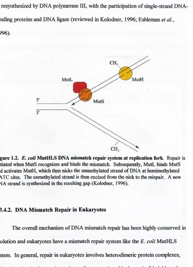
is methylated at GATC sites by the Dam methylase. However, after DNA replication the daughter strand is transiently unmethylated. The MutHLS system utilizes this modification asymmetry to direct the repair to the unmethylated strand via the activity and involvement o f several proteins.Repair is initiated by the recognition and binding o f MutS to a mismatched DNA base pair. MutL subsequently binds to MutS and activates MutH, which then nicks the unmethylated strand o f DNA immediately 5' to the guanine residue o f a hemi-methylated GATC site (Figure 1.2). The incised strand is then displaced by DNA helicase II and excised from the nick to the mispaired base by one o f the single
stranded DNA exonucleases (Exonuclease I [3' exonuclease activity]. Exonuclease VII [both 3' and 5' exonuclease activities] or R eel exonuclease [5' exonuclease activity]) depending on whether the nicked is 5' or 3' to the mispair. The DNA strand is resynthesized by DNA polymerase III, with the participation o f single-strand DNA- binding proteins and DNA ligase (reviewed in Kolodner, 1996; Eshleman
et a i,
1996).
Figure 1.2.
E. coli
MutHLS DNA mismatch repair system at replication fork. Repair is initiated when MutS recognizes and binds the mismatch. Subsequently, MutL binds MutS and activates MutH, which then nicks the unmethylated strand of DNA at hemimethylated GATC sites. The unmethylated strand is then excised from the nick to the mispair. A new DNA strand is synthesized in the resulting gap (Kolodner, 1996).I.3.4.2. DNA Mismatch Repair in Eukaryotes
The overall mechanism o f DNA mismatch repair has been highly conserved in evolution and eukaryotes have a mismatch repair system like the
E. coli
MutHLS system. In general, repair in eukaryotes involves heterodimeric protein complexes, rather than the single proteins or homodimers employed by bacteria (Lindahlet a l.
1997). The initial steps o f correction can be classed as primary and secondary recognition events (Lindahl
et a i,
1997). Primary recognition and binding o f mismatched DNA is carried out by homologues o f theE. coli
MutS protein, MSH2, MSH6 (GTBP) and MSH3, which can associate to form two different heterodimers (Figure 1.3). The M utS a heterodimer [MSH2*MSH6] recognizes single base mispairs, single base loops and two base loops in repeated dinucleotide sequences. The MutSP heterodimer [MSH2*MSH3] preferentially binds two, three and four base loops. a. .G. T'Primary Recognition
-
__ m ]____
GT GTC GTACSecondary Recognition
hMutLa
Removal and Restoration
C. G* T ■T - ■ A-•OT · CA ■OT ■CA ■OTC ■ CAG GTAC CATG Figure 1.3. Model for eukaryotic mismatch recognition, a. Primary recognition: M utSa heterodimer preferentially recognizes single base mispairs, single base loops, and two base loops in repeated dinucleotide sequences, while MutSp heterodimer preferentially binds two, three and four base loops,
b.
Secondary recognition: M utLa heterodimer is recruited byM u tS a mismatch recognition is regulated by adenosine nucleotide binding that acts as a molecular switch (Gradia
et a l,
1997; Gradiaet a i,
1999). The M utSa complex is ON (binds mismatched nucleotides) in the ADP-bound form and OFF in the ATP-bound form. This ADP —>^ATP exchange converts the M utS a complex into a “sliding clamp” that diffuses along the DN A. Stochastic loading o f multiple M utSa sliding clamps onto mismatch-containing DNA leads to activation o f the repair machinery (Gradia a/., 1999).The secondary recognition event involves the M utL a heterodimer, which is composed o f two homologues o f the
E. coli
MutL protein, M LH 1 *PMS2. The M utL a heterodimer binds both the M utSa-D N A and M utSp-DNA complexes (Figure 1.3). Cells with inactivating mutations in either o f these components are completely mismatch repair-deficient. Following mismatch recognition, the DNA strand with the incorrect base is excised and new DNA synthesized by DNA polymerase using the nonmutated strand as a template. Finally, DNA ligase seals the gaps and completes repair.Eukaryotic DN A is not methylated at GATC sites and no eukaryotic MutH homologue has been identified, so some other method o f strand discrimination must exist. One hypothesis is that gaps between the Okazaki fragments on the daughter strand may specify the strand for mismatch repair (Boland, 1998). The DNA replication protein, proliferating cell nuclear antigen (PCNA), has been shown to associate with M LHl and MSH2 and participate in an early step o f mismatch repair, possibly strand discrimination (Umar
et a i,
1996). Recently, researchers cloned ahuman gene encoding a protein that complexes with M L H l, binds to methyl-CpG- containing DNA, and displays endonuclease activity (Bellacosa
et a i,
1999). Deletion mutants lacking the methyl-CpG-binding domain exhibited microsatellite instability, suggesting that cytosine méthylation may have a role in human DNA MMR and that this protein could be a eukaryotic homologue o f the MutH endonuclease.1.3.5. Mouse Models for HNPCC
Loss o f DNA MMR is a rate-limiting step in the etiology o f tumors associated with HNPCC in humans (Lynch and Smyrk, 1996). Mutations
m M LH l màM SH2
account for the majority o f HNPCC kindreds with germ line mutations (Bronner
et
a l,
1994;Fishel^/uf/., 1993; Leach e /a /., 1993; Papadopoulos e /a /., 1994;Nicolaides
et a i,
1994). Studies using mouse models have been designed to define the role o f individual MMR genes in cancer susceptibility. They aim to address, first, whether constitutional DNA MMR deficiency in mice results in high levels o f DNA replication errorsin vivo
and, second, whether the high level o f spontaneous mutation correlates with spontaneous tumor development (Reitmairet a l,
1996; Prollaet a l,
1998; de Wind
et a l,
1998; Edelmannet a l,
1999).In a study using Ms7i2-deficient mice (de Wind
et a l,
1998), it was observed that allMsh2'''
mice died before 1 year o f age. Most o f those animals had succumbed to lymphoma (mostly o f T cell origin) before 30 weeks (Reitmairet a l,
1996; de Windet a l,
1998). M i/ti-deficient mice exposed to the exogenous mutagen ethyl- nitroso-urea (ENU) succumbed to lymphoma much earlier (14 weeks) than wild-type control mice (27 weeks). The early onset and 100% incidence o f tumorigenesis inMs’/?2-deficient mice corroborated the increased risk for MMR-deficient cells to undergo oncogenic transformation (de Wind
et a l,
1998). M//?7-deficient mice also showed high levels o f microsatellite instability and developed lymphomas, intestinal adenomas and sarcomas (Prollaet al.,
1998; Edelmannet a l,
1999), similar toMsh2-
deficient mice.
Pms2-
and Pw75/-deficient mice display a different pattern o f tumordevelopment.
Pms2'''
mice showed frequent alterations in both mononucleotide and dinucleotide repeats (Prollaet al.,
1998; Edelmannet a l,
1999) and succumbed to lymphoma and sarcoma, but did not develop adenomas or adenocarcinomas. On the other hand, mice deficient for thePmsl
gene showed only a small effect on mutations in mononucleotide repeats, did not show any defects in dinucleotide repeats, and did not develop tumors (Prollaet al.,
1998).1.3.6. Mechanism of Tumorigenesis in HNPCC
Defects o f MMR genes in tumors from HNPCC patients lead to a genetic instability in which short repeated sequences (microsatellites) are characteristically altered (Aaltonen
et al.,
1993; Thibodeauet al.,
1993; Peltomakiet a l,
1993b; Lindblomet al.,
1993). This instability is indicative o f a high mutation rate, which is believed to result in a rapid accumulation o f somatic mutations in genes that direct pathways involved in tumor progression. Mutations in critical genes are accelerated and HNPCC patients develop carcinomas at an average age o f 42, more than 20 years earlier than the general population (Lynchet al.,
1993).Remarkably, the tissue distribution o f tumorigenesis in HNPCC patients is limited to the colon, even though DNA M MR genes have a ubiquitous role in limiting mutations. This tissue specificity may be related to the fact that M LH l and MSH2 proteins are highly expressed in the epithelium o f the digestive tract (Fink
et al.,
1997). Defective DNA MMR in such a highly proliferative tissue could lead to more rapid accumulation o f mutations in those genes with mono- or di-nucleotide repeats (de Wind
et al.,
1998). Such frameshift mutations, associated with alterations in normal mismatch repair, have been reported in several downstream genes, includingTGFpRII
(Markowitzet a l,
1995),IGFIIR
(Souzaet a l,
1996),Β Α Χ
(Rampinoet
a l,
1997) andM.S773m áM SH ó
(Yamamotoé ta l,
1998).1.4. Neurofibromatosis Type 1
Neurofibromatosis type 1 (N Fl), also known as von-Recklinghausen's disease or peripheral neurofibromatosis, is a common autosomal dominant disorder. The hallmark o f N Fl is the presence o f multiple benign cutaneous neurofibromas consisting o f a mixture o f Schwann cells, fibroblasts, and mast cells (Lott and Richardson, 1981). Other clinical features include café au lait spots, freckling o f the axilla and groin, and pigmented nodules o f melanocytic origin in the iris, known as Lisch nodules. While these are the most commonly noted signs in N F l, the disease is characterized by a variable number o f diverse pathologies throughout the body, including cutaneous, osseous, hematological, developmental, and nervous system abnormalities (Riccardi, 1992). Despite recent advances in the understanding o f the molecular basis o f N F l, these clinical criteria continue to be the most reliable means
for making the diagnosis (Grifa
el al.,
1995). The two major life-threatening complications o f N Fl are hypertension and a significantly higher rate o f malignancy (Zoller cifa/., 1995; Sorensen a/., 1986).1.4.1. Incidence of Neurofibromatosis Type 1
Several studies were carried out in an attempt to ascertain the frequency o f N F l. Littler and M orton (1990) reviewed data from four studies and found a carrier incidence for N Fl at birth o f 1 in 2500. The gene frequency was estimated to be 0.0002. New mutations were found to be responsible for approximately 56% o f cases. Lazaro
el al.
(1994) gave the incidence o f N Fl as approximately 1 in 3,500 and estimated that about half o f NFl cases are due to new mutations. Gartyet al.
(1994) found an unusually high frequency o f NFl in young Israeli adults. They surveyed 374,440 17-year-old Jewish recruits for military service and found 390 with signs and symptoms o f N Fl — an incidence 2 to 5 times greater than previously reported. In this group, N Fl was more common in recruits whose parents were o f North African and Asian origin (1.81 per 1,000 and 0.95 per 1,000, respectively) and less common in those o f European and North American origin (0.64 per 1,000). All o f these differences were statistically significant. The epidemiologists suggested that the increased incidence o f N Fl in this group might be due to advanced age o f the recruits’ parents and/or a founder effect.
1.4.2. N F l Gene Structure
The
NFl
gene was isolated by positional eloning (Wallaceet a i,
1990; Cawthonet al.,
1990; Viskochilet aL,
1990) and maps to chromosome region17ql 1.2 (van Tuinen
et a l,
1987). It consists o f at least 59 exons spanning over 350 kb o f genomic DNA (Wallaceet a l,
1990; Liet a l,
1995; Goldberg and Collins,1991). In the eentral region o f neurofibromin
{NFl
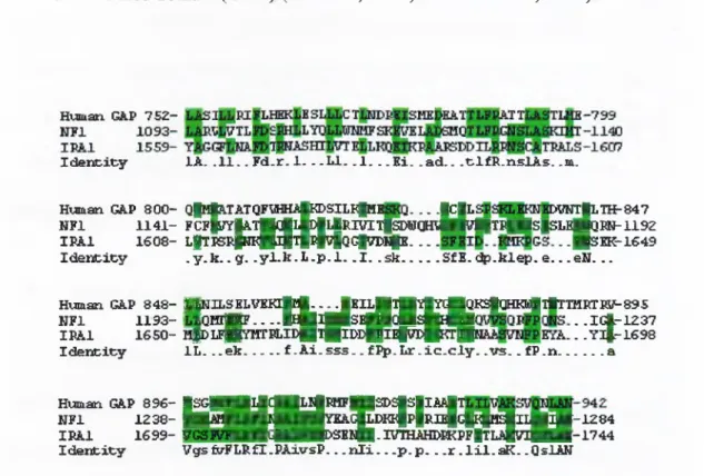
gene product) there is a 360 amino acid region, encompassing exons 21-27a, that shows high homology (Figure1.4) to the catalytic domain o f the mammalian GTPase activating protein (GAP) (Xu
et a l,
1990; Ballesteret a l,
1990). This region o f theNFl
gene product is called theN Fl GAP-related domain (GRD) (Xu
et a l,
1990; Ballesteret a l,
1990).Human GA.P 75Z
-I d e n c x c y lA . . 1 1 . . F d .r . 1. . . L I . . 1 . . .K i . . a d . . . t l f R . n s l A s . .m.
- - J C iL >VHT|LTH-847 Human GAP 8 0 0 - Q|Hfe.TATQFlH№ ^8IL^pIE
N F l 1 1 4 1 - F C F |^ |A T |||G |l№ |lJ R B rtlp D N Q m iH ta |T P iH S |S L E |n 3 R N -1 1 9 Z IP A l 1 6 0 8 - L |T P S I # I l4 ||4 4 4 ^ Q C f f n > l№ ____. .■ s E K -1 6 4 9
I d e n t i f y . y . k . . g. . y l . b . L . p . l . . 1 . . s k ... SfE . d p .k le p . e ___e N . . .
Hviman GAP 8 4 8 - IjffiH ILSBLV BK ljM .^^jK IlJglgYlYdBQ K SlaH ii^llT IH R TSlf-O gS N F l 1 1 9 3 - ^ Q B l |j № .. . . U | 4 P S E | a « & | | ( M . .IG |.-1Z 37 IP A l 1 6 5 0 - B fD L F Q p m B L ID B T fllD D p p i^ C iB H T llj^ ^ . . y i f - 1 6 9 8 I d e n c ic y IL . . . e k ... f . A i . s s s . . f ^ . L r . i c . c l y . . v s . . f P .n ...a Human GAP 8 9 6 - |S G H |[ |d ( ||| |^ R H F ||S D S |S |X ^ N F l 1 2 3 8 - 1 | | 5 ^ B B i^ M I ™ ^ ^ P K I 1p|rIB 1G P 1k^ I I B ^ -1 2 8 4 IP A l 1 6 9 9 - p 3 K i v H v H B > S E l 4 i - t V I E N ^ P K P F № L A |v i B H r - t ' 7 4 4 I d e n t i t y V gs& 'F L R fl.P A xvsP .. . n i i — p . p . . .r . l i l . a K . .QslAN
Figure 1.4. Sequence alignment of
GAP, NFl
andIR41
gene products. Shown is the region (190 residues) with higliest homology between the human GAPa, NFl, and yeast IRAl proteins. Gaps were introduced to allow tlie best alignment. Aligned identical residues are shaded. Consensus sequence summarizes conserved residues; upper case letters indicate residues identical in all three proteins, and lower case residues indicate residues shared in any two of tlie tliree proteins (Xu et a l,
1990).Three genes,
EVI2A, EVI2B,
andOMGP,
transcribed in the opposite orientation toNFI,
are embedded within intron 27 (Cawthonet a i,
1991). A K3 pseudogene, transcribed in the same orientation toN F l,
lies in intron 37 (Xuet al.,
1992),
1.4.3. NFI Gene Expression
NFI
is transcribed as an mRNA o f 11 -13 kb. This transcript encodes a protein (neurofibromin) o f 2818 amino acids with an estimated molecular weight o f 327 kDa (Wallaceei a i,
1990).NFI
is widely expressed in a variety o f human and mammalian tissues (Danglotet a i,
1995; Marchuket a i,
1991; Suzukiet a i,
1991; Takahashiet a i,
1994).NFl
gene expression is regulated by three differentmechanisms, including alternative splicing o f the mRNA transcript, mRNA editing, and unequal allelic expression (reviewed in Skuse and Cappione, 1997). Aberrations
NFI
RNA processing might regulate intracellular neurofibromin levels and, thus, explain the great variation in expressivity o f disease traits.1.4.3.1. Alternative Splicing
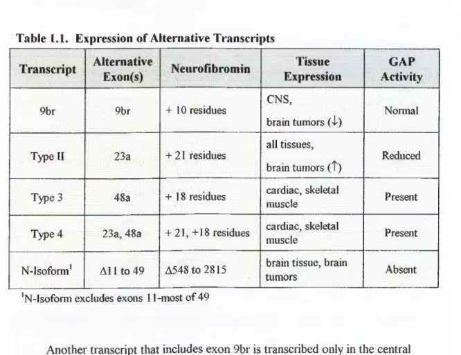
The multiple
NFI
transcripts are summarized in Table 1.1 (Skuse and Cappione, 1997). The differentNFI
isoforms can be placed into two main groups: first, the transcripts differing in GRD (type I, type II, and type 4) and second, the transcripts differing in other coding regions. The two best-characterized transcripts differ by the inclusion or exclusion o f exon 23a within theNFI
GRD. The type I transcript excludes exon 23a, while the type II transcript includes exon 23a (63 bp)and introduces 21 amino acids into the resulting neurofibromin. Both transcripts are expressed in normal tissues; however, in brain tumors, there is an increased expression o f the type II transcript (Suzuki
et a l,
1991; Flatten et al., 1996).Table 1.1. Expression of Alternative Transcripts
Transcript
Alternative
Exon(s)
NeuroBbromin
Tissue
Expression
GAP
Activity
9br 9br + 10 residues CNS, brain tumors (n1^) NomialType
II
23a + 21 residuesall tissues, brain tumors ( t)
Reduced
Type 3 48a + 18 residues cardiac, skeletal
muscle Present
Type 4 23a, 48a + 21, +18 residues cardiac, skeletal
muscle Present
N-Isoform* A ll t o 49 A548to2815 brain tissue, brain
tumors Absent
'N-lsofomi excludes exons 11-most of 49
Another transcript that includes exon 9br is transcribed only in the central nervous system. In brain tumors, its expression is reduced (Danglot
et al.,
1995). Flatten and coworkers reported the absence o f the 9br transcript in optic nerve astrocytomas as well (Flattenet al.,
1996).The type 3 transcript includes exon 48a and is expressed in cardiac and
skeletal muscles (Gutmann
et al.,
1993). The type 4 transcript includes both exon 23a and exon 48a (Gutmannet al.,
1995) and is also expressed in skeletal and cardiacmuscles. The N-isoform excludes exon 11 and most o f exon 49; it is expressed both in normal brain and in brain tumors (Takahashi
et a l,
1994).I.4.3.2. mRNA Editing
In addition to the expression o f alternative transcripts, the
NFl
mRNA is a subject to another mechanism o f RNA processing, namely mRNA editing. Editing is a form o f post-transcriptional modifications by which the sequence o f the mRNA is changed from that prescribed by the encoding DNA. These changes in the mRNA sequence may involve either base modifications or base substitutions (Smith and Sowden, 1996; Ashkenas, 1997).Base modification mRNA editing involves chemical modification o f existing nucleotides to convert them into different ones. For example, the cytidine at position 3916 in the
NFl
mRNA transcript maybe deaminated to uracil (Skuseei al.,
1996). This modification introduces an in-frame stop codon into the 5' portion o f the N Fl GRD, which is expected to result in a truncated neurofibromin completely lacking GTPase activating function. Thus, mRNA editing can result in loss o f tumor suppressor activity without the involvement o f mutations in theNFl
gene itself.Base substitution mRNA editing involves the addition, deletion, or
replacement o f specific nucleotides within the target transcript. Site-specific editing o f the
NFl
transcript was identified by sequence homology o f a mooring sequence inNFl
to the m otif responsible for editing in the apolipoprotein B mRNA (Skuseet al.,
consists o f three motifs: regulator, spacer, and mooring sequence. The mooring sequence was shown to be the critical element for site-specific editing (Backus and Smith, 1991; Skuse
ei al.,
1996). The editing process involves the assembly o f an “editosome” composed o f two RNA binding proteins and a cytidine deaminase at the editing site. The assembled editosome chemically converts cytidine to uracil via deamination.Cappione and coworkers (1997) investigated whether elevated levels
oiN F I
mRNA editing could inactivate the
NFl
tumor-suppressor fimction by comparing the levels o f editedNFl
mRNA in tumor and nontumor tissue samples removed from the same N F 1 patients. Tumor tissue contained higher levels o f editedNFl
mRNA than normal tissue from the same individual. Comparison o f nontumor tissue from NFl individuals with tissue from normal individuals revealed no significant difference in the level o fNFl
editing. These results suggest that low levels o fNFl
mRNA editing may appropriately regulateNF'l
gene expression but that high levels o f mRN A editing may contribute to the development o f benign neurofibromas and possible progression toward malignant tumors (Cappioneet al.^
1997). Thus, mRNA editing provides another level by which gene expression can be regulated and the resulting protein diversity can be expanded.1.4.4. NFl Gene Product: Neurofibromin
Using antibodies raised against both fiasion proteins and synthetic peptides, a specific protein o f about 250 kDa was identified by both immunoprécipitation and immunoblotting (Gutmann
ei a i,
1991). The protein was detected in human, rat, andmice tissues and appears to be expressed in all tissues. Based on the observed homology between the
NFl
gene product and members o f the GTPase-activating protein (GAP) superfamily, the name NFl-GAP-related protein (N Fl-G RP) was suggested.DeClue and coworkers (1991) immunized rabbits with a bacterially
synthesized peptide corresponding to the GAP-related domain o f N Fl (NFl-GRD). In HeLa cell lysates, these antisera detected a 280-kDa protein, corresponding to the
NFl
gene product. Neurofibromin was present in a large molecular mass complex in fibroblast and Schwannoma cell lines and appeared to associate with a very large protein (400-500 kDa) in both cell lines.1.4.4.1. Neurofibromin Structure
Neurofibromin, a 250 kDa hydrophilic protein, consists o f 2818 amino acids
(Xu
et a l,
1990). Several studied have shown that neurofibromin is structurallyrelated to the Ira (inhibitors o f Ras activators) proteins o f yeast and to the G AP protein o f mammalian cells (Xu
et a i,
1990; Buchberget al.,
1990). Other studies showed the functional relationships between these proteins (Ballesteret a i,
1990). The high homology between neurofibromin and GAP is limited to the catalytic domain; however, the homology between neurofibromin and Ira l/ Ira2 extendsbeyond the catalytic domain, suggesting that these structurally conserved regions have conserved biological functions as well (Ballester
et a l,
1990).The neurofibromin domain encoded by exons 21-27b shows strong homology to the GTPase-activating proteins (GAP) superfamily (Scheffzek
et a l,
1998). This domain is referred to as the functional GAP-related domain (N Fl-G R D ) and thus far, represents the only defined segment o f neurofibromin. X-ray crystallography o f the N Fl-G R D catalytic fragment comprising residues 1198-1530 (N Fl-333) revealed N Fl-G R D as a helical protein (Figure 1.5) that structurally resembles thecorresponding fragment derived from pl20G A P (GAP-334) (Scheffzek
et al.,
1998).Figure 1.5. NFl-GRD interaction with Ras.
Hypothetical complex between Ras (above) andNFl-333
(below) modeled according to the crystal structure of the Ras-GAP complex. Point mutations that affect the interaction with Ras are indicated by small spheres.(Scheffzek et a/., 1998)
This helical domain consists o f two domains: a central domain (N Fl J comprised o f segments 1253-1304,1331-1403 and 1412-1463, and an external
domain (NFlex) comprised o f segments 1206-1252, 1485-1503 and 1514-1530. A groove on the surface o f NFU has been identified as the Ras-binding groove based on structural comparisons o f GAP-334 and N Fl-333. This central domain represents the minimum catalytic domain o f the Ras-GAP modules (Scheffzek
et al.,
1998).I.4.4.2. Neuroflbromin Functions
Neurofibromin appears to act as a tumor suppressor since loss o f heterozygosity at the
NFl
locus has been observed in DNA isolated from N Fl patients (Sawadaet a i,
1996). The identification o f a domain with structural homology to the mammalian GTPase activating protein pl20G A P (Ballesteret a l,
1990) has helped in understanding the function o f the
W l
gene product.1.4.4.2.1. Tnmor Suppressor
N Fl patients have multiple benign tumors, which are predisposed to malignant transformation. The presence o f a domain in neurofibromin with high homology to the catalytic domain o f mammalian GAP and yeast Iral/Ira2 proteins, which can downregulate p2T“ activity, suggested that
F Fl
might function as a tumor suppressor gene. Loss o f heterozygosity observed inNFl
appears to confirm neurofibromin’s role as a tumor suppressor (Sawadaet al.,
1996).Over 80% o f germline mutations appear to result in severe truncation o f neurofibromin (reviewed in Shen
et al.,
1996). Skuse and Cappione (1997) reviewed the possible molecular basis for the wide clinical variability observed in N Fl, even among affected members o f the same family (Husonet al
., 1989) and suggested thatalternative splicing and mRNA editing may be involved. They proposed that the classical 2-hit model for tumor suppressor inactivation (Knudson, 1993) be expanded to include post-transcriptional mechanisms that regulate
NFl
gene expression,reasoning that aberrations in these mechanisms may play a role in the observed clinical variability (Skuse and Cappione, 1997).
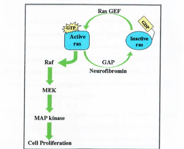
I.4.4.2.2. GTPase Activating Protein (GAP)
The GAP-related domain (GRD) encoded by exons 20-27b is the only region o f neurofibromin to which a biologic function has been ascribed. Biochemical analysis has shown that the GAP-related domain o f neurofibromin can accelerate the
conversion o f the active GTP-bound Ras to inactive GDP-bound Ras and thus, downregulate Ras activity (Shen
el a i,
1996).Ras acts as a molecular switch (Figure 1.6): switched ON when bound to GTP and SAvitched OFF when bound to GDP. This switch involves conformational changes in Ras that are controlled by two classes o f enzymes, the guanine nucleotide exchange factors (GEF) and the GTPase activating proteins (GAP). GEF proteins bind Ras and promote the dissociation o f GDP, thus, allowing Ras to bind GTP and switch on. GAP proteins, such as neurofibromin, accelerate the slow intrinsic GTPase activity o f Ras-GTP, switching Ras off.
In its active form, Ras triggers a kinase signal cascade first binding Raf, a serine threonine kinase, which phosphorylâtes and activates MEK, a tyrosine /serine protein kinase, that phosphorylâtes and activates MAP kinase. This MAP kinase
phosphorylâtes many different proteins, including transcription factors, and regulates expression o f cell cycle and differentiation proteins.
Figure 1.6. Regulation of Ras-induced cell proliferation by neurofibromin.
Neurofibromin downregulates Ras activity by catalyzing the conversion of active Ras»GTP to inactive Ras»GDP. In this model, loss of neurofibromin would result in high levels of
activated Ras»GTP, increased cell proliferation and tumor formation.
Two models involving neurofibromin and Ras have been proposed to explain the various clinical symptoms o f N Fl (McCormick, 1995). In the upstream
regulatory model, loss o f neurofibromin would result in decreased GAP activity, which would lead to high levels o f activated Ras*GTP and associated increased cell proliferation and tumor formation. In the downstream model, neurofibromin acts as
an effector o f Ras*GTP in signaling pathways controlling cell proliferation or differentiation.
I.4.4.2.3. Other Functions
The GTPase-activating activity o f both the N Fl-G R D peptide and the full- length neurofibromin can be inhibited by tubulin binding (Bollag
el ciL,
1993). The neurofibromin - tubulin interaction was demonstrated by their copurification. Tubulin binding determinants were localized to an 80-residue segment immediately N-terminal to the GAP related domain o f neurofibromin (Bollaget ah,
1993). Theseobservations suggest that neurofibromin serve as a link between tubulin and the growth regulator, Ras (Bollag
et a i,
1993).The N Fl-G R D structural domain comprises only 13 % o f the neurofibromin protein. The vast majority o f mutations identified in the
NFl
gene, to date, do not disrupt the NFl-GRD. Furthermore, an isoform o f neurofibromin, which lacks the N Fl-G R D domain, has been identified (Suzukiet al.,
1992). These observations suggest that neurofibromin may have additional biological functions that remain to be identified.1.4.5. NFl Mutation Rate
Mammalian gene mutagenesis can be influenced by genomic position (Wolfe
et
a i,
1989). Mutations in human genes have been found to occur non-randomly with respect to the surrounding DNA sequence (reviewed in Shenet al.,
1996).NFJ
has a mutation rate about 100-fold higher than the average mutation rate per generation perlocus (Shen
et a l,
1996). The mutations observed so far inNFl
are not randomly distributed with respect to the DNA sequence, suggesting that the sequencecomposition o f
NFl
also influences the mutation formation (Shenet a l,
1996). The highNFl
mutation rate might be explained by the following three observations. First, theNFl
gene spans large region (> 350 kb) o f genomic DNA (Liet a l,
1995), which increases the probability for a random hit. Second, theNFl
coding sequence contains107 CpG islands and 40 microsatellites, both o f which are known mutation hotspots (Rodenhiser
et a l,
1997). Third, some o f theNFl
exons show high homology to pseudogenes, which may contribute inN Fl
mutagenesis by gene conversions (Cummingset a l,
1993).1.4.5.1. Location of Mutations in NFl
Mutations in
NFl
occur non-randomly, with respect to the DNA sequence, throughout the coding region (Shenet a l,
1996). CpG dinucleotides appear to be a target for point mutations in exons 10a, 29, 31, and 42 (Valeroet a l,
1994;Purandare
et a l ,
1994; Heimet a l ,
1995). Particular sequence patterns in the human genome, including direct repeats, palindromes, symmetrical elements, and runs o f identical bases, have been associated with insertions and deletions (Shenet a l,
1996). Examination o f the flanking nucleotide sequences o f definedNFl
deletions and insertions has provided similar examples for such local environmental effects (reviewed by Shenet a l,
1996).To date, 171 mutations have been reported in the Human Genome Mutation Database (Krawczak and Cooper, 1997; Human Genome Mutation Database,
http://www.uwcm.ac.uk/uwcm/mg/search/120231.html, July 1999). These mutations (Figure 1.7) include 48 missense or nonsense nucleotide substitutions, 23 nucleotide substitutions that affect splicing, 46 small deletions, 23 small insertions, one small insertion/deletion, and 24 gross deletions /duplications /complex rearrangements. The vast majority o f
NFl
mutations lead to severe truncation o f neurofibromin. Some nonsense and frame shift mutations have been associated with low levels o f mRNA (reviewed in Shen et al, 1996).1.4.5.2. Types of Mutations in NFl
Figure 1.7.
NFl
mutation map. Mutations within the coding regionoiNFl
are designated by arrowheads above each exon (not to scale). Mutation types: missense and nonsense nucleotide substitutions (blue); small deletions (red); small insertions (yellow); splice site nucleotide substitutions (green). NFl-GRD exons are depicted as yellow boxes.Mutation screening and detection has become a valuable tool for the early diagnosis o f cancer and for identifying the cause o f the observed phenotype. Several screening protocols have been developed, including protein truncation test (PTT), reverse transcriptase PCR (RT-PCR), heteroduplex analysis (HA), single-strand conformation polymorphism (SSCP), DNA sequence analysis, and restriction enzyme analysis. The optimal mutation detection technique would (1) be fast, (2) be able to screen large stretches o f DN A with high sensitivity and specificity, (3) not involve expensive or elaborate instrumentation, (4) not require toxic or dangerous compounds and (5) provide information about the location and nature o f the mutation.
Unfortunately, no single procedure yet describe possesses all o f these attributes.
1.5.1. Protein Truncation Test
The protein truncation test (PTT) rapidly detects frameshift and nonsense mutations that interrupt the reading frame (Roest
et a l,
1993). Templates are generated by PCR, using cDNA synthesized by reverse transcription o f mRNA (RT- PCR). A promoter is incorporated into forward primer. Afterin vitro
transcription and translation assays are performed, the protein product is analyzed by gelelectrophoresis. Truncating mutations are detected by comparing the molecular weight o f the test protein to wild-type control protein.
1.5.2. RT-PCR
mRNA is isolated from blood and used as a template for RT-PCR, a PCR protocol utilizing an RNA template, exonic primers and reverse transcriptase (Guatelli
et al.,
1990). The PCR products are then analyzed by gel electrophoresis. If amutation is present, the PCR product may be smaller reflecting the smaller transcript. Alternatively, the PCR product may not be detected at all if there is no mRNA
transcript. This method is very useful for detecting exonic deletions that might not be detected using genomic DNA screening protocols (Hutter
et al.,
1994).1.5.3. Heteroduplex Analysis (HA)
Complementary single-stranded DNA derived from alleles that differ in sequence will include mismatched base pairs (heteroduplexes) if allowed to anneal. Heteroduplex molecules are formed by denaturing the normal and mutant DNA molecules at 95"C and then allowing the single stands to anneal by cooling to room temperature slowly. Double-stranded heteroduplex molecules may show an altered electrophoretic migration in non-denaturing gels compared to homoduplexes o f either allele (White
et a l,
1992). The main advantage o f HA is its simplicity, but itssensitivity is about 80-90% (Grompe, 1993).
1.5.4. Single Strand Conformation Polymorphism (SSCP)
Single strand conformation polymorphism or PCR-SSCP is one o f the simplest methods for mutation detection. In this method, the target sequence o f interest is amplified by PCR using labeled deoxynucleotide triphosphates. The PCR products
are then denatured in formamide loading buffer by heating at 95°C for 2 minutes. The single-stranded molecules are then loaded on a non-denaturing polyacrylamide gel and size-fractionated at constant power using low voltage. The gel is then exposed to an X-ray film, which is developed after overnight exposure. DNA sequence variants often exhibit different electrophoretic mobility depending on the tertiary structures o f the single-stranded DNA molecules (Hayashi and Yandell, 1993). Thus, mutant sequences usually appear as new bands in the autoradiograms. If bands with altered mobility are detected, DNA sequence analysis is performed for confirmation.
Typically, a PCR-SSCP autoradiogram o f one amplification product should give two bands (one for each o f the separated strands) or a single band if the two strands are not resolved. Sometimes, however, more than two bands appear in the SSCP analysis o f PCR products representing a single molecular species (a
homozygous locus, for example). This is because the sequence may have more than one stable conformation. The appearance o f bands o f such iso-conformers is usually reproducible, both to position in the gel and relative abundance. Therefore, iso- conformer bands can be easily distinguished from bands o f mutant alleles, since control samples analyzed in parallel in the same gel also have these bands (Hayashi, 1996).
The tertiary structure o f a single-stranded DN A molecule changes under different physical conditions, like temperature and ionic environment, and accordingly, the sensitivity o f SSCP depends on these conditions. However, the mutation