Reaction-Based Sensing of Fluoride
Ions Using Built-In Triggers for
Intramolecular Charge Transfer and
Photoinduced Electron Transfer
O. Altan Bozdemir,†Fazli Sozmen,‡Onur Buyukcakir,†Ruslan Guliyev,† Yusuf Cakmak,†and Engin U. Akkaya*,†,§
UNAM-Institute of Materials Science and Nanotechnology, Bilkent UniVersity, Ankara 06800, Turkey, Department of Chemistry, Faculty of Arts and Sciences, Akdeniz UniVersity, Antalya 07058, Turkey, and Department of Chemistry, Bilkent UniVersity, Ankara 06800, Turkey
Received December 10, 2009
ABSTRACT
Two Bodipy derivatives with silyl-protected phenolic functionalities signal fluoride concentrations both in solution and in a poly(methyl methacrylate) matrix. The exact location of the “nascent” phenolate group is important. If it is at the meso position, photoinduced electron transfer is triggered; however, if it is in full conjugation via a styryl moiety to the Bodipy core, strong intramolecular charge transfer is triggered, resulting in a large red shift in the absorbance peak. In either case, a selective methodology for fluoride sensing is the invariable result.
Anion sensing is a challenging task.1This is due to large variations in size, shape, charge distribution, and strong solvation in polar and/or H-bond donor solvents. While
continuous monitoring and real time imaging of anionic species require reversible chemosensors, for some anions, a reaction-based sensor,2a “chemical dosimeter”. would be equally useful. For practical applications, one can easily envision a “dipstick” for selected anions such as cyanide or fluoride considering how and where it could be relevant to measure the concentrations of these ions. Fluoride, being a strong base in organic solvents can be considered to be an easy target for sensing,3as many different types of
fluores-†UNAM-Institute of Materials Science and Nanotechnology, Bilkent
University.
‡Akdeniz University.
§Department of Chemistry, Bilkent University.
(1) (a) Schmidtchen, F. P.; Berger, M. Chem. ReV. 1997, 97, 1609– 1646. (b) Sesler, J. L.; Gale, P. A.; Cho, W. S. Anion Receptor Chemistry; Royal Society of Chemistry: Cambridge, U.K., 2006. (c) Beer, P. D.; Gale, P. A. Angew. Chem., Int. Ed. 2001, 40, 486–516. (d) Marshall, W. J.; Bangert, S. K.; Clinical Chemistry, 5th ed.; Elsevier: Edinburgh, 2004. (e) Martinez-Manez, R.; Sancenu¨n, F. Coord. Chem. ReV. 2006, 250, 3081–
3093. (b) Sanceno´n, F.; Martinez-Ma´n˜ez, R. Chem. ReV. 2003, 103, 4419–4476(2) (a) Sessler, J. L.; Cho, D.-G Chem. Soc. ReV. 2009, 38, 1647–1662..
ORGANIC
LETTERS
2010
Vol. 12, No. 7
1400-1403
10.1021/ol100172w 2010 American Chemical Society
cence signals can be produced by the changes in acid-base equilibria and via strong hydrogen bonding interactions in noncompetitive media.4However, in aqueous solution fluo-ride sensing or signaling has proved to be very difficult, and there are few molecular systems that can reversibly sense fluoride anions in aqueous systems.5 Each system suffers certain complications such as the requirement for large fluoride concentrations, signal generation, or dependence on ternary systems with complex equilibria. Reaction-based sensors, although irreversible in nature, can be coerced to work in aqueous solution. There are a few reactions involving fluoride anions that could take place in aqueous solutions, and most of them exploit the extraordinary affinity of fluoride to silicon. Considering this particular affinity and literature examples6 for fluoride sensing based on removal of silyl protective groups on alcohols or phenols, we designed compounds 1 and 2 as potential fluoride responsive mol-ecules. Previous work in our group7 and by others8 has demonstrated that a phenoxy substituent at the meso (8) position of the Bodipy core is a very strong photoinduced electron transfer (PET) donor. The spectral signature of PET is quenching of fluorescence without any significant changes in the emission wavelength.9So, using probe compound 1, fluoride anions should be signaled by a decrease in the emission intensity as a consequence of the removal of the triisopropylsilyl (TIPS) protective group. In aprotic organic solvents, the immediate product of deprotection is a phenolate anion, which as stated previously will quench the emission via PET.
In the second design (Scheme 1), we made use of a
Knoevenagel-type reaction of Bodipy methyl groups to place a styryl-linked, TIPS-protected phenol moiety. (Methyl groups at 3 and 5 were previously shown to be reactive in condensation with aldehydes.10 Very recently, we demon-strated that methyl groups at the 1 and 7 positions11can also react under similar, but somewhat forcing conditions.) Here, the expectation is that the deprotection reaction facilitated by fluoride anions will generate strong intramolecular charge transfer (ICT) donor phenoxide ion in full conjugation with Bodipy dye, which would raise the HOMO level, reduce the energy gap for S0-S1 transition, and thus result in a large
red shift in the major peak in absorbance. In most instances, such large red shifts result in a decrease in the emission intensity.
With these expectations, we set out to synthesize target molecules 1 and 2. The syntheses are shown in Scheme 1. TIPS-protected hydroxybenzaldehyde is the key reagent, which is easily obtained from commercially available materi-als (Supporting Information). Target 1 was obtained follow-ing a routine Bodipy formation procedure in a reasonable yield. The second fluoride probe, 2, was obtained by condensation reaction of the aldehyde with a known Bodipy derivative, 1,3,5,7-tetramethyl-8-phenyl-Bodipy.
The absorbance spectra of compounds 1 and 2 show peaks at 498 and 560 nm, respectively. For compound 1, the addition of fluoride ions in the form of a tetrabutylammonium salt results in minimal changes to the absorption spectrum (10 nm blue shift, Figure 1).
(3) (a) Lin, Y.-C.; Chen, C.-T. Org. Lett. 2009, 11, 4858–4861. (b) Bhosale, S. V.; Bhosale, S. V.; Kalyankar, M. B.; Langford, S. J. Org.
Lett. 2009, 11, 5418–5421. (c) Xu, Z.; Kim, S. K.; Han, S. J.; Lee, C.;
Kociok-Kohn, G.; James, T. D.; Yoon, J. Eur. J. Org. Chem. 2009, 3058– 3065. (d) Lam, S.-T.; Zhu, N.; Yam, V. W.-W. Inorg. Chem. 2009, 48, 9664–9670. (e) Sun, Y.; Wang, S. Inorg. Chem. 2009, 48, 3755–3767. (f) Agou, T.; Sekine, M.; Kobayashi, J.; Kawashima, T. Chem. Commun. 2009, 1894–1896. (g) Chawla, H. M.; Shrivastava, R.; Sahu, S. N. New J. Chem.
2008, 32, 1999–2005. (h) Hundnall, T. W.; Gabbai, F. P. Chem. Commun. 2008, 4596–4597. (i) Kim, H. J.; Kim, S. K.; Lee, J. Y.; Kim, J. S. J. Org. Chem. 2006, 71, 6611–6614. (j) Melaimi, M.; Gabbai, F. P. J. Am. Chem. Soc. 2005, 127, 9680–9681. (k) Peng, X.; Wu, Y.; Fan, J.; Tian, M.; Han,
K. J. Org. Chem. 2005, 70, 10524–10531. (l) Kim, S. K.; Yoon, J. Chem.
Commun. 2002, 770–771.
(4) Cametti, M.; Rissanen, M. Chem. Commun. 2009, 2809–2829.
(5) (a) Cooper, C. R.; Spencer, N.; James, T. D. Chem. Commun. 1998, 1365–1366. (b) Hudnall, T. W.; Gabbai, F. P. J. Am. Chem. Soc. 2007,
129, 11978–11986.
(6) (a) Kim, S. K.; Hong, J.-I. Org. Lett. 2007, 9, 3109–3112. (b) Kim, T.-H.; Swager, T. M. Angew. Chem., Int. Ed. 2003, 42, 4803–4806. (c) Jiang, X.; Vieweger, M. C.; Bollinger, J. C.; Dragnea, B.; Lee, D. Org.
Lett. 2007, 9, 3579–3582. (d) Yang, X. F. Spectrochim. Acta, Part A 2007, 67, 321–326. (e) Yang, X. F.; Ye, S. J.; Bai, Q.; Wang, X.-Q. J. Fluoresc. 2007, 17, 81–87. (f) Zhu, C.-Q.; Chen, J.-L.; Zheng, H.; Wu, Y.-Q.; Xu,
J.-G. Anal. Chim. Acta 2005, 539, 311–316. (g) Kim, S. Y.; Park, J.; Koh, M.; Park, Hong, J.-I. Chem. Commun. 2009, 4735–4737.
(7) (a) Coskun, A.; Deniz, E.; Akkaya, E. U. Org. Lett. 2005, 7, 5187– 5189. (b) Baki, C. N.; Akkaya, E. U. J. Org. Chem. 2001, 66, 1512–1513. (c) Turfan, B.; Akkaya, E. U. Org. Lett. 2002, 4, 2857–2859.
(8) (a) Baruah, M.; Qin, W.; Basaric, N.; De Borggraeve, W. M.; Boens, N. J. Org. Chem. 2005, 70, 4152–4157. (b) Yujiang, M.; Bentley, P. A.; Wanga, W. Tetrahedron Lett. 2006, 47, 2447–2449. (c) Kennnedy, D. P.; Kormos, C. M.; Burdette, S. C. J. Am. Chem. Soc. 2009, 131, 8578–8586. (d) Ulrich, G.; Ziessel, R.; Harriman, A. Angew. Chem., Int. Ed. 2008, 47, 1184–1201. (e) Loudet, A.; Burgess, K. Chem. ReV. 2007, 107, 4891–4932.
(9) (a) de Silva, A. P.; Gunaratne, H. Q. N.; Gunnlaugsson, T.; Huxley, A. J. M.; Rademacher, C. P.; Rice, T. E. Chem. ReV. 1997, 97, 1515–1566. (b) Czarnik, A. W. Acc. Chem. Res. 1994, 27, 302–308. (c) Fabbrizzi, L.; Poggi, A. Chem. Soc. ReV. 1995, 24, 197–202.
(10) (a) Rurack, K.; Kollmannsberger, M.; Daub, J. New J. Chem. 2001,
25, 289–292. (b) Deniz, E.; Isbasar, G. C.; Bozdemir, O. A.; Yildirim, L. T.;
Siemiarczuk, A.; Akkaya, E. U. Org. Lett. 2008, 10, 3401–3403. (c) Dost, Z.; Atilgan, S.; Akkaya, E. U. Tetrahedron 2006, 62, 8484–8888. (d) Guliyev, R.; Coskun, A.; Akkaya, E. U. J. Am. Chem. Soc. 2009, 131, 9007– 9013. (e) Coskun, A.; Akkaya, E. U. Tetrahedron Lett. 2004, 45, 4947– 4949. (f) Yu, Y.-H.; Descalzo, A. B.; Shen, Z.; Rohr, H.; Liu, Q.; Wang, Y.-W.; Spieles, M.; Li, Y.-Z.; Rurack, K.; You, X.-Z. Chem.-Asian J. 2006,
1, 176–187. (g) Lee, J.-S.; Kang, N.-Y.; Kim, Y. K.; Samanta, A.; Feng,
S. H.; Kim, H. K.; Vendrell, M.; Park, J. H.; Chang, Y.-T. J. Am. Chem.
Soc. 2009, 131, 10077–10082.
(11) Buyukcakir, O.; Bozdemir, O. A.; Kolemen, S.; Erbas, S.; Akkaya, E. U. Org. Lett. 2009, 11, 4644–4647.
Scheme 1.Synthesis of Target Compounds 1 and2
The most remarkable change is in the emission spectrum, because the emission at 506 nm is quenched by a factor of 20 in the presence of 0.5 mM fluoride ions (Figure 2).
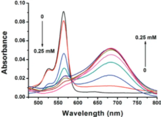
In accordance with our design expectations, probe 2 shows a large bathochromic shift on exposure to fluoride ions. The absorption peak (Figure 3) at 560 nm gradually decreases, and a new peak at 682 nm emerges. The transition has an identifiable isosbestic point at 581 nm suggesting the existence of two chromophores related by a chemical transformation. A bathochromic shift of 120 nm looks quite spectacular in solution and corresponds to a color change from purple to green.
The emission spectrum (Figure 4), on the other hand, shows the presence of one emissive species, and it is the probe compound 2. Deprotection leads to the formation of nonemissive phenolate. These reactions, as expected, are highly selective for fluoride. Comparative studies of the effects of a series of anions are shown in Figures 5 for compounds 1 and 2 in the form of bar graphs. In addition, the insets are digital photographs of acetonitrile solutions under the indicated conditions. The fluoride selectivity is impressive for both of the probe compounds.
The deprotection reaction is very fast for compound 1. Essentially, the reaction seems to reach an equilibrium during the time that is needed for mixing of the fluoride solution and taking the measurement at all concentrations. Naturally, the position of the equilibrium is different at each fluoride concentration, allowing quantification of a concentration-dependent response.
In compound 2, however, the deprotection is slower. To accommodate for the time dependence, we allowed 5 min of equilibration period following the addition of fluoride before we obtained absorption and emission spectra. A time course of deprotection as assessed by spectrophotometry is available in Supporting Information. A pseudo-first-order rate constant can be calculated relatively easily; at 0.25 mM F -concentration and 5 × 10⁻⁶M probe concentration (probe 2) the rate constant was determined to be 2.3× 10⁻²s-1at 25°C in acetonitrile.
Thus, we were able to demonstrate ion sensing in a polar organic solvent. Since the silyl group is unreactive in neutral aqueous solutions, (pOH ) 7), the silyl-deprotection-based chemodosimetry concept is fully transferable to aqueous solutions with minor modifications to improve water solubil-ity. However, much larger concentrations of fluoride would Figure 1.Absorbance spectra of compound 1 + F-in acetonitrile
in the presence of increasing F-concentrations (0, 0.025, 0.05, 0.075, 0.1, 0.125 0.15, 0.2, 0.25, 0.375, 0.5 mM). Probe concentra-tion is 5.0× 10⁻⁶M.
Figure 2.Emission spectra of compound 1 + F-in acetonitrile in the presence of increasing F-concentrations (0, 0.025, 0.05, 0.075, 0.1, 0.125 0.15, 0.2, 0.25, 0.375, 0.5 mM). Probe concentration is 5.0× 10⁻⁶M. Excitation wavelength is 480 nm.
Figure 3.Absorbance spectra of compound 2 + F-in the presence of increasing F-concentrations (0, 0.025, 0.05, 0.075, 0.1, 0.125 0.15, 0.2, 0.25 mM) after 5 min. Probe concentration is 5.0× 10⁻⁶ M.
Figure 4.Emission spectra of compound 2 + F-in acetonitrile in the presence of increasing F-concentrations (0, 0.025, 0.05, 0.075, 0.1, 0.125 0.15, 0.2, 0.25 mM) after 5 min. Probe concentration is 5.0× 10⁻⁶M. Excitation wavelength is 550 nm.
be required to overcome inactivation by strong hydrogen bond donor H2O. Instead of creating the illusion of relevance
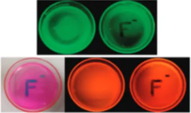
by swamping the cells with ions, at concentrations orders of magnitude higher than any realistic situation could account for, we opted for the creation of ion-responsive polymer films. To emphasize the practical potential of these probe compounds, we prepared poly(methyl methacrylate) (PMMA) films impregnated (0.5 mg of probe compounds and 500 mg PMMA) with the probe compounds 1 and 2. The probes in the polymer matrix responded to tetrabutylammonium fluo-ride solution as expected. Figure 6 shows the appearance of the fluoride-exposed polymer films in the ambient light conditions and under UV illumination at 360 nm. Quenching of the emission is clear on the fluoride-exposed regions in both cases, and for the polymer film containing fluoride probe 2, color is visibly changed. The polymer films are stable under ambient light and temperature over weeks, with no
discernible change in color or in response. The dyes do not wash off from the films as well (Supporting Information). Both compounds respond apparently much faster in the polymer matrix (instantaneously) than in solutions and do not wash off in the aqueous solutions. This simple demon-stration shows the viability of polymer-strip design for fluoride monitoring in environmental samples.
In conclusion, in this work we were able to demonstrate that the designs based on selective chemical reactions, especially when parameters that influence triggering of certain photophysical processes are incorporated, can be quite successful. In the probes or dosimetric reagents reported above, this has been achieved for PET and ICT. Thus, it is clear that depending on the exact requirements for a particular application, either an emission change or visible color change can be reproducibly produced. We believe other rationally designed reaction-based probes, with built-in triggers for photophysical and even photochemical processes, will appear in due course. Work to that end is in progress in our laboratory.
Acknowledgment. The authors gratefully acknowledge support from TUBA (Turkish Academy of Sciences). R.G. thanks TUBITAK for a scholarship.
Supporting Information Available: Experimental pro-cedures, structural proofs, and spectral data for all new compounds. This material is available free of charge via the Internet at http://pubs.acs.org.
OL100172W
Figure 5. Normalized emission ratios (“probe + anions” to free probe). Normalization was achieved by setting the maximal value to 1. The probe was excited at 490 nm, and the emission data at 507 nm were collected. The insets show the appearance of solutions under ambient light (top) and under a hand-held UV lamp (360 nm). Probe concentrations were 5× 10⁻⁶M, and the anions were added at 0.5 mM (1) or 0.25 mM (2) concentrations, all in acetonitrile. Top: data for probe 1. Bottom: data for probe 2.
Figure 6.Digital photographs of PMMA polymer sheets doped with chemosensor 1 (top) and 2 (bottom) under UV irradiation. Fluoride solution in aqueous acetonitrile (% 20 pH 10.0 buffer in acetonitrile) was applied using an appropriate mask (right). UV irradiation was achieved using a hand-held UV lamp at 360 nm.