Andrologia. 2020;52:e13739. wileyonlinelibrary.com/journal/and | 1 of 9 https://doi.org/10.1111/and.13739
© 2020 Wiley-VCH GmbH
1 | INTRODUCTION
46,XX testicular disorder of sex development (46,XX TDSD) (MIM 400045) was first described by De la Chapelle, Hortling, Niemi, and Wennström (1964) and generally characterised by a male phenotype despite having a female karyotype. The incidence is estimated to be one in 20,000 male newborns. Patients with sex-determining region Y (SRY)-positive 46,XX TDSD are usually asymptomatic males diagnosed in puberty or adulthood because of hypergonadotropic hypogonad-ism, microorchidhypogonad-ism, and infertility due to azoospermia and Sertoli cells only (Delot & Vilain, 1993; Ropke & Tuttelmann, 2017; Zenteno-Ruiz, Kofman-Alfaro, & Mendez, 2001). Until the early stages of puberty,
Sertoli and Leydig cells are functional in these patients. Sertoli cells produce anti-mullerian hormone (AMH), which is responsible for the regression of müllerian ducts, and Leydig cells synthesise testosterone and dihydrotestosterone to drive the differentiation of derivatives of the Wolffian duct in developing male fetuses. Therefore, these pa-tients have normal internal and external male genitalia with an aver-age gonadal size during childhood. Germ cell proliferation and growth in seminiferous tubules are triggered by the onset of puberty. During the adolescence and adulthood with the effect of the extra X chro-mosome, testicles remain smaller than normal and cause to spermato-genic failure because of the dramatic germ cell loss during meiosis. Absence of azoospermia factor (AZF)a, AZFb and AZFc regions results Received: 6 March 2020
|
Revised: 16 May 2020|
Accepted: 4 June 2020DOI: 10.1111/and.13739 O R I G I N A L A R T I C L E
Multiscale analysis of SRY-positive 46,XX testicular disorder of
sex development: Presentation of nine cases
Omer Salih Akar
1| Sezgin Gunes
2| Ummet Abur
1| Engin Altundag
1|
Ramazan Asci
3| Onur Emre Onat
4| Tayfun Ozcelik
4| Gonul Ogur
1 1Department of Medical Genetics, Facultyof Medicine, Ondokuz Mayis University, Samsun, Turkey
2Department of Medical Biology, Faculty of Medicine, Ondokuz Mayis University, Samsun, Turkey
3Department of Urology, Faculty of Medicine, Ondokuz Mayis University, Samsun, Turkey
4Department of Molecular Biology and Genetics, Bilkent University, Ankara, Turkey Correspondence
Sezgin Gunes, Department of Medical Biology, Faculty of Medicine, Ondokuz Mayis University, 55139 Samsun, Turkey. Email: [email protected]
Abstract
46,XX testicular disorder of sex development (46,XX TDSD) is a relatively rare con-dition characterised by the presence of testicular tissue with 46,XX karyotype. The present study aims to reveal the phenotype to genotype correlation in a series of sex-determining region Y (SRY)-positive 46,XX TDSD cases. We present the clinical find-ings, hormone profiles and genetic test results of six patients with SRY-positive 46,XX TDSD and give the details and follow-up findings of our three of previously published patients. All patients presented common characteristics such as azoospermia, hyper-gonadotropic hypogonadism and an SRY gene translocated on the terminal part of the short arm of one of the X chromosomes. Mean ± standard deviation (SD) height of the patients was 164.78 ± 8.0 cm. Five patients had decreased secondary sexual characteristics, and three patients had gynaecomastia with varying degrees. Five of the seven patients revealed a translocation between protein kinase X (PRKX) and inverted protein kinase Y (PRKY) genes, and the remaining two patients showed a translocation between the pseudoautosomal region 1 (PAR1) of X chromosome and the differential region of Y chromosome. X chromosome inactivation (XCI) analysis results demonstrated random and skewed XCI in 5 cases and 1 case, respectively. In brief, we delineate the phenotypic spectrum of patients with SRY-positive 46,XX TDSD and the underlying mechanisms of Xp;Yp translocations.
K E Y W O R D S
in Sertoli cell-only syndrome (SCOS; Kamp et al., 2001), maturation ar-rest and azoospermia, respectively (Krausz & Riera-Escamilla, 2018). The probability of sperm retrieval is virtually zero in patients with com-plete AZFa and AZFb microdeletions; therefore, assisted reproductive techniques are not recommended to these patients (Grinspon & Rey, 2019). Although all patients generally have small testicular tissues, the external genitalia varies from normal virilised male to ambiguous gen-italia depending on the presence and length of SRY gene. Hence, SRY-negative patients tend to be more frequently having genital ambiguities compared to SRY-positive ones. Approximately 80%–90% of 46,XX TDSD patients have SRY gene which is usually translocated on the dis-tal portion of the short arm of an X chromosome (Chen et al., 2019; Dauwerse, Hansson, Brouwers, Peters, & Breuning, 2006; Gunes & Esteves, 2020; Queralt et al., 2008; Ropke & Tuttelmann, 2017; Zenteno-Ruiz et al., 2001). Typical features of SRY-positive 46,XX TDSD patients are female karyotype with completely normal male phenotype and virilised male external genitalia, small testes, azoosper-mia and hypergonadotropic hypogonadism (Akinsal, Baydilli, Demirtas, Saatci, & Ekmekcioglu, 2017; Ropke & Tuttelmann, 2017). Patients usually come to attention after puberty because of hypogonadism and infertility (Zenteno-Ruiz et al., 2001). Gynaecomastia, sparse body and/or pubic hair, cryptorchidism and/or hypospadias could also be de-tected in these patients (Majzoub et al., 2017).
SRY-positive 46,XX TDSD results from an aberrant Y to X trans-location during the paternal meiosis. Either the nonallelic homolo-gous recombination (NAHR) between the identical sequences X- and Y chromosome or spontaneous errors during the replication-based mechanisms could mediate the Xp;Yp translocations in 90% of cases (Giglio et al., 2002).
Clinical pictures of the patients with 46,XX TDSD are hetero-geneous ranging from infertile men seeking fertility with normal male internal and external genitalia to the child attending to urology and child health clinics at an early age due to the ambiguous gen-italia or micropenis (Majzoub et al., 2017). Therefore, the present study aimed to reveal the clinical and genetic findings of this het-erogeneous group of patients in the view of our nine patients with SRY-positive 46,XX-DSD. Additionally, we aimed to evaluate the mo-lecular basis of this disorder based on the results of array compara-tive genomic hybridisation (array-CGH) analysis.
2 | MATERIALS AND METHODS
2.1 | Clinical evaluation of patients
Nine patients with SRY-positive 46,XX TDSD out of 1,300 consec-utive infertile men (548 severe oligozoospermic and 752 azoosper-mic patients) attending to Urology Clinics of the Ondokuz Mayis University between 2004 and 2017 were enrolled in the present study. The study was approved by the Ondokuz Mayıs University Clinical Research Ethical Committee (Approval No: 2018/353), and patients signed informed consent before participating in the study. The medical and family history, detailed physical examination
including measuring of height, weight and body mass indexes, an inspection of the external genitalia and assessment of secondary sex characteristics were evaluated. Testicular volumes of these patients were calculated using the formula [length (L) × width (W) × height (H) × 0.52 with the dimensions] obtained by testicular ultrasound.
Serum levels of follicle-stimulating hormone (FSH), luteinising hormone (LH), prolactin (PRL), oestradiol (E2) and total testosterone (TT) were measured using a radioimmunoassay in all patients. Semen samples were processed within 1 hr after the collection and liquefac-tion and then analysed according to the World Health Organizaliquefac-tion (WHO) guidelines in the year of investigation (1999 or 2010) by the same laboratory technician.
The clinical data, karyotype and fluorescence in situ hybridi-sation (FISH) findings, and X chromosome inactivation (XCI) pat-terns of three of our patients had been reported previously (Gunes et al., 2013). However, array-CGH data have not been analysed and published in our previous publication. Therefore, array-CGH and follow-up findings of these cases after publication have also been included in the present study.
2.2 | Cytogenetic analysis
Peripheral blood samples were obtained from all patients for chro-mosomal analysis. Blood lymphocytes were cultured using modi-fied methotrexate–thymidine synchronisation method and GTG banding as previously described (Abur et al., 2019; Rooney, 2001). Chromosomal analyses were assessed with CytoVision software (version 3.93; Applied Imaging).
2.3 | Fluorescence in situ hybridisation analysis
FISH analyses were conducted on both metaphase spreads and inter-phase nuclei with (SRY/CEPX; Vysis; Gunes et al., 2013). Image analyses were evaluated using CytoVision software (version 3.93; Applied Imaging) with Olympus BX51 microscope equipped with Progressive Scan Video Camera.2.4 | DNA extraction
The genomic DNA was extracted from peripheral blood lympho-cytes using QIAamp DNA Blood Mini Kit (Qiagen GmbH) according to the manufacturer's instruction (Abur et al., 2019).
2.5 | Array comparative genomic
hybridisation analysis
Array-CGH analysis was carried out in 7/9 patients using a 60K oligonucleotide microarray (Agilent Technologies) as described
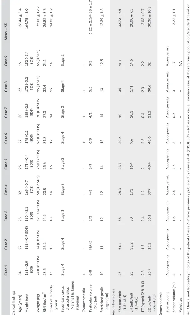
T A B LE 1 C lin ic al , h or m on al a nd s em en a na ly si s r es ul ts o f p at ie nt s w ith S RY -p os iti ve 4 6, X X T D SD C as e 1 C as e 2 C as e 3 C as e 4 C as e 5 C as e 6 C as e 7 C as e 8 C as e 9 Mea n ± SD C lin ic al fi ndin gs A ge (y ea rs) 34 27 25 32 25 27 30 22 16 26 .4 4 ± 5 .4 H ei gh t ( cm ) 16 1 (− 2. 0 SD S) 16 8 (− 0. 9 SD S) 16 0 (− 2. 1 SD S) 16 9 (− 0. 7 SD S) 17 1 (− 0. 4 SD S) 17 5 ( 0. 2 SD S) 15 5 (− 2. 9 SD S) 172 (− 0. 2 SD S) 15 2 (− 3. 4 SD S) 16 4. 78 ± 8 .0 W ei gh t ( kg ) 74 ( 0. 8 S D S) 74 ( 0. 8 S D S) 62 (− 0. 4 SD S) 68 ( 0. 2 S D S) 75 ( 0. 9 S D S) 96 ( 0. 8 S D S) 70 ( 0. 4 S D S) 95 ( 3 S D S) 65 ( 0 S D S) 75 .0 0 ± 12 .2 B M I ( kg /c m 2) 28 .5 26 .2 24 .2 23 .8 25 .6 31 .3 27. 3 32 .4 24 .1 26 .8 2 ± 3. 3 O ns et o f p ub er ty (y ea rs) 15 15 13 14 16 12 14 15 14 14 .3 3 ± 1. 2 Se co nd ar y se xu al cha ra ct er is tic s (M ar sh al l & T an ne r st ag ein g) St ag e 4 St ag e 4 St ag e 3 St ag e 3 St ag e 3 St ag e 4 St ag e 3 St ag e 4 St ag e 2 G yn ae co m as tia + − − − − + + + − Te st icula r v ol um e (R /L ) (m l) 8/8 N A /5 3/3 4/8 3/3 6/8 4/1 5/5 3/3 5. 22 ± 2 .5 /4 .8 8 ± 1 .7 St ret ch ed p eni le le ng th (c m ) 10 11 12 12 14 13 14 13 12 .5 12 .3 9 ± 1. 3 Se ru m h or m one s FS H ( m IU /m l) (1 .5 –1 2. 4) 28 51 .1 38 28 .3 23 .7 20 .6 40 35 41 .1 33 .7 3 ± 9 .5 LH ( m IU /m l) (1 .7– 8. 6) 23 33 .2 30 17. 1 16 .4 9. 6 20 .1 17. 1 14 .6 20.0 0 ± 7. 5 TT ( ng /m l) ( 2. 8–8 .0 ) 2. 8 1. 5 2.4 1.9 2 2. 8 0. 6 2. 3 2. 2 2. 03 ± 0 .7 E2 ( pg /m l) (7 .6 –4 3. 0) 20 .9 15 .1 36 .1 39 .9 40 .4 40. 6 21 .2 30. 6 32 30 .3 8 ± 1 0.1 Semen a na ly si s Sp er m c ou nt A zo os pe rm ia A zo os pe rm ia A zo os pe rm ia A zo os pe rm ia A zo os pe rm ia A zo os pe rm ia A zo os pe rm ia A zo os pe rm ia A zo os pe rm ia Semen v ol ume (m l) 4 2 1. 6 2. 8 2. 5 2 0. 2 3 1.7 2. 22 ± 1 .1 Pe lle t te st − − − − − − − − NA N ote : C lin ic al a nd l ab or at or y f in di ng s o f t he p at ie nt s C as es 7 –9 h av e p re vi ou sl y p ub lis he d b y G un es e t a l. (2 01 3) . S D S = (o bs er ve d va lu e − m ed ia n va lu e of th e re fe re nc e po pu la tio n) /s ta nd ar d de vi at io n va lu e o f r ef er enc e p op ul at io n. A bb re vi at io ns : B M I, b od y m as s i nd ex ; E 2, o es tr ad io l; F SH , f ol lic le -s tim ul at in g h or m on e; L , l ef t; L H , l ut ei ni si ng h or m on e; N A , n ot a va ila bl e; N O A , n on ob st ru ct iv e a zo os pe rm ia ; R , r ig ht ; SD , s ta nd ar d de vi at io n; SD S, s ta nd ar d de vi at io n s co re ; T D SD , t es tic ul ar d is or der o f s ex de ve lo pmen t; T T, to ta l t es to st er on e.
elsewhere (Pinkel et al., 1998). DNA samples of Case 4 and Case 9 were inadequate; therefore, we could not perform array-CGH analy-sis to two of these patients. Human male genomic DNA was used as reference DNA for the array-CGH analysis. Labelling with Cy3 and Cy5, washing and scanning steps were performed following a stand-ard protocol. Data extraction was carried out using Agilent Feature Extraction Software, and the data analysis was assessed on Agilent Cytogenomics Software (v.2.0.6.0; Agilent Technologies). Data in-cluded imbalances with at least three consecutive probes with ab-normal log2 ratios. The probe sequences and gene annotations were based on GRCh37/hg19 assembly
Karyotype results, FISH and array-CGH analyses were described according to the International System for Human Cytogenetic Nomenclature 2016.
2.6 | X chromosome inactivation analysis
The polymorphic region of exon 1 of the androgen receptor (AR) gene was used to assess the XCI patterns of the patients. The genomic DNA was cleaved using methylation-sensitive restriction enzyme
HpaII (MBI Fermentas) which digests the active X chromosome
(unmethylated) fragments. After amplification of AR gene exon 1 using PCR, amplicons were separated on 8% denaturing acrylamide: bisacrylamide (29:1) gel for 4 hr. Then, the densitometric analysis of the alleles was carried out with MultiAnalyst version 1.1 software (Bio-Rad; Gunes et al., 2013).
2.7 | Data analysis
Data were analysed using SPSS Statistics Version 22.0. Results were expressed as mean and standard deviation (SD) for continuous vari-ables. Standard deviation scores (SDS) of height and weight were calculated using the following formula:
3 | RESULTS
3.1 | Clinical findings
Nine male patients were diagnosed with SRY-positive 46,XX TDSD. All cases demonstrated male phenotype with similar clinical find-ings, including small testes, hypergonadotropic hypogonadism and pellet-negative azoospermia. Clinical and laboratory findings of our six new patients and three of our previously published patients were presented in Table 1. Mean ± SD height of the patients was calcu-lated to be 164.78 ± 8.0 cm, and these mean adult heights are ap-parently shorter than the normal adult Turkish men (164.78 ± 8.0 vs. 174.3 ± 4.9 cm) (Aslan et al., 2011; Soylemez et al., 2012). Five out of 9 (55%) patients had decreased secondary sex characteristics.
SDS = (observed value − median value of the reference population)∕standard deviation value of reference population
T A B LE 2 C yt og en et ic , m ol ecu la r c yt og en et ic a nd m ol ecu la r re su lts o f t he p at ie nt s C as e 1 C as e 2 C as e 3 C as e 4 C as e 5 C as e 6 C as e 7 C as e 8 C as e 9 K ar yo ty pe 46 ,X X 46 ,X ,der (X ) a [2 9] /4 7, X ,der (X ) a ,der (X ) a [1 ] 46 ,X ,der (X ) a 46 ,X X 46 ,X X 46 ,X X 46 ,X X 46 ,X ,der (X ) a 46 ,X X FI SH SR Y-C EP X b SR Y*1 , C EP X *2 SR Y* 1, C EP X *2 [2 43 ]/ SR Y* 2, C EP X *3[ 4] / SR Y* 0, C EP X *1[ 6] / SR Y*1 , C EPX *2 SR Y*1 , C EPX *2 SR Y*1 , C EPX *2 SR Y*1 , C EPX *2 SR Y*1 , C EPX *2 SR Y*1 , C EPX *2 SR Y*1 , C EPX *2 X i nac tiv at io n pat te rn N I N I Ra ndo m Ra ndo m N I Ra ndo m Ra ndo m Sk ew ed Ra ndo m A rr ay -CG H X p br ea kp oi nt W ithi n PR K X W ith in P A R1 ( D H RSX ) W ith in P A R1 (C RL F2 ) NA W ithi n PR K X W ithi n PR K X W ithi n PR K X W ithi n PR K X NA Yp br ea kp oi nt W ith in i nv er te d PR K Y Pr ox im al t o RS P4 Y1 W ithi n PC D H 11 Y NA W ithi n in ve rt ed PR K Y W ithi n in ve rt ed PR K Y W ithi n in ve rt ed PR K Y W ithi n in ve rt ed PR K Y NA N ote : K ar yo ty pe , F IS H a nd a rr ay -C G H f in di ng s o f t he p at ie nt s. K ar yo ty pe a nd F IS H a na ly si s f in di ng s o f C as es 7 –9 h av e p re vi ou sl y p ub lis he d b y G un es e t a l. (2 01 3) . A bb re vi at io ns : C EP , c hr om os ome en umer at io n p ro be ; C G H , c om pa ra tiv e g eno m ic h yb rid is at io n; C RL F2 , c yt ok in e r ec ep to r-lik e f ac to r 2 ; N A , n ot a va ila bl e; N I, n ot i nf or m at iv e; P A R1 , t he X p/ Yp pseu doa ut oso m al re gi on -1 ; P CD H 11 Y, p ro to ca dh er in 1 1 Y -li nk ed ; PR K X, p ro te in k in as e X ; PR K Y, p ro te in k in as e Y ; S RY , s ex -d et er m in in g r eg io n Y ; S TS , s eq ue nc e t ag ge d s ite . ader (X ): der (X )t (X ;Y )(p 22 .3 ;p 11 .3 ). bC as es 1 ,3 ,4 ,5 ,6 ,7 ,8 & 9 :4 6, X X .is h d er (X )t (X ;Y )(p 22 .3 ;p 11 .3 ) ( D X Z1 x2 )(S RY + ); C as e 2 :4 6, X X . n uc i sh (D X Z1 x1 )[6 ]/ (D X Z1 x2 ,S RY x1 )[2 43 ]/ (D X Z1 x3 ,S RY x2 )[4 ].
F I G U R E 1 Representative results of the array CGH analysis. The segments highlighted with red and blue rectangles represent the decreased and increased copy numbers, respectively. PAR1 regions of X and Y chromosomes are indicated with the lines.
Stretched penile length mean ± SD of our patients was found to be 12.39 ± 1.3 cm.
Additionally, clinical follow-up of our three previously published cases (Case 7–9) was included in the present study. Case 9 has been diagnosed 12 years ago. After his diagnosis with 46,XX TDSD, he started to receive intermittent testosterone replacement treatment. His height increased 12 cm after starting of testosterone replace-ment therapy. He is still single, and his gynaecomastia increased depending on the testosterone treatment. His erectile function is normal with testosterone treatment. Case 8 is still single as well and his erectile function, libido and ejaculation are normal under testos-terone therapy. He received antidepressant therapy for a time to overcome the depression symptoms. Case 7 is still married, and his sexual functions are normal.
3.2 | Karyotype and FISH analysis
Six out of nine patients revealed 46,XX karyotype and the remain-ing three showed 46,X,der(X)t(X;Y)(p22.3;p11.3) karyotype. The karyotype of one out of these three patients with derivative X chro-mosome demonstrated low level somatic chromosomal mosaicism [46,X,der(X)(29)/47,X,der(X),der(X)(1)]. FISH analysis of this patient showed that SRY gene region was translocated on the distal ter-minal of the short arm of one of the X chromosomes and mosai-cism of 45,X/46,X,der(X)/47,X,der(X),der(X) cell lines. Low level of 45,X-bearing cell lines detected by cytogenetic analysis in this pa-tient, but could not be in FISH analysis (Table 2).
3.3 | Array-CGH analysis
Array-CGH analysis findings of seven out of nine patients were presented in Figure 1, and these results are summarised in Table 2. Five out of seven patients had two copies of the pseudoautosomal
region 1 (PAR1), an approximately 1.0 Mb deletion in X-differential region with breakpoints on protein kinase X (PRKX), gene and ~7 Mb deletion in Yp-differential region, including an ~0.6-Mb interstitial deletion with the breakpoint on protein kinase Y (PRKY). These findings were compatible with an Yp to Xp translocation resulting from a common ~3.5 Mb paracentric Yp inversion. The remaining two patients had three copies of the proximal and two copies of the distal part of the PAR1, indicating Xp terminal deletion in the PAR1 region and presence of the Yp terminal segments including PAR1 re-gion and a very distal portion of Y-differential rere-gion. These results were in concordance with the Xp;Yp translocation associated with the breakpoints inside of the X-PAR1 and inside of the Y-differential region (Figure 2). Additionally, array-CGH analysis also revealed the absence of AZFa, AZFb and AZFc in the patients.
3.4 | XCI analysis
The methylation analysis of the AR gene yielded random and skewed XCI in five patients in one patient respectively (Figure 3). A single polymorphic allele was observed in exon 1 of AR gene for Case 1, Case 2 and Case 5; therefore, the XCI analyses of these patients were accepted noninformative. These findings could be explained by the presence of two alleles with the same repeat number or alleles do not amplify under the same conditions (Table 2).
4 | DISCUSSION
In the present study, seven of our patients applied to our clinic with infertility complaints, one with intermittent scrotal pain in adulthood and one with hypogonadism in puberty. All our patients had hypo-gonadism and azoospermia with 46,XX or 46,X,der(X) karyotypes with translocated SRY locus onto Xp. None of them had genital am-biguity consistent with literature data.
F I G U R E 2 Schematic representation of the aberrations. The yellow, the pink, the blue and the green segments denote the PAR1, the X-differential region, the Y-differential region and the Y-differential inverted region, respectively.
Testosterone synthesis was found to be impaired and de-creased with an increase in FSH and LH levels after puberty (Anik, Catli, Abaci, & Bober, 2013; Vorona, Zitzmann, Gromoll, Schuring, & Nieschlag, 2007). It was proposed that testosterone levels may be normal during adolescence but decreased in adult-hood (Velasco, Savarese, Sandorfi, Jimenez, & Jabbour, 2011). Additionally, the testosterone-to-oestradiol ratio was observed to be related to gynaecomastia, and poor axillary and pubic hair after puberty. Variations were observed in the secondary sexual char-acteristics among the patients. These variations have been sug-gested to be associated with the length of the translocated SRY gene region (Nakashima et al., 2014; Sharp et al., 2005). However, we have not found any significant correlation between the length of translocated SRY region and secondary sexual characteristics. Testosterone replacement therapy has been reported to cause the gynaecomastia as a result of the aromatisation of exogenous an-drogen (Rhoden & Morgentaler, 2004). Therefore, the increase in gynaecomastia with testosterone replacement treatment in Case 9 may be explained with the aromatisation of exogenous androgen. We did not observe under-masculinised external genitalia among the patients, but the stretched penile length mean of our patients was found to be slightly shorter than normal healthy Turkish men (12.39 ± 1.3 vs. 13.7 ± 1.6 and 13.98 ± 1.6) (Aslan et al., 2011; Soylemez et al., 2012).
One of the characteristic clinical findings of the patients was short stature which is compatible with the literature. The mean height of our patients was apparently shorter than the mean height of nor-mal adult Turkish men (164.78 ± 8.0 cm vs. 173.2 cm, 174.79 ± 5.44, 174.3 ± 4.9) (Aslan et al., 2011; Ozer, 2008; Soylemez et al., 2012), and the height of the patients has closely been resembled the mean height Turkish women (164.78 ± 8.0 cm vs. 158.9 ± 6.4 cm and 161.4 cm) (Ozer, 2008). The impaired testosterone-driven pubertal growth is proposed to be the primary cause of short stature in these patients. Case 9 had been diagnosed at puberty, although his height increased 12 cm with the testosterone replacement therapy, he still is shorter than normal adult Turkish men (162.0 cm vs. 173.2 cm, 174.79 ± 5.44, 174.3 ± 4.9) (Aslan et al., 2011; Ozer, 2008; Soylemez et al., 2012). Despite the fact that the patients with Klinefelter syn-drome also show both hypogonadism and testosterone deficiency, they present tall stature compared to those of SRY-positive 46,XX
TDSD patients (Kirsch, Weiss, Zumbach, & Rappold, 2004). This dis-crepancy could partly be explained by the absence of Y chromosome material except SRY gene in these patients. Therefore, the Y chro-mosome could be suggested to play a role in the control of growth and development of men and have an impact on the male stature. Furthermore, it has also been hypothesised that the expression of short stature homeobox (SHOX) gene has an impact on the growth of these patients (Rappold et al., 2002).
SRY-positive 46,XX TDSD results from translocation of Yp to Xp with various mechanisms and several breakpoint locations. The array-CGH analysis showed two main mechanisms lead-ing to Xp;Yp translocations in our patients. The first mechanism is NAHR, which occurs between the homologous sequences of short arms of X- and Y chromosomes, and the second one is rep-lication-based mechanisms takes place between the nonhomol-ogous sequences. NAHR causes to Xp;Yp translocation in Cases 1, 5, 6, 7 and 8. The most common paracentric inversion of the Yp, about 3.5-Mb length, results in the displacement of PRKY and PRKX genes in the same direction. These genes are located outside of the PAR1 of both in X- and Y chromosomes and show a high sequence similarity, but they are orientated in two oppo-site directions. Therefore, the homology of these sequences may lead to the crossover errors between the X- and Y chromosomes and creates an ectopic recombination during paternal meiosis (Jobling et al., 1998; Nakashima et al., 2014; Wang et al., 1995). Replication-based mechanism seems to be responsible for the translocations between the nonhomologous parts of Xp and Yp in Case 2 and Case 3. Uniform translocations of the terminal part of the Yp-differential region to Xp-PAR1 have been reported previously (Jobling et al., 1998; Nakashima et al., 2014; Rouyer, Simmler, Page, & Weissenbach, 1987; Sharp et al., 2005). The fork stalling, template switching, microhomology-mediated break-in- duced replication and nonhomologous end-joining (NHEJ) mech-anisms could be the cause of such translocations and deletions (Giglio et al., 2002; Nakashima et al., 2014; Simmons, Carvalho, & Lupski, 2012). There was no significant variation between these mechanisms with respect to the clinical findings of the patients. Also, the loss of very distal part of Xp-differential region in Cases 1, 5, 6, 7, 8 and duplication of the proximal PAR1 in Case 2 and Case 3 seem to have no significant effect on the clinical picture. F I G U R E 3 XCI patterns of Cases 1–6, PCR products of undigested and HpaII-digested DNA from peripheral blood. Line 1: marker 242- and 331-bp fragments are visible; line 2 and 3: normal male control; line 4 and 5: normal female control; line 5 and 6: case 1; line 6–17 shows the results of the cases as the first line is ‘undigested’ and the second line as ‘HpaII-digested’ DNA for each patient.
The results of the XCI analysis of patients with 46,XX TDSD are controversial in the literature. Although some authors found skewed XCI patterns among these patients, the others could not confirm these results (Bouayed Abdelmoula et al., 2003; Gunes et al., 2013; Nakashima et al., 2014; Vorona et al., 2007). Dispate one of our patients had skewed XCI pattern, the remaining five patients had random XCI pattern in the present study. The patient with skewed XCI pattern had paracentric Yp inversion-mediated Xp;Yp translocation. There were no significant differences be-tween the patients with skewed XCI and the others based on clin-ical findings, especially regarding to the external genitalia. It has been proposed that XCI pattern is essential for the formation of external genitalia. Nakashima et al. (2014) reported that patients with random XCI had a normal male external genitalia; however, the patients with skewed XCI pattern that lead to inactivation of the expression of derivative X chromosome-bearing SRY gene cause to hypogenitalismus including micropenis, microphallus, cryptorchi-dism and penoscrotal hypospadias. Our single patient with skewed XCI pattern had normal external male genitalia. However, we could not be able to identify whether the derivative X chromosome was inactivated. Additionally, no correlation was found between the XCI status and the mechanism of translocation among our patients. These data are consistent with the findings of Nakashima and col-leagues (Nakashima et al., 2014).
Case 8 demonstrated low level of X-chromosomal mosaicism, 46,X,der(X)/47,X,der(X),der(X)/45,X, and the clinical picture was not distinct from the diploid cases. X-chromosomal mosaicism in SRY-positive 46,XX TDSD has been reported in the literature previously, and mitotic nondisjunction has been suggested to be responsible for mosaicism (Chernykh et al., 2009; Macia Bobes, Alonso Troncoso, Botas Cervero, Castano Fernandez, & Fau Cubero, 2002). Patients with mosaicism have a clinical variability ranging from the true hermaphrodites to complete masculinisa-tion based on the propormasculinisa-tion of cell lines (Chernykh et al., 2009). In our patient, the 46,XX cell line was predominant, and very low level of 45,X and 47,X,der(X),der(X) mosaicism was detected and having complete masculinisation is consistent with the previous studies.
5 | CONCLUSION
In brief, this study summarises the clinical and molecular data of a rare but important cause of male infertility. This report serves us to delineate the complex structure of the Xp;Yp translocations caused by NAHR or replication-based mechanisms and the outcomes of the derived chromosome. Further studies on this clinically and ge-netically heterogeneous disorder could clarify the genomic basis of Xp;Yp translocations in 46,XX TDSD and their refined conse-quences. Finally, testicular sperm extraction is not recommended in these patients and adoption or in vitro fertilisation with a sperm donor should be considered.
ACKNOWLEDGEMENT
The authors thank all the staff of the andrology, medical biology and medical genetics laboratories for providing technical assistance in routine infertility test.
CONFLIC T OF INTEREST
The authors declare no conflict of interests. ORCID
Omer Salih Akar https://orcid.org/0000-0001-5686-2185
Sezgin Gunes https://orcid.org/0000-0002-3103-6482
Ummet Abur https://orcid.org/0000-0002-4811-9321
Engin Altundag https://orcid.org/0000-0001-8841-1426
Ramazan Asci https://orcid.org/0000-0002-2119-8963
Onur Emre Onat https://orcid.org/0000-0002-7105-1572
Tayfun Ozcelik https://orcid.org/0000-0001-5937-1082
Gonul Ogur https://orcid.org/0000-0002-9944-4423 REFERENCES
Abur, U., Gunes, S., Ascı, R., Altundag, E., Akar, O. S., Ayas, B., … Ogur, G. (2019). Chromosomal and Y chromosome microdeletion analysis in 1,300 infertile males and the fertility outcome of patients with AZFc microdeletions. Andrologia, 51, e13402. https://doi.org/10.1111/ and.13402
Akinsal, E. C., Baydilli, N., Demirtas, A., Saatci, C., & Ekmekcioglu, O. (2017). Ten cases with 46, XX testicular disorder of sex devel-opment: Single center experience. International Brazilian Journal of Urology, 43(4), 770–775. https://doi.org/10.1590/S1677 -5538. IBJU.2016.0505
Anik, A., Catli, G., Abaci, A., & Bober, E. (2013). 46, XX male disorder of sexual development:A case report. Journal of Clinical Research in Pediatric Endocrinology, 5(4), 258–260. https://doi.org/10.4274/ Jcrpe.1098
Aslan, Y., Atan, A., Omur Aydin, A., Nalcacioglu, V., Tuncel, A., & Kadioglu, A. (2011). Penile length and somatometric parameters: A study in healthy young Turkish men. Asian Journal of Andrology, 13(2), 339– 341. https://doi.org/10.1038/aja.2010.109
Bouayed Abdelmoula, N., Portnoi, M.-F., Keskes, L., Recan, D., Bahloul, A., Boudawara, T., … Rebai, T. (2003). Skewed X chromosome in-activation pattern in SRY positive XX maleness: A case report and review of literature. Annales de Genetique, 46(1), 11–18. https://doi. org/10.1016/S0003 -3995(03)00011 -X
Chen, T., Tian, L., Wu, F., Xuan, X., Ma, G., Tang, R., & Lu, J. (2019). Clinical and genetic analysis in males with 46, XX disorders of sex develop-ment: A reproductive centre experience of 144 cases. Andrologia, 51(4), e13232. https://doi.org/10.1111/and.13232
Chernykh, V. B., Kurilo, L. F., Shilova, N. V., Zolotukhina, T. V., Ryzhkova, O. P., Bliznetz, E. A., & Polyakov, A. V. (2009). Hidden X chromo-somal mosaicism in a 46,XX male. Sexual Development, 3(4), 183–187. https://doi.org/10.1159/00022 8718
Dauwerse, J. G., Hansson, K. B., Brouwers, A. A., Peters, D. J., & Breuning, M. H. (2006). An XX male with the sex-determining region Y gene in-serted in the long arm of chromosome 16. Fertility and Sterility, 86(2), 463.e1–463.e5. https://doi.org/10.1016/j.fertn stert.2005.12.062 De la Chapelle, A., Hortling, H., Niemi, M., & Wennström, J. (1964). XX Sex
chromosomes in a human male. First case. Acta Medica Scandinavica, 175(Suppl 412), 25–38. https://doi.org/10.1111/j.0954-6820.1964. tb046 30.x
Delot, E. C., Vilain, E. J.,(1993), Nonsyndromic 46, XX Testicular Disorders of Sex Development. In: Adam, M. P., Ardinger, H. H.,
Pagon, R. A., Wallace, S. E.,Bean, L. J. H., Stephens, K., & Amemiya, A. (eds). GeneReviews((R)). Seattle (WA): University of Washington Giglio, S., Calvari, V., Gregato, G., Gimelli, G., Camanini, S., Giorda, R.,
… Zuffardi, O. (2002). Heterozygous submicroscopic inversions in-volving olfactory receptor-gene clusters mediate the recurrent t(4;8) (p16;p23) translocation. American Journal of Human Genetics, 71(2), 276–285. https://doi.org/10.1086/341610
Grinspon, R. P., & Rey, R. A. (2019). Molecular Characterization of XX Maleness. Int J Mol Sci, 20(23), 6089.
Gunes, S., Asci, R., Okten, G., Atac, F., Onat, O. E., Ogur, G., … Bagci, H. (2013). Two males with SRY-positive 46, XX testicular disorder of sex development. Systems Biology in Reproductive Medicine, 59(1), 42–47. https://doi.org/10.3109/19396 368.2012.731624
Gunes, S., & Esteves, S. C. (2020). Role of genetics and epigenetics in male infertility. Andrologia, e13586. https://doi.org/10.1111/and.13586 Jobling, M. A., Williams, G., Schiebel, K., Pandya, A., McElreavey, K.,
Salas, L., … Tyler-Smith, C. (1998). A selective difference between human Y-chromosomal DNA haplotypes. Current Biology, 8(25), 1391–1394. https://doi.org/10.1016/s0960 -9822(98)00020 -7 Kamp, C., Huellen, K., Fernandes, S., Sousa, M., Schlegel, P. N., Mielnik,
A., Kleiman, S., Yavetz, H., Krause, W., Kupker, W., Johannisson, R., Schulze, W., Weidner, W., Barros, A., & Vogt, P. H. (2001). High dele-tion frequency of the complete AZFa sequence in men with Sertoli-cell-only syndrome. Mol Hum Reprod, 7, 987–994.
Kirsch, S., Weiss, B., Zumbach, K., & Rappold, G. (2004). Molecular and evolutionary analysis of the growth-controlling region on the human Y chromosome. Human Genetics, 114(2), 173–181. https://doi. org/10.1007/s0043 9-003-1028-z
Krausz, C., & Riera-Escamilla, A. (2018). Genetics of male infertility. Nat Rev Urol, 15, 369–384.
Macia Bobes, C., Alonso Troncoso, I., Botas Cervero, P., Castano Fernandez, G., & Fau Cubero, C. (2002). Clinical and genetic study of a 46, XX man with occult mosaicism. Archivos Espanoles de Urologia, 55(8), 952–954.
Majzoub, A., Arafa, M., Starks, C., Elbardisi, H., Al Said, S., & Sabanegh, E. (2017). 46 XX karyotype during male fertility evaluation; case se-ries and literature review. Asian Journal of Andrology, 19(2), 168–172. https://doi.org/10.4103/1008-682X.181224
Nakashima, S., Ohishi, A., Takada, F., Kawamura, H., Igarashi, M., Fukami, M., & Ogata, T. (2014). Clinical and molecular studies in four patients with SRY-positive 46, XX testicular disorders of sex development: Implications for variable sex development and genomic rearrange-ments. Journal of Human Genetics, 59(10), 549–553. https://doi. org/10.1038/jhg.2014.70
Ozer, B. (2008). Secular trend in body height and weight of Turkish adults. Anthropological Science, 116(3), 191–199. https://doi.org/10.1537/ ase.061213
Pinkel, D., Segraves, R., Sudar, D., Clark, S., Poole, I., Kowbel, D., … Albertson, D. G. (1998). High resolution analysis of DNA copy num-ber variation using comparative genomic hybridization to microar-rays. Nature Genetics, 20(2), 207–211. https://doi.org/10.1038/2524 Queralt, R., Madrigal, I., Vallecillos, M. A., Morales, C., Ballescá, J.-L.,
Oliva, R., … Margarit, E. (2008). Atypical XX male with the SRY gene located at the long arm of chromosome 1 and a 1qter microdeletion. American Journal of Medical Genetics. Part A, 146A(10), 1335–1340. https://doi.org/10.1002/ajmg.a.32284
Rappold, G. A., Fukami, M., Niesler, B., Schiller, S., Zumkeller, W., Bettendorf, M., … Ogata, T. (2002). Deletions of the homeobox gene SHOX (short stature homeobox) are an important cause of growth failure in children with short stature. Journal of Clinical Endocrinology
and Metabolism, 87(3), 1402–1406. https://doi.org/10.1210/ jcem.87.3.8328
Rhoden, E. L., & Morgentaler, A. (2004). Treatment of testosterone-in-duced gynecomastia with the aromatase inhibitor, anastrozole. International Journal of Impotence Research, 16(1), 95–97. https://doi. org/10.1038/sj.ijir.3901154
Rooney, D. E. (2001). Human cytogenetics: A practical approach (3rd ed.). Oxford, UK and New York, NY: Oxford University Press.
Ropke, A., & Tuttelmann, F. (2017). Mechanisms in endocrinology: Aberrations of the X chromosome as cause of male infertility. European Journal of Endocrinology, 177(5), R249–R259. https://doi. org/10.1530/EJE-17-0246
Rouyer, F., Simmler, M. C., Page, D. C., & Weissenbach, J. (1987). A sex chromosome rearrangement in a human XX male caused by Alu-Alu recombination. Cell, 51(3), 417–425. https://doi.org/10.1016/0092-8674(87)90637 -4
Sharp, A., Kusz, K., Jaruzelska, J., Tapper, W., Szarras-Czapnik, M., Wolski, J., & Jacobs, P. (2005). Variability of sexual phenotype in 46, XX(SRY+) patients: The influence of spreading X inactivation versus position effects. Journal of Medical Genetics, 42(5), 420–427. https:// doi.org/10.1136/jmg.2004.022053
Simmons, A. D., Carvalho, C. M., & Lupski, J. R. (2012). What have stud-ies of genomic disorders taught us about our genome? Methods in Molecular Biology, 838, 1–27. https://doi.org/10.1007/978-1-61779 -507-7_1
Soylemez, H., Atar, M., Sancaktutar, A. A., Penbegul, N., Bozkurt, Y., & Onem, K. (2012). Relationship between penile size and somato-metric parameters in 2276 healthy young men. International Journal of Impotence Research, 24(3), 126–129. https://doi.org/10.1038/ ijir.2011.53
Swerdloff, R. S., & Ng, C. M. (2000). Gynecomastia: Etiology, diagnosis, and treatment. In K. R. Feingold, B. Anawalt, A. Boyce, G. Chrousos, K. Dungan, A. Grossman, … D. P. Wilson (Eds.), Endotext. South Dartmouth, MA: MDText.com, Inc.
Velasco, G., Savarese, V., Sandorfi, N., Jimenez, S. A., & Jabbour, S. (2011). 46, XX SRY-positive male syndrome presenting with primary hypo-gonadism in the setting of scleroderma. Endocrine Practice, 17(1), 95–98. https://doi.org/10.4158/EP101 84.CR
Vorona, E., Zitzmann, M., Gromoll, J., Schuring, A. N., & Nieschlag, E. (2007). Clinical, endocrinological, and epigenetic features of the 46, XX male syndrome, compared with 47, XXY Klinefelter patients. Journal of Clinical Endocrinology and Metabolism, 92(9), 3458–3465. https://doi.org/10.1210/jc.2007-0447
Wang, I., Weil, D., Levilliers, J., Affara, N. A., de la Chapelle, A., & Petit, C. (1995). Prevalence and molecular analysis of two hot spots for ec-topic recombination leading to XX maleness. Genomics, 28(1), 52–58. https://doi.org/10.1006/geno.1995.1105
Zenteno-Ruiz, J. C., Kofman-Alfaro, S., & Mendez, J. P. (2001). 46, XX sex reversal. Archives of Medical Research, 32(6), 559–566. https://doi. org/10.1016/S0188 -4409(01)00322 -8
How to cite this article: Akar ÖS, Gunes S, Abur Ü, et al. Multiscale analysis of SRY-positive 46,XX testicular disorder of sex development: Presentation of nine cases. Andrologia. 2020;52:e13739. https://doi.org/10.1111/and.13739
![TABLE 2 Cytogenetic, molecular cytogenetic and molecular results of the patients Case 1Case 2Case 3Case 4Case 5Case 6Case 7Case 8Case 9 Karyotype46,XX46,X,der(X)a [29]/47,X,der(X)a ,der(X)a [1]46,X,der(X)a 46,XX46,XX46,XX46,XX46,X,der(X)a 46,XX FISH SRY-CE](https://thumb-eu.123doks.com/thumbv2/9libnet/5894420.121868/4.892.87.368.70.1090/table-cytogenetic-molecular-cytogenetic-molecular-results-patients-karyotype.webp)
