HETEROGENEITY OF HEPATOCELLULAR MALIGNANT PHENOTYPE
A THESIS SUBMITTED TO
THE DEPARTMENT OF MOLECULAR BIOLOGY AND GENETICS AND THE INSTITUTE OF ENGINEERING AND SCIENCE OF
BILKENT UNIVERSITY
IN PARTIAL FULFILLMENT OF THE REQUIREMENTS FOR THE DEGREE OF DOCTOR OF PHILOSOPHY
BY
NURİ ÖZTÜRK AUGUST, 2006
I certify that I have read this thesis and that in my opinion it is fully adequate, in scope, and in quality, as a thesis for the degree of Doctor of Philosophy.
_____________________ Prof. Dr. Mehmet Öztürk I certify that I have read this thesis and that in my opinion it is fully adequate, in scope, and in quality, as a thesis for the degree of Doctor of Philosophy.
____________________
Prof. Dr. Hakan Bozkaya I certify that I have read this thesis and that in my opinion it is fully adequate, in scope, and in quality, as a thesis for the degree of Doctor of Philosophy.
_____________________
Prof. Dr. Ediz Demirpençe I certify that I have read this thesis and that in my opinion it is fully adequate, in scope, and in quality, as a thesis for the degree of Doctor of Philosophy.
__________________________ Assist. Prof. Dr. Uygar Tazebay I certify that I have read this thesis and that in my opinion it is fully adequate, in scope, and in quality, as a thesis for the degree of Doctor of Philosophy.
_______________________ Assist. Prof. Dr. Tamer Yağcı Approved for the Institute of Engineering and Science
____________________
Prof. Dr. Mehmet Baray
ABSTRACT
Heterogeneity of Hepatocellular Malignant Phenotype
Nuri Öztürk
Ph.D. in Molecular Biology and Genetics Supervisor: Prof. Dr. Mehmet Öztürk
August, 2006, 155 Pages
Hepatocellular carcinoma (HCC) is one of the most wide-spread carcinomas throughout the world - responsible for more than 600,000 deaths annually - and is strongly associated with several etiological factors; including aflatoxin B1, alcohol, and Hepatitis virus B and C. In HCC, many genes undergo somatic aberrations with a tendency to cluster at genes involved in cell cycle regulation, in the p53 and canonical Wnt signaling pathways, and in the TGF-β/IGF axis. Almost a third of HCCs display mutations affecting canonical Wnt signalling. However, the role of canonical Wnt signaling aberrations in HCC is not known in detail, since transgenic mice which express mutant β-catenin (an integral component of canonical Wnt signaling) do not develop liver tumours.
To study the heterogeneity of hepatocellular malignancies, we have concentrated on canonical Wnt signaling in HCC cell lines. We have found that canonical Wnt signaling was active in 80% of well-differentiated, and 14% of poorly-differentiated cell lines respectively. Furthermore, ectopic expression of a mutant β-catenin resulted in strong canonical Wnt activity in well-differentiated, but not in poorly-differentiated HCC cells. Our findings suggested that heterogeneity in HCC exists even in the same pathway as exemplified by differential canonical Wnt signaling activity in well- and poorly-differentiated HCC cell lines. During this study, we produced monoclonal antibodies against β-catenin to distinguish between the pools of nuclear/cytoplasmic and membrane-associated β-catenin in cells, since it is believed that the nuclear β-catenin pool is more potent in tumorigenesis. Monoclonal antibody (MAb), 4C9 recognised β-catenin out of adherens junctions, while another MAb, 9E10 recognised all β-catenin forms even though their epitopes were adjacent.
Differential Wnt signaling activity in HCC cell lines prompted us to investigate the interactions between β-catenin and other molecules, which have important functions in hepatocytes and may affect β-catenin/TCF transcriptional activity. C/EBPα is a potent inhibitor of cell proliferation in HCC cell lines and is involved in liver-specific gene expression, and some somatic alterations of it have been observed in AML and HCC. We investigated the effect of C/EBPα on β-catenin signalling. We have found that C/EBPα inhibits mutant catenin-TCF transcriptional activity, and physically interacts with β-catenin in HCC cells.
While we were analyzing some stably mock-transfected Huh7 clones to use as controls, we observed heterogeneity in their proliferation rates. Further analysis of these clones revealed that some clones ceased to proliferate when passaged extensively. One of these clones (C3) was not tumorigenic in immunodeficient mice. Based on these observations, we hypothesized that some cancer cells could produce senescencent progeny in cell culture. Indeed, we showed that breast- and liver-cancer-derived cells display senescencent phenotypes at variable ratios. By using our experimental system, we also showed that replicative senescence program may work independently of functional p53 and p16 pathways, and the SIP1 gene is partially responsible for replicative senescent phenotypes in our Huh7-derived senescent clone C3. Overexpression of mouse SIP1 in p53- and Rb-deficient Hep3B cells induced partial senescent phenotypes at early passages. However, stable Hep3B cells repressed mouse SIP1 expression by an unknown mechanism and escaped senescent arrest in late passages.
Our results suggest that Wnt pathway may have a dual role in hepatocellular malignancy, as it is active/easily inducible in well-differentiated HCC cells and inactive/repressed in poorly-differentiated ones. The further study of β-catenin in tumor samples by using our monoclonal antibodies may reveal new aspects in β-catenin signaling. However, the mechanism of these phenomena and the inhibition of β-catenin-TCF signaling by C/EBPα require more study to reach a more comprehensive conclusion. The study of reprogramming of replicative senescence in HCC-derived cells indicated that senescence program may work independent of p53 and p16 pathways and heterogeneity of hepatocellular malignancy exists even within the established HCC derived cell lines.
ÖZET
Hepatoselüler Malignansi Fenotipinin Heterojenliği
Nuri Öztürk
Moleküler Biyoloji ve Genetik Doktorası Tez Yöneticisi: Prof. Dr. Mehmet Öztürk
Ağustos, 2006, 155 sayfa
Hepatoselüler karsinoma (HCC), en yaygın karsinomlardan olup dünya genelinde her yıl 600.000’den fazla ölüme sebep olmaktadır ve aflatoksin B1, alkol ve Hepatit B ve C gibi etiyolojik faktörlerle ilişkilendirilmektedir. HCC’de bir çok gen somatik değişime uğramakla birlikte hücre döngüsünde, p53 ve kanonik Wnt yolaklarında ve TGFβ/IGF ekseninde bulunan genler daha çok etkilenmektedir. HCC’lerin yaklaşık üçte birinde kanonik Wnt yolağını etkileyen mutasyonlara rastlanmaktadır. Bununla birlikte, mutant β-katenin (kanonik Wnt sinyalinin temel öğelerinden biri) anlatımı yapan transgenik fareler karaciğer tümörü geliştirmediği için, bu yolak değişimlerinin karaciğer tümörlerindeki etkileri ayrıntılı bilinmemektedir.
Karaciğer tümörlerindeki heterojenliği çalışmak amacıyla, HCC hücre hatlarında kanonik Wnt yolağı üzerine yoğunlaştık. Kanonik Wnt yolağının %80 oranında iyi farklılaşmış ve %14 oranında az farklılaşmış HCC hücre hatlarında etkin olduğunu tespit ettik. Bu bulguya ek olarak, mutant β-katenininin ektopik anlatımı iyi farklılaşmış HCC hücre hatlarında az farklılaşmış olanların aksine kuvvetli kanonik Wnt aktivitesine sebep oldu. Elde ettiğimiz sonuçlar HCC’deki heterojenliğin aynı sinyal yolağı içinde bile olabileceğine işaret etmektedir ve bu olgu, kanonik Wnt yolağının iyi ve az derecede farklılaşmış HCC hücrelerinde farklı olmasıyla örneklendirilmiştir. Bu çalışma sırasında, nükleer/sitoplazmik β-katenin havuzunun tümör gelişiminde daha önemli olduğu varsayıldığından, bu havuzu zardaki β-kateninden ayırt edebilmek amacıyla monoklonal β-katenin antikorları ürettik. 4C9 ve 9E10 epitopları birbirine yakın olmakla birlikte, 4C9 monoklonal antikoru (MAb) sadece yapışma kavşakları dışındaki β-katenini tanımaktayken diğer MAb, 9E10, tüm β-katenin formlarını tanımıştır.
Kanonik Wnt yolağının HCC hücrelerinde farklı aktivite göstermesi, bizi HCC’de önemli fonksiyonları olan ve β-katenin ile etkileşerek β-katenin/TCF yazılım aktivesini değiştirebilecek molekül adaylarını araştırmaya yöneltti. C/EBPα karaciğer hücrelerinde
hücre büyümesini durdurmakta ve karaciğere özgü genlerin anlatımını düzenlemektedir. Bu genin somatik değişimleri AML ve hepatoselüler karsinomada gözlenmiştir. Bu sebeple, C/EBPα’nın β-katenin sinyali üzerine etkisini araştırdık. Hepatoselüler karsinoma hücre hatlarında, C/EBPα’nın mutant β-katenin-TCF yazılım aktivitesini inhibe ettiğini ve β-katenin proteini ile fiziksel olarak etkileştiğini bulduk.
Kontrol olarak kullanmak amacıyla elde edilen kararlı eşdeğer Huh7 hücre hattı klonlarında büyüme hızlarının farklı olduğunu gözlemledik. Bu hücrelerin ayrıntılı analizlerinde, kültürde uzun dönem üretildiklerinde büyümeyi kestikleri gösterilmiştir. Bu klonlardan birisi (C3) yabancı dokuya cevap veremeyen farelerde tümör oluşturamamıştır. Bu gözlemlere dayanarak bazı kanser hücrelerinin kültür ortamında kendiliğinden yaşlanmış yavru hücreler verebilecekleri tezini oluşturduk. Gerçekten de, meme ve hepatoselüler kanser hücrelerinin değişen oranlarda yaşlanmış hücreler ürettiğini tespit ettik. Buna ek olarak, elimizdeki modeli kullarak replikatif yaşlanmanın fonksiyonel p53 ve p16 yolakları olmadan da gerçekleşebildiğini ve SIP1 geninin Huh7’den türetilmiş bir klonda (C3) gözlemlenen replikatif yaşlanmada kısmen sorumlu olduğunu gösterdik. Bunlara ek olarak, p53 ve Rb bakımından eksik Hep3B hücre hattında fare SIP1 geninin anlatımı kısmi yaşlanmaya yol açmıştır. Fakat, bu hücreler uzun süre kültürde tutulduğunda bilinmeyen bir mekanizma ile fare SIP1 geninin anlatımını baskıladılar ve yaşlanma bariyerinden kaçtılar.
Kanonik Wnt sinyalinin iyi derecede farklılaşmış hücrelerde aktif/kolayca indüklenebilir olması ve az derecede farklılaşmış hücrelerde inaktif/baskılanmış olması sebebiyle bu yolak karaciğer hücrelerinde çift yönlü rol oynamaktadır. Tümör örneklerinde β-kateninin bizim antikorlarımızla incelenmesi β-katenin sinyalinin yeni yönlerini açığa çıkarabilir. Bu bulguların mekanizmasının açıklanması ve β-katenin-TCF sinyalinin C/EBPα tarafından baskılanmasının net olarak yorumlanması için daha fazla çalışma yapılmalıdır. Hepatoselüler karsinomadan türetilmiş hücrelerde yaşlanmanın yeniden programlanabilmesi çalışması, replikatif yaşlanmanın p53 ve p16 yolaklarından bağımsız gerçekleşebileceğini ve hepatoselüler karsinomada heterejonliğin yıllardır kültürde tutulmuş HCC hücre hatlarının kendi içinde bile olduğuna işaret etmektedir.
ACKNOWLEDGMENTS
Thank you …
…Dr. Mehmet Öztürk, my supervisor, for all that I have learnt from you and for your patience.
…Dr. Rengül Çetin-Atalay, for the knowledge and experience you have shared, and the support you have given.
…Dr. Can Akçalı for the experience you have shared and for your patience. …Dr. Tamer Yağcı, for the experience you have shared and for your patience. …Ayça Arslan-Ergül, for the two-year-internship, the friendship and the knowledge you have shared.
…Haluk Yüzügüllü for the experience you have shared and for your friendship. …Şerif Şentürk, for the experience you have shared and for your friendship. …Dr. Esen Oktay, for your patience and friendship.
…Nilgün Taşdemir for your patience and friendship.
…Oğuz Kanca and Alper Tunga for your patience and friendship.
…Hani Al-Otaibi, for the friendship and the experience you have shared. …Dr. Esra Erdal, for the friendship and the experience you have shared.
…Dr. Tolga Çağatay, Dr. Emre Sayan, Dr. Berna Sayan for the friendship and the experience you have shared.
…Molecular Oncology Group, Mine Mumcuoğlu, Sevgi Bağışlar, Özge and Guvanch for the friendship and the experience you have shared.
…MBG supervisors, Dr. Özlen Konu, Dr. Uygar Tazebay, Doç. Dr. Işık Yuluğ, Prof. Dr. Tayfun Özçelik, Dr. İhsan Gürsel, Dr. Cengiz Yakıcıer for the knowledge and experience you have shared.
… The department personnel for your cooperation and reciprocal friendship.
…My family.
TABLE OF CONTENTS SIGNATURE PAGE………...….i ABSTRACT...ii ÖZET...iv ACKNOWLEDGEMENTS ...vi TABLE OF CONTENT...vii
LIST OF TABLES ...xiv
LIST OF FIGURES ...xv
ABBREVIATIONS...xviii
CHAPTER 1. INTRODUCTION……….………...1
1.1 Hepatocellular malignancy...1
1.2 Pathogenesis of hepatocellular carcinoma...3
1.2.1 Significance of viral hepatitis in hepatocellular carcinoma…………...3
1.2.2 Alcohol and cirrhosis...6
1.2.3 Hepatotoxic chemicals...6
1.2.4 Hemochromatosis and iron...6
1.2.5 Obesity and HCC……….7
1.3 Liver cirrhosis and senescence...7
1.3.1 Cellular senescence... …....8
1.3.2 Replicative senescence and telomere shortening...9
1.3.3 The p53 and p16 pathways in replicative senescence………...10
1.3.4 SIP1 and telomerase...12
1.4 Genetics of HCC...12
1.4.1 Allelic imbalance and microsatellite instability...13
1.4.2 Cell cycle regulation...13
1.4.3 Alterations of the TGFβ /IGF- axis...15
1.4.4 Wnt signaling...16
1.4.4.1 Wnt signaling pathways...16
1.4.4.2 Canonical Wnt signaling pathway ...18
1.4.4.3 Major components of the canonical Wnt signaling pathway....22
1.4.4.3.1 Wnts...22
1.4.4.3.3 LRP/Arrow co-receptors...23 1.4.4.3.4 Extracellular inhibitors ...24 1.4.4.3.5 Dishevelled...24 1.4.4.3.6 Axin...25 1.4.4.3.7 APC...26 1.4.4.3.8 GSK3β...26
1.4.4.3.9 Protein phosphatase 2A………...….27
1.4.4.3.10 β-catenin ………....…..27
1.4.4.3.11 T-Cell Factor/Lymphocyte enhancer binding factor (TCF/LEF) family of transcriptional factors and other components in nucleus ……….…...29
1.4.4.4 On/Off states of the canonical Wnt signaling pathway...31
1.4.4.4.1 Off state...31
1.4.4.4.2 On state...32
1.4.4.5 Canonical Wnt signaling in HCC...33
1.4.4.6 β-catenin mutations in well-differentiated and poorly-differentiated HCC……….……….34
1.5 C/EBPα………...35
1.5.1 C/EBPα isoforms...37
1.5.2 Physiological roles of C/EBPα...40
1.5.3 C/EBPα knock-out and knock-in mice………...……….…..40
1.5.4 The role of C/EBPα in adipocyte differentiation……….…..41
1.5.5 The role of C/EBPα in lung development and lung cancer………...42
1.5.6 The role of C/EBPα in granulopoiesis and in acute myeloid leukemia………42
1.5.7 The role of C/EBPα in hepatocyte proliferation and differentiation………….43
CHAPTER 2. OBJECTIVES AND RATIONALE………...47
CHAPTER 3. MATERIALS AND METHODS…………...……….49
3.1 MATERIALS...49
3.1.2 Bacterial Strains………....…….49
3.1.3 Enzymes……….………...……..49
3.1.4 Nucleic Acids………...………..49
3.1.5 Oligonucleotides……….50
3.1.6 Electrophoresis, photography, luciferase assay, ELISA readings, and spectrophotometry ………...50
3.1.7 Tissue culture reagents and cell lines ………...……50
3.1.8 Antibodies and chemiluminescence………...51
3.1.9 TRAP and telomere length assays kits……….……..51
3.2 SOLUTIONS AND MEDIA...51
3.2.1 General Solutions………51
3.2.2 Solutions for plasmid DNA isolation……….51
3.2.3 Microbiological media, reagents and antibiotics…...………52
3.2.4 Tissue culture solutions………..53
3.2.5 SDS (Sodium Deodecyl Sulfate)-PAGE (Polyacrylamide Gel Electrophoresis) solutions………..55
3.2.6 Immunoblotting solutions……….………..56
3.2.7 RNA Study solutions………..56
3.2.8 Immunofluorescence solutions……….………..57
3.2.9 ELISA solutions………57
3.3 METHODS………..……58
3.3.1 General Methods………...58
3.3.1.1 Transformation of E .coli……….58
3.3.1.2 Preparation of competent cells……….58
3.3.1.2.1 Conventional “Calcium Chloride” Method…………..58
3.3.1.2.2 Super competent E. Coli preparation………58
3.3.1.3.1 Conventional “Calcium Chloride” Transformation…..59
3.3.1.3.2 Super Competent E.coli Transformation………..59
3.3.1.3 Long term storage of bacterial strains………..59
3.3.1.4 Purification of plasmids………...60
3.3.1.4.2 Purification of plasmid DNA using MN
(Macherey-Nagel) miniprep kit………...61
3.3.1.4.3 Large-scale plasmid DNA purification………61
3.3.1.5 Preparation of genomic DNA from cultured cells for “TeloTAGGG Telomere Length Assay”……….………..61
3.3.1.6 Quantification and qualification of nucleic acids ………...61
3.3.1.7 Restriction enzyme digestion of DNA………....61
3.3.1.8 Gel electrophoresis of nucleic acids ………...62
3.3.1.8.1 Horizontal agarose gels of DNA samples…...……….62
3.3.1.8.2 Gel electrophoresis of total RNA………..……...62
3.3.2 Computer analyses………...………….63
3.3.3 Production and characterization of anti-β-catenin monoclonal antibodies……….…...………...63
3.3.3.1 Immunization of mice with recombinant β-catenin protein..…..63
3.3.3.2 Fusion………...……...63
3.3.3.3 Recloning (Subcloning)……….………...64
3.3.3.4 Maintenance of hybridomas and collection of hybridoma supernatant……….64
3.3.3.5 Characterization of Ig subtypes of the anti-β-catenin antibodies………...65
3.3.3.6 Production of anti-β-catenin ascites in BALB/c mice………….65
3.3.4 Epitope mapping of home-made monoclonal anti-β-catenin antibodies...65
3.3.4.1 Bacterial expression constructs………...65
3.3.4.2 Induction and batch purification of GST fusion proteins………66
3.3.5 Tissue culture techniques……...………66
3.3.5.1 Cell lines and stable clones………..66
3.3.5.2 Thawing cell lines..………..…67
3.3.5.3 Growth conditions of cells………...68
3.3.5.4 Cryopreservation of cell lines………..68 3.3.5.5 Transient transfection of eukaryotic cells using BES method… 69
3.3.5.6 Transient transfection of eukaryotic cells using “Lipofectamine
Reagent”………..…...70
3.3.6 Extraction of total RNA from tissue culture cells and tissue samples …..70
3.3.7 First strand cDNA synthesis………..……70
3.3.8 Primer design for expression analysis by semi-quantitative PCR……….70
3.3.9 Fidelity and DNA contamination control in first strand cDNAs………...71
3.3.10 Expression analysis of a gene by semi-quantitative PCR………..71
3.3.11 Crude total protein extraction………73
3.3.12 Western blotting………73
3.3.13 Immunofluorescence………..74
3.3.14 Immunoprecipitation and SAP treatment………..75
3.3.15 BrdU incorporation assay………..76
3.3.16 Low-density clonogenic assay………...76
3.3.17 In vivo studies……….……...76
3.3.18 SABG Assay………..76
3.3.19 TRAP and Telomere Length Assays……….76
3.3.20 shRNA………...76
3.3.21 ELISA (Enzyme-linked immunosorbant assay)………77
3.3.22 Luciferase assay……….78
CHAPTER 4. RESULTS………..79
4.1 Dual Role of canonical Wnt (Wnt/β-Catenin) signaling in hepatocellular carcinoma………79
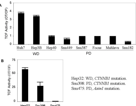
4.1.1 Differential activity of canonical Wnt signaling in well- and poorly- differentiated hepatocellular carcinoma cells………79
4.1.2 Canonical Wnt signaling activity in hepatocellular carcinoma cells……...80
4.1.3 Canonical Wnt signaling is repressed in poorly-differentiated hepatocellular carcinoma cells………...82
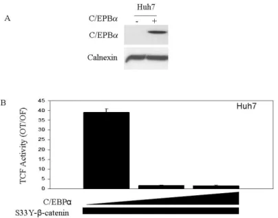
4.2 CEBPα inhibits mutant β-catenin/TCF transcriptional activity in hepatocellular carcinoma cells………...86
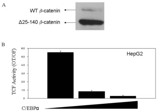
4.2.1 CEBPα inhibits transfected mutant β-catenin/TCF transcriptional activity in Huh7 cells………..86 4.2.2 C/EBPα inhibits endogenous mutant β-catenin/TCF transcriptional activity in HepG2 cells………...…………87 4.2.3 C/EBPα inhibits an inducible truncated β-catenin driven TCF/LEF activity………...…………88 4.2.4 Overexpressed C/EBPα and mutant β-catenin proteins interact in Huh7 cells………89 4.3 Monoclonal anti-β-catenin antibodies as a novel tool to distinguish cellular pools of β-catenin………91
4.3.1 Production of home-made anti-β-catenin antibodies……….91 4.3.2 Epitope mapping and immunoglobulin subtyping for monoclonal antibodies 9E10 and 4C9………..……….92 4.3.3 9E10 and 4C9 recognize different pools of β-catenin………...93 4.4 Reprogramming of replicative senescence in hepatocellular carcinoma derived cells………95
4.4.1 Senescent phenotype in cancer cell lines………...95 4.4.2 A model for cancer cell derived replicative senescent progeny: C3 clone………...98 4.4.3 Senescence in human tissues ……….……….106 4.4.4 Analysis of HCC cells for SIP1, TGF-β1, hTERT and E-cadherin expressions..……….………108 4.4.5 Overexpression of SIP1 in Hep3B cells……….………..109
CHAPTER 5. DISCUSSION………..…………..……….115
5.1 Canonical Wnt signaling in well- and poorly-differentiated HCC cell lines..115 5.1.1 Canonical Wnt signaling is active in well-differentiated and inactive in poorly-differentiated HCC cell lines………115 5.1.2 Canonical Wnt signaling is easily activated in well-differentiated HCC
5.2 C/EBPα inhibits mutant β-catenin/TCF transcriptional activity in
hepatocellular carcinoma cells……….118
5.3 Monoclonal anti-β-catenin antibodies as novel tools to differentiate cellular pools of β-catenin……….119
5.4 Reprogramming of replicative senescence in hepatocellular carcinoma derived cells………120
CHAPTER 6. FUTURE PERSPECTIVES………..124
REFERENCES………127
LIST OF TABLES
Table 1.1: Mutations in tumor suppressor genes and oncogenes in HCC………...12
Table 3.1: The sequences of primers used for cloning and sequencing ………..66
Table 3.2: The amounts of reagents and media used in BES- transfection method…...69
Table 3.3: RT-PCR primer list……….72
Table 3.4: Antibodies used for immunoblot assays and their working dilutions……….74
Table 4.1: The differentiation properties and the mutations of Wnt signaling components in HCC cell lines ………...80
Table 4.2: Spontaneous senescence in epithelial cancer cell lines ………..…96
LIST OF FIGURES
Figure 1.1: Multistage process of hepatocarcinogenesis……….………….…………...2
Figure 1.2:.Simplified model of molecular changes during hepatocellular carcinogenesis……….………….….….……5
Figure 1.3: Cell physiological stimuli provoking a cell to enter senescence…..……...9
Figure 1.4: Wnt signaling pathways………..…….17
Figure 1.5: Wnt-dependent stabilization of β-catenin ………...….19
Figure 1.6: Overview of the canonical Wnt signaling pathway ………...20
Figure 1.7: β-catenin in adherens junctions……….27
Figure 1.8: Primary β-catenin structure and protein interactions………...29
Figure 1.9: Schematic representation of the C/EBP family members ………...….36
Figure 1.10: Functional domains of C/EBPα isoforms ………...…….…...38
Figure 1.11: Generation of C/EBPα isoforms ………...………...39
Figure 1.12: The involvement of C/EBPα in diverse physiological contexts ………….40
Figure 4.1.1: TCF/LEF activity in WD and PD HCC cell lines ………..82
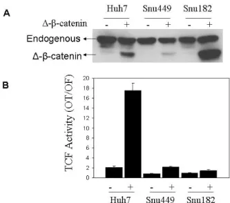
Figure 4.1.2: Ectopic expression of a mutant β-catenin (S33Y) results in high canonical Wnt activity in well-differentiated, but not in poorly-differentiated hepatocellular carcinoma cell lines ………...83
Figure 4.1.3: Ectopic expression of mutant β-catenin (∆N-β-catenin) results in high canonical Wnt activity in well-differentiated, but not in poorly-differentiated hepatocellular carcinoma cell lines ………...84
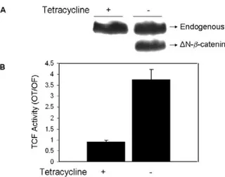
Figure 4.1.4: Ectopic expression of mutant β-catenin (∆N-β-catenin) results in low canonical Wnt activity in PD Snu449 derived stable clone ………..85
Figure 4.2.1: C/EBPα inhibits mutant S33Y-β-catenin driven TCF/LEF activity in Huh7 cell line ………..87
Figure 4.2.2: C/EBPα inhibits endogenous mutant β-catenin driven TCF/LEF activity in HepG2 cells ………..88
Figure 4.2.3: C/EBPα inhibits truncated β-catenin driven TCF/LEF activity in tetracycline regulated Snu449.8 stable clone ………89
Figure 4.2.4: The overexpressed C/EBPα and β-catenin interacts in Huh7 cells: Huh7
cells were transfected with mutant S33Y-β-catenin and C/EBPα……….90
Figure 4.2.5: Screening of Huh7-pAUCT-C/EBPα clones for inducible C/EBPα expression ……….90
Figure 4.3.1: Home-made monoclonal anti-β-catenin antibodies recognize wild-type and mutant β-catenin forms differentially ………...91
Figure 4.3.2: Restriction of 9E10 and 4C9 binding sites………..92
Figure 4.3.3: Epitopes of 4C9 and 9E10………...…93
Figure 4.3.4: 4C9 and 9E10 can differentiate β-catenin forms in HCC cells …………..93
Figure 4.3.5: 4C9 immunoreactivity did not change when β-catenin is treated with SAP to remove phosphorylation ………..…………94
Figure 4.4.1: Huh7 derived clone C3 displays senescent phenotype when cultured extensively ………95
Figure 4.4.2: Established human cancer cell lines generate senescence-associated β-galactosidase (SABG)-expressing progeny………...97
Figure 4.4.3: Proliferation rates of C1 and C3 clonal cells at different passages ………99
Figure 4.4.4: p53-and p16INK4a-deficient Huh7 cells generate progeny that undergo in vitro and in vivo replicative senescence resulting in loss of tumorigenicity …………..100
Figure 4.4.5: Complementary information on presenescent and senescent C3 cells, as compared to immortal C1 cells ………...101
Figure 4.4.6: C3 clonal cells undergo telomere-dependent replicative senescence associated with SIP1 expression and hTERT repression. SIP1 expression is lost, while hTERT is repressed in primary HCC tumors………..102
Figure 4.4.7: Complementary information on the expression of hTERT regulatory genes………....103
Figure 4.4.8: Data showing replicative senescence in Huh7-derived G12 clone……...104
Figure 4.4.9: ShRNA-mediated downregulation of endogenous SIP1 transcripts releases hTERT repression and rescues C3 cells from senescence arrest……….………...106
Figure 4.4.10: SABG staining in pathological liver tissues ………..107
Figure 4.4.11: SABG staining in primary liver tissues ……….107
Figure 4.4.13: Sip1 and E-cadherin expression is conversely correlated ……….109 Figure 4.4.14: Ectopic expression of SIP1 in p53- and pRb-negative Hep3B
hepatocellular carcinoma cells results in the repression of hTERT expression, and confers the ability to generate senescent progeny………111
Figure 4.4.15: Ectopic expression of SIP1 in p53- and pRb-negative Hep3B
hepatocellular carcinoma cells results in telomere shortening………112
Figure 4.4.16: Telomere lengths in Hep3B clones at early and late passages………..113 Figure 4.4.17: Telomerase activity in Hep3B clones at early and late passages………113 Figure 4.4.18: Exogenous SIP1 expression and repression of E-cadherin in early and late
ABBREVIATIONS
AD activation domain
AFB1 aflotoxinB1
AFP alpha-feto protein
AI allelic imbalance
Amp ampicillin
ALT alternative mechanism of telomere lengthening
AP alkaline phosphatase
ARF Alternative Reading Frame BMP bone morphogenetic protein BRCA breast cancer gene, early onset
BrdU Bromodeoxyuridine
Brm Brahma
bZIP basic region leucine zipper C/EBP CCAAT enhancer binding protein
CaMK calmodulin-dependent protein kinase CDK Cyclin Dependent Kinase
CDP CAAT displacement protein CFA Freund's complete adjuvant
CK Casein Kinase
CK19 Cytokeratin 19
Co-IP Co-Immunoprecipitation
CO2 carbon dioxide
CRC colorectal cancer
CRD cysteine-rich domain
C-terminus carboxy terminus
Dkk Dickkopf
DNA Deoxyribonucleic acid
Dsh Dishevelled (in Drosophila) Dvl Dishevelled (in vertebrates)
ED embriyonal day
EDTA ethylenediaminetetraacetic acid
EtBr ethidium bromide
FAP familial adenomatous polyposis FBS fetal bovine serum
Fz Frizzled
g gram g gravity
GAPDH glyceraldehyde-3-phosphate dehydrogenase GSK3β glycogen synthase kinase 3 beta
HAT Hypoxanthine Aminopterin Thymidine
HB Hepatoblastoma
HBV Hepatitis B Virus
HbX hepatitis B virus X protein HBXAg Hepatitis B virus X antigen
HCC Hepatocellular Carcinoma
HCV Hepatitis C Virus
HDAC histone deacetylase
HH hereditary hemochromatosis
HMG high mobility group hMSH2 Human Mut S homolog-2 HRP horse radish peroxidase
HT Hypoxanthine-Thymidine
hTERT human Telomerase Reverse Transcriptase hTR human telomerase RNA
I immortal
IFA incomplete Freund's adjuvant
Ig immunoglobulin
i.p intraperitoneal
IP immunoprecipitation
IGF2R1 IGF2 Receptor IGFBP IGF-binding protein
JNK Jun Kinase
Kan kanamycin
Kb Kilo base
kDa kilo Dalton
LAP Latent Associated Protein
LB Luria-Bertani media
LCD large cell-dysplasia
LEF lymphocyte enhancer-binding factor LOH loss of heterozygosity
LRP5 LDL-receptor related protein 5 LRP6 LDL-receptor related protein 6 LTBP Latent TGF- β Binding Protein LTR long terminal repeat
M6P/IGF2R mannose-6-phosphate/insulin-like growth factor 2 receptor
MAb monoclonal antibody
MAP/ERK mitogen activated protein/extracellular signal-regulated kinase MAPK Mitogen Activated Protein Kinase
MDM2 Mouse Double Minute 2
mg milligram
µg microgram
MgSO4 Magnesium Sulfate
MHBSt carboxyterminal truncated middle hepatitis B surface protein
ml milliliter
µl microliter
MMLV Murine Maloney Leukemia Virus
MQ MilliQ water
NaCl Sodium Chloride
NaF sodium fluoride
Na3VO4 sodium ortho-vanadate
NEAA Non-essential Amino Acid nm nanometer (1/109 of a meter)
NS3 Nonstructural Protein 3
NS5A Nonstructural Protein 5A N-terminus amino terminus
O/N over night
OD Optical Density
PAb polyclonal antibody
PAGE Polyacrylamide gel electrophoresis PBS Phosphate Buffered Saline
PBS-T Phosphate Buffered Saline with Tween-20 PCP planar cell polarity
PCR Polymearase chain reaction
PD poorly-differentiated
PD Population doubling
PI-3 kinase Phosphatidylinositol 3-kinase PKR double-stranded RNA protein kinase PMSF Phenylmethylsulphonylfluoride
PP2A protein phosphatase 2A
PPARγ peroxisome proliferators antigen receptor γ
pRb Retinoblastoma protein
PS presenescent
RD regulatory domains
RNA Ribonucleic acid
ROS reactive oxygen species rpm revolutions per minute RTK receptor tyrosine kinases
S senescent
s.c subcutaneous
SABG senescence associated beta galactosidase SAP shrimp alkaline phosphatase
SCD small cell-dysplasia
SDS Sodium Dodecyl Sulfate
SDS-PAGE SDS- Polyacrylamide Gel Electrophoresis sFRPs secreted Frizzled-related proteins
shRNA short hairpin RNA
SIP1 Smad interacting protein 1
siRNA Small interfering RNA
Smad homolog of mothers against decapentaplegic (MAD) SWI/SNF switching-defective and sucrose nonfermenting TBS Tris Buffered Saline
TBS-T Tris Buffered Saline with Tween-20 TCF T-Cell Factor (T-cell specific, HMG-box) TEMED N, N, N, N-tetramethyl-1, 2 diaminoethane TGFα transforming growth factor alpha
TGF-β Transforming growth factor TNF Tumor necrosis factor
Tris Tris (hydroxymethyl)-methylamine
UV Ultraviolet
WD well-differentiated
WIF-1 Wnt-inhibitory factor-1
X-Gal 5-bromo-4-chloro-3-indolyl-b-D-galactoside ZFHX1B Zinc finger homeobox 1B
CHAPTER 1. INTRODUCTION
1.1 Hepatocellular malignancy
The liver is the largest internal organ in the body, filling the upper right side of the abdomen and protected by the rib cage. The physiological functions of the liver can be summarized as follows: 1) Bile production and excretion. 2) Excretion of bilirubin, cholesterol, hormones, and drugs. 3) Metabolism of fats, proteins, and carbohydrates. 4) Enzyme activation. 5) Storage of glycogen, vitamins, and minerals. 6) Synthesis of plasma proteins, such as albumin and globulin, and clotting factors 7) Blood detoxification and purification.
A malignancy is cancerous growth, in which a mass of cells grows in an uncontrolled fashion, with a tendency to create daughter cells that break away and grow elsewhere, invading and damaging surrounding tissue. Primary liver cancer is different from cancers that have spread from other places in the body to the liver (liver metastases). There are two main kinds of primary liver cancer, hepatoma and cholangiocarcinoma. Hepatoma originates from the hepatocytes, the main functioning liver cells, while cholangiocarcinoma originates in the bile ducts.
Hepatoblastoma (HB) is the most common pediatric liver malignancy, comprising approximately 1% of all pediatric cancers (Schnater JM. et al., 2003). Liver angiosarcoma is a very rare type of primary liver cancer developing from the cells of blood vessels within the liver.
Hepatocellular carcinoma (HCC), the most frequent primary liver cancer, is the fifth most common cancer world-wide and one of the most deadly cancers with approximately 600,000 yearly deaths (Llovet JM., 2003). Hepatocarcinogenesis nearly always develops in the setting of chronic hepatitis or cirrhosis; conditions in which many hepatocytes are killed, inflammatory cells invade the liver and connective tissue (Thorgeirsson SS. and Grisham GW., 2002). Infection with hepatitis B virus (HBV), hepatitis C virus (HCV) and chronic exposure to aflatoxin B1 (AFB1) are responsible for about 80% of all HCCs in humans (Bosch FX. et al., 1999). Development of HCC is slow process requiring 10-30 years from the initiation of to the fully malignant
phenotype. The preneoplastic liver is often the site of chronic hepatitis, cirrhosis, or both. Hepatocyte cycling leads to the production of monoclonal populations of aberrant and dysplastic hepatocytes that have telomere erosion and telomerase re-expression, sometimes microsatellite instability, and occasionally structural aberrations in genes and chromosomes. Development of dysplastic hepatocytes in foci and nodules and emergence of hepatocellular carcinoma are associated with the accumulation of irreversible structural alterations in genes and chromosomes, however the genomic basis of the malignant phenotype is heterogeneous (Thorgeirsson SS. and Grisham GW., 2002). The sequential events leading to HCC may be summarized in five steps: chronic liver injury that produces inflammation, cell death, cirrhosis and regeneration, DNA damage, dysplasia, and finally HCC (Figure 1.1) (Tannapfel A. and Wittekind C., 2002). More than 60–80% of HCC cases arise in cirrhotic liver.
Figure 1.1: Multistage process of hepatocarcinogenesis (Tannapfel A. and Wittekind C., 2002) (See text for details).
1.2 Pathogenesis of hepatocellular carcinoma
1.2.1 Significance of viral hepatitis in hepatocellular carcinoma
The epidemiologic association of chronic HBV or HCV infection with HCC has been well established. The availability of cloned HBV and HCV genomes made it possible to detect hepatitis viruses in hepatocellular carcinomas, and their involvement in hepatocarcinogenesis. Oncogenic mechanism of HBV and HCV infection may be simply defined as releasing the growth control of hepatocytes by coding for a factor like the X protein of HBV (HbX) that activates otherwise dormant genes or activates proto-oncogenes or silences anti-proto-oncogenes; by inserting its DNA sequences that can activate and influence the transcription of cellular genes; by causing chronic inflammation with cell death and hepatocyte regeneration and with fibrosis; and by activation of the immune system liberating cytokines at the wrong time at the wrong place. Following liver cell necrosis, inflammation, regeneration, and fibrosis, quiescent hepatocytes start to proliferate. During this physiological process, irregular regeneration of hepatocytes through clonal expansion may lead to loss of control over growth and development of HCC (Ueno Y. et al., 2001).
Regarding the contribution of HBV to hepatocarcinogenesis, the role of integration of HBV DNA into host chromosomes and the subsequent chromosomal instabilities is an interesting issue. Integration of HBV DNA into HepG2 cells or into transgenic mouse chromosomes has resulted in chromosomal instability that may lead to loss of heterozygosity (LOH) in many loci during chronic infection (Hino O. et al., 1991; Livezey KW. and Simon D., 1997). It has also been shown that HBV chronic carriers display a higher incidence of chromosomal instabilities than the corresponding the uninfected population (Simon D. et al., 1991; Laurent-Puig P. et al., 2001).
Transcriptional activation of a wide range of viral, as well as cellular genes such as c-fos , c-myc, insulin-like growth factor 2 (IGF2), insulin-like growth factor I receptor (IGFR1) and β-interferon , were shown to be induced by HBV encoded X antigen
(HBxAg) (Twu JS. and Schloemer RH., 1987; Colgrove R. et al., 1989; d'Arville CN. et al., 1991; Caselmann WH., 1996; Kim SO. et al., 1996). In chronic HBV infection, it has been shown that HBxAg binds and functionally inactivates the tumor suppressor p53 (Ueda H. et al., 1995; Huo TI. et al., 2001) and the negative growth regulator p55sen
(Feitelson MA., 1999), both of which are involved in senescence related pathways. Inactivation of the retinoblastoma (Rb) tumor suppressor by hyperphosphorylation resulting in the activation of E2F1 and the trigger of the cell cycle has been reported in HbxAg-positive HCC cells (Sirma H. et al., 1999). It has also been shown that HBxAg can down regulate the expression of translational factor sui1, and cyclin-dependent kinase inhibitor p21WAF1/CIP1/SDI1 (Sirma H. et al., 1999). As with HbxAg, carboxyterminal truncated middle hepatitis B surface protein (MHBSt) can activate various viral and cellular gene promoters (Caselmann WH. et al., 1990; Kekule AS. et al., 1990). Recent data suggests that HbxAg contributes to HCC development also by mechanisms other than transactivation. HBxAg binds to the X-associated protein 1 and possibly disturbs its function in nucleotide excision repair mechanism (Becker SA. et al., 1998). It has also been shown that HBxAg-stimulated cell growth is associated with constitutive activation of the ras/raf/MAPK and NFκ-B signal transduction pathways (Lucito R. and Schneider RJ., 1992; Benn J. and Schneider RJ., 1994; Shirota Y. et al., 2001).
Studies have recently begun to clarify the molecular mechanisms of HCV-induced carcinogenesis. Studies with HCV proteins showed that viral proteins interact with various cellular proteins, including 14-3-3 protein, apolipoprotein AII, tumor necrosis factor (TNF) receptor, lymphotoxin-ß receptor, DEAD domain of RNA helicase, nuclear ribonucleoprotein, double-stranded RNA protein kinase (PKR), p53 and SNARE-like protein (Ghosh AK. et al., 1999; Shimotohno K., 2000; Ray RB. and Ray R., 2001).
In chronic HCV infection, inactivation of p53 can be achieved either by HVC core protein which transcriptionally represses the p53 promoter (Ray RB. et al., 1997; Pontisso P. et al., 1998), or by nonstructural protein 3 (NS3) and NS5A which bind and most likely inactivates p53 (Ishido S. and Hotta H., 1998; Majumder M. et al., 2001). Recently, stable expression of HCV core protein in HepG2 cells was shown to result in constitutive activation of the mitogen activated protein kinase/extracellular signal-regulated kinase (MAPK/ERK) pathway (Hayashi J. et al., 2000).
Chronic infection by HBV, HCV and chronic hepatitis lead to the activation of cytokines and growth factors such as transforming growth factor-α (TGF-α) and insulin-like growth factor-2 (IGF2), and the simultaneous decrease in IGF-binding proteins (IGFBPs), which increases the bioavailability of the insulin-like growth factors (IGFs).
IGF2 and TGFα in preneoplastic hepatocytes conbribute to the increase in the rate of proliferation (Grisham JW., 2001). Structural genomic alterations accumulate slowly during the preneoplastic phase (Thorgeirsson SS. and Grisham JW., 2002; Suriawinata A. and Xu R., 2004). However, once the hepatocytes acquire a dysplastic phenotype, characterized by small cell size, increased nuclear to cytoplasmic ratio, basophilia and abnormal nuclear morphology the rate at which these cells acquire additional disruptions to the genome increases dramatically (Hytiroglou P., 2004) (Figure 1.2).
Figure 1.2: Simplified model of molecular changes during hepatocellular carcinogenesis. Normal hepatocytes proliferate at a very slow rate. Chronic infection by HBV, HCV and chronic hepatitis lead to the activation of cytokines and growth factors such as TGFα and IGF2, and the simultaneous decrease in IGFBPs, which increases the bioavailability of the IGFs. These changes lead to an increased rate of proliferation, which for many years is balanced by loss of hepatocytes through apoptosis. During the preneoplastic period, additional changes are acquired, including multiple chromosomal abnormalities, loss of tumor suppressors, decreased expression of EGR1 and C/EBPα, and increased expression of FOXM1b. At later stages, increased expression of TGF-β is thought to promote angiogenesis and metastasis (Greenbaum LE, 2004).
1.2.2 Alcohol and cirrhosis
Beside for the HBV and mostly HCV-associated cirrhosis, non-viral induced cirrhosis is also coupled with elevated risk of HCC (Furuya K. et al., 1988; Floreani A. et al., 1999).
It has been shown that alcoholic cirrhosis is associated with both HCC and cholangiocarcinoma. In the absence of cirrhosis, however, alcohol has not been shown to be carcinogenic in animal studies. It appears that, in large part, the relationship of alcoholic cirrhosis to liver cancer may be due to concomitant infection with HCV (Farber E., 1996). The effects of ethanol metabolism on DNA integrity and repair mechanisms are likely to play a significant role in the process of transformation. However, it is becoming increasingly apparent that ethanol, the metabolism of ethanol, or both also affect cell signaling pathways that regulate normal and abnormal hepatocyte function, proliferation, and apoptosis (McKillop IH. and Schrum LW., 2005).
1.2.3 Hepatotoxic chemicals
Aflatoxin B1 (AFB1), benzo(a)pyrene and vinly chloride were shown to have a
well-defined genotoxic effect in hepatocarcinogenesis. Consumption of food contaminated with aflatoxins, toxic metabolites of some species of Aspergillus fungi, has been allied with both human and animal HCCs. Aflatoxin metabolism produces aflatoxin B1-8,9-epoxide, a toxic product that induces a G to T mutation of the p53 gene at codon 249 up-regulating insulin-like growth factor II , which leads to a reduction of apoptosis (Bressac B. et al., 1991; Hsu IC. et al., 1991; Lee YI. et al., 2000).The genotoxic effects of benzo(a)pyrene and vinly chloride on p53 gene has been supported by human and animal data (Puisieux A. et al., 1991; Barbin A. et al., 1997).
1.2.4 Hemochromatosis and iron
Alteration in iron absorption and deposition in the liver and other organs occurs in hereditary hemochromatosis (HH), which is an autosomic recessive disease (Bailey MA. et al., 2002). Iron overload has been associated with a high risk of HCC in patients with untreated hemochromatosis and dietary iron intake in patients without hereditary iron overload (Hann HW. et al., 1989; Fargion S. et al., 1992). Iron overload as a cofactor for
hepatocarcinogenesis is the iron deposition which occurs in hepatocytes, Kupffer cells and bile ducts. It results in increased production of oxygen free radicals, peroxidation of membrane lipids, and other reactive oxygen species (ROS)-mediated damages (Kicic A. et al., 2001).
1.2.5 Obesity and HCC
Recently, several epidimological observations have implicate obesity as a risk factor for certain malignancies such as endometrium cancer, breast cancer etc. Obesity and diabetes mellitus were suggested as important risk factors for cryptogenic chronic liver disease in patients with HCC is supported by the analysis of surgically treated patients (Regimbeau JM. et al., 2004).
1.3 Liver cirrhosis and senescence
Liver cirrhosis, the irreversible terminal stage of chronic liver disease, characterized by widespread fibrous scarring, is a major cause of morbidity and mortality worldwide, with no effective therapy. Independent of its cause, cirrhosis is considered a major clinical and histopathological risk factor for HCC development. A variety of factors induce liver cirrhosis, including viral hepatitis, alcohol abuse, prolonged biliary obstruction, genetically transmitted metabolic disorders, and others. Serious complications of liver cirrhosis includes those: accumulation of fluid in the abdomen (ascites), bleeding disorders (coagulopathy), increased pressure in the blood vessels (portal hypertension), and confusion or a change in the level of consciousness (hepatic encephalopathy). Regenerative nodules are characteristic lesions of the cirrhotic liver. They exhibit a lack of bile ducts and poorly organized hepatocytes surrounded by fibrosis and proliferating cholangiocytes. Dysplastic foci, which are smaller than 1 mm, can be found in regenerative nodules. There are two types of dysplastic foci in cirrhotic livers, small cell-dysplasia (SCD) and the large cell-dysplasia (LCD), according to the nuclear/cytoplasmic ratio. SCD are thought to be HCC precursor lesions that result from the proliferation of hepatocytes and oval cells. On the other hand, LCD apparently arise from persistent necroinflammation-induced senescent hepatocytes and are therefore not considered to be HCC precursor lesions (Libbrecht L. et al., 2005).
Hepatocyte telomere shortening and senescence are general markers of human liver cirrhosis, and correlates with progression of fibrosis in cirrhosis samples (Wiemann SU. et al., 2002). Additionally, Paradis V. et al. observed an increasing percentage of replicative senescent liver cells from normal liver to chronic hepatitis and HCC (Paradis V. et al., 2002).
1.3.1 Cellular senescence
In 1961, Leonard Hayflick and Paul Moorhead discovered that human cells derived from embryonic tissues can only divide a finite number of times when cultured (Hayflick and Moorhead, 1961). This phenomenon is now known as replicative senescence. Hayflick and Moorhead worked with fibroblasts, a cell type found in connective tissue, but replicative senescence has been found in other cell types: keratinocytes, endothelial cells, lymphocytes, adrenocortical cells, vascular smooth muscle cells, chondrocytes, etc.
Senescence is now understood to be the final phenotypic state adopted by a cell in response to several distinct cell physiological processes. A variety of cell physiological stimuli can provoke a cell to enter senescence, telomere uncapping, DNA damage, oxidative stress, oncogenic activation, improper cell contacts, and lack of growth stimulating factors (Figure 1.3).
Senescent cells have a large, flat morphology, senescence associated β-galactosidase (SABG) activity, cease to proliferate. Even though, different stimuli result similar phenotype in senescent cells, they use distinct but not mutually exclusive effectors of senescence. For example, the replicative senescence is accompanied by telomere shortening, but stress-induced senescence caused by ectopic expression of the Ras oncoprotein is not accompanied by telomere shortening.
Figure 1.3: Cell physiological stimuli provoking a cell to enter senescence. (Ben-Porath I. and Weinberg RA., 2005)
1.3.2 Replicative senescence and telomere shortening
Telomeres are specialized nucleoprotein structures at the end of eukaryotic chromosomes (Blackburn EH., 1991). They serve as protective caps to distinguish the natural ends from random breaks, preventing the activation of DNA repair mechanisms and the resultant chromosome fusions (de Lange T., 2004). Expression of telomerase reverse transcriptase (TERT) is not detected in most human cell types (Ducrest AL. et al., 2002). This ensures that tumor suppression, by the telomere counting mechanism, is adequately enforced in the soma. However telomere erosion is counter-balanced by the de novo synthesis of telomeric repeats in germline cells, some stem cells, and most tumor cells. In other words, chromosome ends are stabilized by telomerase, a specialized reverse transcriptase that adds telomeric repeats (TTAGGG) to these ends in these cells. The main components of this telomerase complex are a reverse transcriptase (hTERT, human telomerase) and an integral RNA component (hTR). On the other had,
noncanonical functions of hTERT have also been described. To name a few, Sharma GG. et al. suggested that hTERT associates with human telomeres and enhances genomic stability and DNA repair (Sharma GG. et al., 2003). It has been recently shown that hTERT has a novel role in the modulation of cyclin D1 expression (Jagadeesh S. and Banerjee PP., 2006). Telomerase activity is not the sole means by which telomere maintenance may be achieved in human cells. An alternative to the telomerase-dependent lengthening of telomeres was discovered when it was observed that telomeres elongated in the absence of detectable telomerase (Bryan TM. et al., 1995). The mechanism of alternative lengthening of telomeres (ALT) resembles homologous recombination and requires many protein factors, such as Rad50, MRE11 and NBS1 (Bechter OE. et al., 2003; Jiang WQ. et al., 2005).
With each round of cell division and DNA replication, loss of telomeric DNA via the end replication problem limits the proliferative capacity of primary cells in vitro at the stage of cellular senescence (Harley CB. et al., 1990; Yu GL. et al., 1990; Wright WE. and Shay JW., 1992). Cells in culture display progressively shorter telomeres as the cells approach replicative senescence (Harley CB. et al., 1990), and cells collected from older organisms, which have presumably undergone more cell divisions in vivo, display both shorter replicative lifespans in culture and shorter telomeric DNA (Hastie ND. et al. 1990). Ectopic expression of hTERT, the catalytic subunit of telomerase, restores telomere length in fibroblasts and results in immortalization of primary human fibroblasts (Bodnar AG. et al., 1998). However, the ectopic expression of hTERT in keratinocytes, mammary epithelial cells, and other cell types restores telomerase enzymatic function and telomere lengths but still does not allow these cells to bypass senescence (Kiyono T. et al., 1998; Dickson MA. et al. 2000). These data suggest that in certain cell types, even when shortened telomeres are restored by the ectopic expression of hTERT, additional signals activate or enforce the senescence program (Lundberg AS. et al., 2000).
1.3.3 The p53 and p16 pathways in replicative senescence
Replicative senescence (permanent growth arrest also called M1 stage) is believed to be initiated by a DNA damage-type signal generated by critically shortened telomeres, or by the loss of telomere integrity, leading to the activation of cell cycle checkpoint
pathways involving p53, p16INK4a and/or retinoblastoma (pRb) proteins (Campisi J., 2005; Dimri GP., 2005). In the absence of functional p53 and/or p16INK4a /pRb pathway responses telomeres continue to shorten resulting in crisis (also called M2 stage). Cells that bypass the M2 stage by reactivating hTERT expression gain the ability for indefinite cell proliferation, also called immortality. (Ben-Porath, I. and Weinberg, RA., 2004; Campisi J., 2005; Shay JW. and Wright WE., 2005). There is accumulating evidence that cancer cells undergo a similar process during carcinogenesis to acquire immortality. Telomerase activity associated with hTERT re-expression is observed in ~80% of human tumors (Shay JW. and Bacchetti S., 1997), and senescence controlling p53 and p16INK4a genes are commonly inactivated in the majority of human cancers (Sherr CJ. and McCormick F., 2002). Moreover, experimental transformation of normal human cells to tumor cells requires hTERT-mediated immortalization, as well as inactivation of p53 and pRb genes (Boehm JS. and Hahn WC., 2005). Another signal implicated in replicative senescence is p21cip1 (Noda A. et al., 1994). p21cip1 has since been well characterized as a downstream effector of p53-mediated growth arrest following DNA damage. Although p53 mediates growth arrest in senescence, the involvement of its p21cip1 effector remains unclear (Bond JA. et al., 1995; Pantoja C. and Serrano M., 1999).
The aberrant expression of hTERT, together with the loss of p53 and p16INK4a/pRB control mechanisms suggests that the replicative immortality is a permanent and irreversible characteristics of cancer cells. Although some cancer cells may react to some extrinsic factors by a senescence-like stress response, this response is immediate, telomere-independent and can not be qualified as replicative senescence (Roninson IB. 2003). Experimental inactivation of telomerase activity in cancer cells mostly results in cell death (Shay JW. and Roninson IB., 2004), whereas ectopic expression of p53,
p16INK4a, or pRb provocate an immediate senescence-like growth arrest, rather than
replicative senescence programming (Roninson IB., 2003). Thus, do date there is no experimental evidence for spontaneous reprogramming of replicative senescence in immortalized cancer cells.
1.3.4 SIP1 and telomerase
The SIP1 gene (Zinc finger homeobox 1B; ZFHX1B) encodes a transcriptional repressor protein that interacts with SMAD proteins of the TGF-β signalling pathway and CtBP co-repressor (Verschueren K. et al., 1999; Postigo AA. et al., 2003). The SIP1 gene is expressed at high levels in almost all human somatic tissues tested, including liver (Cacheux V. et al., 2001). Although the SIP1, as a repressor of E-cadherin promoter, has been suggested to be a promoter of invasion in malignant epithelial tumors (Comijn J. et al., 2001), this gene has recently been implicated in TGF-β-dependent regulation of hTERT expression in breast cancer cells (Lin SY. and Elledge SJ., 2003).
1.4 Genetics of HCC
Cancer proceeds through the accumulation of mutations in genes that govern cell proliferation and death. The long latency period of tumor development indicates at least five different genetic events may be required to reach the fully malignant phenotype (Fearon, ER. and Vogelstein B., 1990). It is believed that hepatocarcinogenesis also shares this common molecular pathogenesis as well as common biological features. The genetic (Table 1.1) and epigenetic alterations generally result in disruption of p53, Wnt or Rb-p16 pathways in HCC.
gene mutation % references
p53 28-50% Puisieux A. and Ozturk M., Oda T..et al., 1992; 1997
M6P/IGF2R 25-55% De Souza AT. et al., 1995; Yamada T. et al., 1997 β-catenin 18-41% Nhieu JT. et al., 1999 ; Terris B. et al., 1999
p16 10% Liew CT. et al., 1999
Axin1&Axin2 5-10% Taniguchi K. et al., 2002
Smad2&Smad4 3-6% Yakicier MC. et al., 1999
BRCA2 5% Katagiri T. et al., 1996
Rb rare Zhang X. et al., 1994
Ras rare Shen HM. and Ong CN., 1996
1.4.1 Allelic imbalance and microsatellite instability
Conventional cytogenetic methods were first used for genetic analysis of hepatoma cell lines and primary liver tumors, and they revealed that most HCCs are aneuploid and harbor multiple different chromosomal abnormalities, with recurrent deletions of the short arm of chromosome 1 (Simon D. et al., 1991). By comparative genomic hybridization, chromosomes 1q, 8q, and 17q show gene dose increase while chromosomes 1p, 4q, 8p, 9p, 13q, 16p, 16q, and 17p show gene dose loss. Frequent LOH, or more comprehensive, allelic imbalance (AI), is consistently observed on chromosomes 1p, 4q, 6p, 8p, 13q, 16q, and 17p by whole-genome allelotyping (Tannapfel A. and Wittekind C., 2002). The chromosome regions with gene dose increase may contain critical oncogenes while those with gene dose loss may contain tumor-suppressor genes. For chromosomes 17p, 13q, 9p, 6q, and 16p, LOH could be related to p53, Retinoblastoma 1(RB1), p16, Insulin-like Growth Factor2 Receptor (IGF2R) inactivation (Feitelson MA. et al., 2002). In dysplastic nodules LOH has been observed with a prevalence of about 50% and 80%, respectively (Thorgeirsson SS. and Grisham JW., 2002). Microsatellite instability is associated with a mutator phenotype at the nucleotide level; it is caused by defects in the DNA mismatch repair genes MSH2 and MLH1 located on chromosomes 2 and 3 (Lengauer C. et al., 1998). In HCC, however, these chromosomal regions are not frequently affected by allelic losses, and microsatellite instability has not been detected by microsatellite marker analysis. But mutations in a mismatch repair gene know as Human Mut S homolog-2 (hMSH2) has been reported at about 30% of HCCs examined (Yano M. et al., 1999). Furthermore, very few mutations have been detected in the mononucleotide repeats in Bax, IGF-IIR, or MLH genes in HCC (Kondo Y. et al., 1999; Salvucci M. et al., 1999).
1.4.2 Cell cycle regulation
Cells respond to proliferative or anti-proliferative signals through the cyclinD1-Rb-Cdk 4/6 pathway or the p53-p14ARF pathway. As cells enter the cycle from quiescence (G0), one or more D-type induced in response to mitogenic signals. These cyclins associate with either Cdk 4 or Cdk 6 subunits and the complex becomes activated by phosphorylation. Active cyclin/CDK complexes drive the cell cycle forward via
phosphorylation of substrates such as Rb in late G1 phase (Weinberg RA., 1995). Rb is thereby inactivated, and its growth repressive functions abolished, resulting in release of a class of associated transcription factors known as E2Fs. Then “free” E2F transactivates cyclin E gene and promote DNA synthesis necessary cell cycle progression. According to this, loss of Rb or its aberrant phosphorylation leads to a loss of growth control at the G1
phase. To maintain Rb protein in its active, anti-proliferative state, p16INK4a inhibit the activity of Cdk 4 by specifically binding thus preventing its association with cyclin D and/or blocking the catalytic activity of kinase (Hirai H. et al., 1995). The p14ARF tumor suppressor, encoded by an alternative reading frame of the INK4a-ARF locus (9q21), senses "mitogenic current" flowing through the Rb pathway and is induced by abnormal growth promoting signals. By antagonizing Mdm2, a negative regulator of the p53 tumor suppressor, ARF triggers a p53-dependent transcriptional response that diverts incipient cancer cells to undergo growth arrest or apoptosis. Although ARF is not directly activated by signals that damage DNA, its loss not only dampens the p53 response to abnormal mitogenic signals but also renders tumor cells resistant to treatment by cytotoxic drugs and irradiation.
Taken together, disturbances in the p16-cyclin D-Cdk 4-Rb and p14-Mdm2-p53 pathways could be a main axis of genetic events in HCC because all "players" in these pathways seem to be altered in HCC. The Rb gene, one of the main players, is localized to chromosome 13q, which is a common deletion site for HCC and Rb mutations are also observed at 15% of HCCs (Ozturk M., 1999). Moreover, Rb protein is a target for ubiquitin dependent degradation and the degradation mechanism was shown to be dysregulated in HCCs by overexpression of a pRb specific ubiquitin ligase, gankyrin (Higashitsuji H. et al., 2000). Additionally, overexpression of cyclin D has been observed in about 10-13% of HCC cases (Ozturk M., 1999). It has recently been shown in a transgene mouse model that overexpression of cyclin D1 is sufficient to initiate
hepatocellular carcinogenesis (Deane NG. et al., 2001). The transduction of antisense cyclin D1 inhibits tumor growth in a xenograft hepatoma model. Correcting alterations
that have occurred in the G1 phase regulatory machinery may therefore provide a novel
weapon to treat and prevent HCC (Deane NG. et al., 2001). Also, it was reported that about 50% of HCC display de novo methylation of INK4a-ARF locus that encodes
p16INK4 and p14ARF and LOH at the same locus with 20 % (Ozturk M. 1999; Liew CT. et al., 1999).
1.4.3 Alterations of the TGF-β /IGF-axis
Transforming growth factor-β (TGF-β) is a cytokine with pleiotropic activity, and is released in an inactive form. To exhibit its biological activity, it requires binding to extracellular matrix proteins and, after that, proteolytic elimination of latent associated protein (LAP) and latent TGF- β Binding Protein (LTBP). Active TGF-β is a homodimeric 25kDa protein. TGF-β initiates signaling through heteromeric complexes of transmembrane type I and type II serine/threonine kinase receptors. Activated TGF-β receptors phosphorylate receptor-regulated Smads which induces both inhibition and apoptosis in hepatocytes. Genetic alterations of the TGF-β pathway are mediated by mutations of the Smad2 and Smad4 gene, which occur in about 10% of HCC cases (Yakicier CM. et al., 1999). Mutations of the TGF-β receptor (TGF-β1RII) gene itself are detected in patients with HCC and may also abrogate TGF-β signaling (Enomoto A. et al., 2001). It has been suggested that Smad7, expressed in tumor cells, is considered to be one of resistance mechanisms to increased TGF-β1 in late stage hepatocarcinogenesis, especially in advanced HCCs without reduced TGF-beta receptor II. Smad4, in stellate cells of HCCs, might be involved in the host resistance to hepatocarcinogenesis (Park YN. et al., 2004).
A potent activator of TGF-β is the mannose-6-phosphate/insulin-like growth factor 2 receptor (M6P/IGF2R) which suppresses cell growth through binding to the insulin-like growth factor (IGF) 2 and latent complex of TGF-β. The deregulation of the IGF axis, including the autocrine production of IGFs, IGF binding proteins (IGFBPs), IGFBP proteases, and the expression of the IGF receptors, has also been identified in the development of HCC. Also, both LOH and mutations of the M6P/IGF2R have been reported in about 30% HCC patients (Oka Y. et al., 2002; Piao Z. et al., 1997, De Souza AT. et al., 1995).
PTEN/MMAC1/TEP1 (PTEN) tumor suppressor gene was shown to block growth-stimulatory and survival signals mediated by PI-3 kinase and converging on the activation of protein kinase B/Akt. Alterations, mainly mutations but also LOH, of PTEN
have been reported in about 27% of HCC cases (Kawamura N. et al., 1999). Recently it has been shown that PTEN expression is down-regulated in HCC cell lines probably due to loss of activity of PTEN promoter (Ma DZ. et al., 2005).
1.4.4 Wnt signaling
Aberrant Wnt/β-catenin (canonical Wnt) signaling has been found in 30%–40% of human hepatocellular carcinomas (de La Coste A. et al., 1998; Giles RH. et al., 2003).
Axin, an important regulator of β-catenin, is mutated in about 10% of HCC cases, leading to an activation of the Wnt pathway. However, mutations in the axin gene have until now been identified only in HCC that lack mutations in the β-catenin gene It was shown that transduction of the wild-type axin gene (AXIN1) induces apoptosis in HCC cells, indicating that axin 1 may be an effective growth suppressor of hepatocytes (Satoh S. et al., 2000).
Somatic APC mutations are rare events in HCC, but it was reported that biallelic inactivation of the APC gene contributed to the development of HCC in a patient with familial adenomatous polyposis (FAP) and a known germline mutation of the APC gene at codon 208 (Su LK. et al., 2001). E-cadherin, a compenet of adherens junctions, which is essential both for maintenance of tissue structure and regulation of free cytoplasmic β-catenin level, is rarely mutated in HCC. However, loss of function of E-cadherin due to LOH or de novo methylation occurs in about 30% of HCC cases (Slagle BL. et al., 1993).
1.4.4.1 Wnt signaling pathways
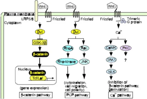
Wnt proteins constitute a large family of cysteine rich secreted ligands that regulate cell growth, motility, and differentiation during embryonic development. Wnts act in a paracrine fashion by activating at least three diverse signaling cascades inside the target cells (Figure 1.4).
1) The canonical Wnt pathway (Wnt/β-catenin signaling): The ‘‘classical’’, also called canonical, Wnt pathway activates target genes through stabilization of β-catenin and relocalization in the nucleus. The function of this pathway during embryonic development has been originally elucidated by experimental analysis of axis development in the frog Xenopus laevis and of segment polarity and wing development in the fly
Drosophila melanogaster. The abnormalities of this pathway lead to several human diseases, including tumor formation and bone abnormalities.
2) The planar cell polarity pathway: This pathway involves RhoA and Jun Kinase (JNK) and controls cytoskeletal rearrangements. Its main role is the temporal and spatial control of embryonic development, as exemplified in the polar arrangement of cuticular hairs in Drosophila or the convergent-extension movements in Xenopus embryos. On a cellular level, this pathway regulates the polarity of cells through effects on their cytoskeletal organization. To date, there is no evidence for the involvement of the planar cell polarity pathway in tumor development (Veeman MT. et al., 2003; Nelson WJ. and Nusse R., 2004; Bejsovec A., 2005).
Figure 1.4: Wnt signaling pathways (Kikuchi A. et al., 2006). Wnts act in a paracrine fashion by activating at least the diverse signaling cascades inside the target cells. The Canonical Wnt signaling regulates gene expression through the stabilization and translocation of β-catenin into nucleus. The Planar cell polarity pathway activates Dvl, small G proteins (Rho or Rac), and Rho kinase or JNK, resulting in regulation of the cytoskeleton, cell migration, and cell polarity. Wnt/Ca2+ pathway activates CaMK, PKC,
TAK, and NLK, resulting in the inhibition of the β-catenin pathway and regulates gastrulation. Dvl, Dishevelled; JNK, Jun N-terminal kinase; CaMK, calmodulin-dependent protein kinase.
3) Wnt/Ca2+ pathway: This pathway is stimulated by Wnt 5a and Wnt 11 and involves an increase in intracellular Ca2+ and activation of Ca2+-sensitive signaling components, such
as calmodulin-dependent kinase, the phosphatase calcineurin, and the transcription factor NF-AT (Veeman MT. et al., 2003; Nelson WJ. and Nusse R., 2004; Bejsovec A., 2005). The Wnt/Ca2+ pathway can counteract the canonical Wnt pathway (Weidinger G. and Moon RT., 2003). It is not clear whether this pathway is conserved in mammals and whether it is implicated in tumorigenesis.
1.4.4.2 Canonical Wnt signaling pathway
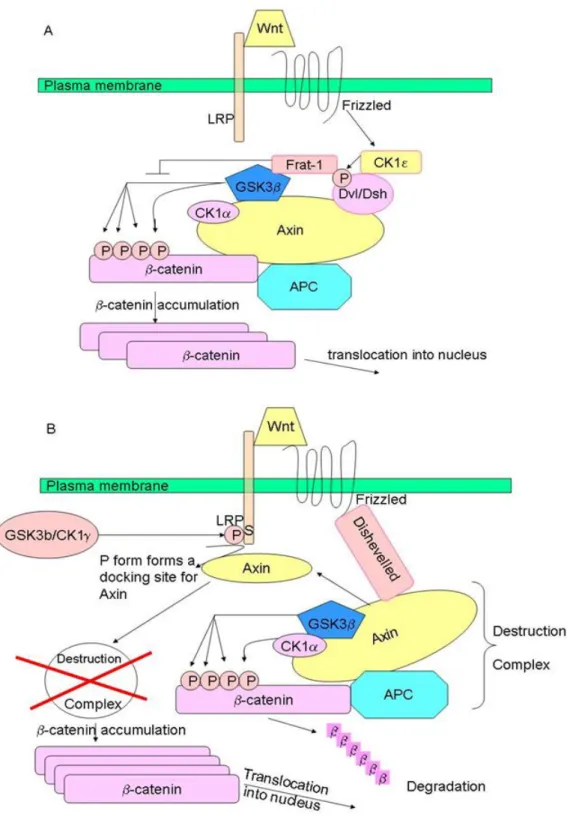
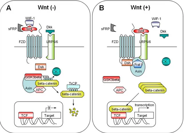
Wnt/β-catenin signaling pathway, named also canonical Wnt pathway, refers to the molecular cascade initiated by a Wnt ligand and culminating in the stabilization and translocation of the β-catenin protein into nucleus. Wnt-1 (originally named int-1, in Drosophila the homologous gene is called wingless, and the combination of both names led to the term) was the first identified as a gene activated by integration of the LTR of the mouse mammary tumor virus resulting in the development of mammary tumors in mice (Nusse R. and Varmus HE., 1982). β-catenin is the main executer of canonical Wnt signaling. In the absence of Wnts, β-catenin is phosphorylated by casein kinases 1α at serine residue 45; this in turn enables glycogen synthase kinase 3β to phosphorylate serine/threonine residues 41, 37, and 33. Phosphorylation of these amino acids triggers ubiquitination of β-catenin by β-transduction repeat containing protein (β-TrCP) and degradation in proteasomes. Phosphorylation of β-catenin occurs in a multiprotein complex containing the scaffold protein axin and/or its homologue axin2/conductin, the tumor suppressor gene product APC and GSK3β. In the presence of Wnts, β-catenin can be stabilized by two mechanisms. According to first model, Wnt signaling triggers CKIε-dependent phosphorylation of Dvl (Dsh in Drosophila) and enhances the binding of Dvl (Dsh) to Frat-1. Frat-1 binds to GSK-3β in the Axin complex, resulting in a reduction of the phosphorylation of β-catenin by GSK-3β. According to second model, Wnt triggers the phosphorylation of LRP6 by CK1γ and GSK-3β, which provides a docking site for Axin and recruits it to the plasma membrane. Wnt also enhances the binding of Dvl (Dsh) to Frizzled, and Dvl (Dsh) bound to Frizzled is necessary for the formation of a complex between Axin and LRP6, resulting in reduced phosphorylation of β-catenin and Axin by GSK-3β (Figure 1.5).
Figure 1.5: Wnt-dependent stabilization of β-catenin . A) Through the Dvl/Frat complex. Wnt signaling inhibits β-catenin phosphorylation by GKS3β through Dvl and Frat1.B) Through the Axin and LRP6 complex. Wnt triggers the phosphorylation of LRP6 by CKγ1 and GSK3β. Axin is recruited to the plasma membrane, which is a component of destruction complex, leading to the inhibition of β-catenin phosphorylation.