29
Geliş(Recevied) :12.11.2019 Araştırma Makalesi/Research Article
Kabul(Accepted) :11.12.2019 Doi: 10.30708.mantar.645828
Diversity and Distribution of Dematiaceous Fungi in Çamaltı
Saltern in İzmir Province, Turkey
Özden ÖZGÖK
1, Semra İLHAN
2* *Sorumlu yazar:[email protected]1Eskişehir Osmangazi University, Graduate School of Natural and Applied Science, Department
of Biology, 26480 Eskişehir, Turkey
Orcid ID: 0000-0003-3787-2737/ [email protected]
2 Eskisehir Osmangazi University, Faculty of Science and Letters, Department of Biology,
TR-26040 Eskisehir, Turkey
Orcid ID: 0000-0002-3787-2449/ [email protected]
Abstract: Dematiaceous fungi (black fungi) are a heterogeneous group of fungi present in diverse environments worldwide. Many species in this group are known for surviving in extreme conditions, especially in tropical and subtropical climates. In this study, a total of 16 water samples which were obtained seasonally from 4 different saltpans predetermined in the Çamaltı Saltern were studied. The isolation of fungi was performed by membrane filtration method using DRBC and DRBC10 media. The isolated fungi were identified based on their morphological characteristics and the identification was supported by internal transcribed spacer (ITS)-based phylogenetic analysis.
The average numbers of colonies were determined to be 15 CFU / 100 ml and 24 CFU / 100 ml on DRBC and DRBC10 media, respectively. Thirty species of 10 genera were identified. The genera are as follows: Alternaria, Arthrinium, Biscogniauxia, Chaetomium, Cladosporium,
Drechslera, Phomopsis, Pithomyces, Stachybotrys and Stemphylium. According to their relative
abundance, the dominant genera isolated on DRBC medium were Cladosporium (52%),
Alternaria (35%) and Chaetomium (6%), while Alternaria (50%) and Cladosporium (47%) on the
DRBC10 medium. The fungi isolated from Çamaltı Saltern water samples were found to belong mainly to Capnodiales and Pleosporales. This study represents the first survey of dematiaceous fungi in İzmir Çamaltı Saltern and provides data on their diversity and distribution.
Key words: Solar Saltern, Hypersaline, Black Fungi, Cladosporium, Alternaria
İzmir İlinde Bulunan Çamaltı Tuzlası
Dematiaceous Fungus Çeşitliliği ve Dağılımı
Öz: Dematiaceous funguslar (siyah funguslar) tüm dünyada çeşitli ortamlarda bulunan heterojen bir mantar grubudur. Bu gruptaki birçok türün, özellikle tropikal ve subtropikal iklimlerde, ekstrem koşullarda yaşadığı bilinmektedir. Bu çalışmada, Çamaltı tuzlasında belirlenen 4 farklı havuzdan mevsimsel olarak alınan toplam 16 su örneği çalışılmıştır. Mikrofungusların izolasyonu DRBC ve DRBC10 besiyerlerinde membran filtrasyon yöntemi ile gerçekleştirilmiştir. İzole edilen mikrofunguslar morfolojik özellikleri esas alınarak tanımlanmış ve ITS’ye dayalı filogenetik analiz ile desteklenmiştir.
Ortalama koloni sayısı DRBC besiyerinde 15 KOB / 100 ml ve DRBC10 besiyerinde 24 KOB / 100 ml bulunmuştur. Sonuç olarak, 10 cinse ait 30 tür belirlenmiştir. Bu cinsler; Alternaria,
Arthrinium, Biscogniauxia, Chaetomium, Cladosporium, Drechslera, Phomopsis, Pithomyces, Stachybotrys ve Stemphylium’dur. Göreceli bolluklarına göre, izole edilen baskın cinsler DRBC
besiyerinde Cladosporium (%52), Alternaria (%35) ve Chaetomium (%6) iken DRBC10 besiyerinde Alternaria (%50) ve Cladosporium (%47) olarak belirlenmiştir. Çamaltı Tuzlası tuzlu su örneklerinden izole edilen mikrofungusların ağırlıklı olarak Capnodiales ve Pleosporales takımlarına ait olduğu görülmüştür. Bu çalışma İzmir Çamaltı Tuzlasındaki Dematiaceous fungus çeşitliliği üzerine yapılan ilk çalışmayı temsil etmektedir.
30
IntroductionDematiaceous or melanized fungi are a heterogeneous group of fungi with dark-colored colonies and pigmented fungal elements. They are typically soil saprophytes, plant and (some of them) human pathogens, and laboratory contaminants with a worldwide distribution in humid environments. Furthermore, they have been known as inhabitants of extreme environments, such as rock surface, deserts, acidic and natural or artificial hypersaline environments (Abdel-Hafez, 1981; Butinar et al., 2005; Selbmann et al., 2015; Yenice Gursu et al., 2017).
For the last two decades, some fungi are known to survive in hypersaline environments and even salt-saturated waters, and they are natural inhabitants of such environments. The first comprehensive study suggesting that fungi are active members of hypersaline environments was carried out by Gunde-Cimerman et al., (2000). Since then, various salt-tolerant fungal strains have been investigated and consistently isolated from saline environments around the world, regardless of geographic location. Previously, Cronin and Post (1977) studied on the isolation and description of Cladosporium sp., a halophilic dematiaceous fungus from the Great Salt Lake, a hypersaline lake in Utah. In addition, the study on the isolation and identification of three fungal species from Dead Sea water samples carried out by Buchalo at al. (1998) is noteworthy.
Salterns may reflect all salinity ranges from sea water to salt saturation. Salterns with properties such as high salt concentrations leading to low water activity, low oxygen concentration, high light intensity, limited nutrient availability and near-neutral pH are unique habitats for microorganisms. They are the preferred environments for revealing the biodiversity in hypersaline waters as they provide easy-to-reach sampling points. The multi-pond coastal solar salterns consist of a series of conjunctive saltpans, with a gradient of salinities ranging from seawater to NaCl precipitation. Therefore, multi-pond solar salterns are model habitats for studying biological communities in hypersaline environments. Hypersaline environments such as the Great Salt Lake in the USA, the Dead Sea in Israel, The Cabo Rojo Solar Salterns in Puerto Rico, and Tuz Lake in Turkey are found in all continents (Cantrell et al., 2006; Gunde-Cimerman and Zalar, 2014). İzmir Çamaltı Saltern is the largest coastal solar saltern located on the Aegean coast of Turkey.
The aim of this study was to assess the diversity of dematiaceous fungi in the hypersaline ecosystem of Çamaltı Saltern, İzmir /Turkey. In order to characterize the spatiotemporal species composition of the
dematiaceous fungal community of the Çamaltı Saltern, it has been investigated whether the diversity and composition of fungal communities varies in the saltpans and seasons. In addition, the diversity and distribution of dematiaceous fungi have been evaluated through the frequency of occurrence and relative abundances.
Material and Method
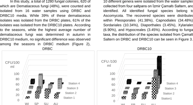
Site Description: İzmir Çamaltı Saltern is the largest coastal solar saltern located on the Aegean coast of Turkey (Figure 1). The saltern, a multi-pond system consisting of 176-180 ponds, covers an area of nearly 58-60 km². These shallow ponds are fed by sea water from the Aegean Sea. The salinity of these ponds ranges from 3.5% to 30%. Since 1863, salt extraction from sea water has been carried out in the Çamaltı saltworks. Annually, 35-40% of the salt produced in Turkey is produced at the saltworks, so it is an important source of salt particularly for the food industry (Koru and Perçin, 2018).
Sampling, Isolation and Enumeration of Fungi: The water sampling was performed compositely from four ponds at Çamaltı Saltern in January, April, July and October 2013 (Figure 1). The total salt concentration of each sample was determined in situ with a hand refractometer (Eclipse) and the pH and temperature were measured with TOA WQC water analyzer at the sampling point. The water samples collected into sterile plastic containers were kept in cold temperature and processed within 24 hours.
In order to isolate and enumerate the fungal species from water, 20 ml of each sample was filtered through the sterile Cellulose Nitrate Membrane Filters (pore size 0.45 µm, Ø 47 mm Sartorius) and the filter papers were placed onto the Petri plates containing DRBC (aw 0.99) and DRBC10 (DRBC+10% NaCI) (aw 0.93) media with chloramphenicol (100 mg/L) (King et al.,1979). Untreated salt harvested from the saltern was used in DRBC10 medium. The plates were incubated for 5 weeks at 25°C. Fungal colony forming unit (CFU) was counted on 3rd, 5th, 7th, 14th and 30th days of incubation, and subcultures were made of all of the morphologically distinct colonies from each sample on Malt Extract Agar (Merck) slants and kept at 4°C. Individual pure strains have been deposited in the culture collection of the Department of Biology, Eskisehir Osmangazi University (Turkey).
The filtration was conducted in parallel from the three aliquots of water samples and the average numbers of colonies were calculated as CFU / 100ml. Water activties of the media have been determined using the water activity meter (Aqualab, Decagon Devices, USA).
31
Figure 1. Çamaltı Saltern and the sampling stations Identification of Fungi: The identification of the
fungal species was performed based on the examination of their micro- and macro-morphological structures using stereomicroscopy (Zeiss Stemi 2000-C) and light microscopy, as well as the upper and lower surface colorations on MEA, PDA and Synthetic Nutrient-Poor Agar (SNA). The genus identifications were carried out by following Barnett and Hunter (1999) and the identifications of the species were executed complying with related literatures, “Dematiaceous Hyphomycetes” (Ellis, 1971), “Biodiversity in the Cladosporium herbarum complex (Davidiellaceae, Capnodiales), with standardization of methods for Cladosporium taxonomy and diagnostics” (Schubert et al., 2007), “Fungi and Food Spoilage” (Pitt and Hocking, 2009), and Alternaria: An Identification Manual (Simmons, 2007). Microscopic measurement of reproductive or vegetative organs, which are of great importance in determination of filamentous fungi, was performed on days 7 and 14 using the Differential Interference Contrast (DIC) microscope with Nikon H550L Imaging System.
After all these processes, their identity was confirmed using the analysis of internal transcribed spacer regions (ITS1and ITS2) of the ribosomal DNA operon (including the 5.8S gene) of the filamentous fungi. For the molecular study, all isolates were grown in Malt Extract broth (Merck) of 2 mL of in 15 mL tubes and incubated at 25°C for 7 days in darkness (Samson et al., 2004). Genomic DNA of the selected pure cultures was extracted using CTAB following the related protocol (Graham et al., 1994; Murray and Thompson, 1980). The extracted DNAs were stored at -20°C in a freezer. The ITS regions were amplified with the universal primers
forward ITS1 (5′-TCCGTAGGTGAACCTGCG G-3′) and reverse ITS4 (5′-TCCTCCGCTTATTGATATGC-3′) (White et al., 1990). The purification and sequencing analyses of amplicons was performed by BM Labosis (Ankara, Turkey).
The gathered sequences were analyzed using Chromas (Mc Carthy, 1996) and the phylogenetic tree was deduced from the neighbor–joining algorithm by MEGA 6 with 1000 bootstrap replicates (Tamura et al., 2011). The consensus sequence was aligned with all sequences from related species retrieved from the NCBI
GenBank database using BLAST
(http://www.ncbi.nlm.nih.gov) (Altschul et al., 1990; Altschul et al., 1997; Geer et al., 2010). The sequences were submitted to GenBank with the accession numbers. Data Analysis: For each fungal species, the frequency of occurrence (%) and relative abundance (%) were calculated (Maria and Sridhar, 2003; Sarma and Hyde, 2001). The frequency of occurrence of fungi was artificially grouped according to the percentage occurrence of fungi as very frequent (>20%), frequent (10-20%), and infrequent (<10%) used by Leong et al. (1991) (Sarma and Hyde, 2001).
Results
The results of the physicochemical analysis of 16 water samples collected from the stations, i.e. 4 different saltpans on Çamaltı Saltern are presented in Table 1. During the year, the temperature varied between 6.0°C and 35.2°C according to in situ measurements at the stations. While the average temperature at the sampling points showed a significant difference according to the seasons, no significant difference was observed between
32
the saltpans. Average temperatures were 10.5°C in winter, 6°C in the spring, 33.3°C in summer and 19°C in autumn. The pH of the water samples ranged from 6.83 to 8.26. It was determined that the average pH value was the lowest in winter whereas it was the highest in spring.The salinity of the water samples varied temporarily and spatially. The highest salinity was determined to be measured as 28% in the saltpan 4 in autumn, while the lowest salinity was 16% in the saltpan 3 in winter (Table 1).
Table 1. The coordinates for each station in the Çamaltı Saltern and analyzed environmental variables
Coordinates Temperature (°C) SU/AU/WI/SP pH SU/AU/WI/SP Salinity (%) SU/AU/WI/SP Station 1 38°29'41.85”N 26°54'57.60"E 32.8/19.0/8.5/6.0 7.91/7.85/7.07/7.85 26.5/24.0/23.0/20.0 Station 2 38°29'47.94”N 26°55'8.06"E 32.0/19.0/11.0/6.0 7.94/7.83/7.04/8.21 24.5/25.0/22.0/20.0 Station 3 38°29'32.45”N 26°55'14.25"E 35.2/19.0/11.0/6.0 7.95/7.89/6.90/8.26 20.5/27.0/16.0/20.0 Station 4 38°29'30.81”N 26°55'30.08"E 33.3/19.0/11.5/6.0 7.87/7.98/6.83/8.20 22.0/28.0/23.5/20.0
SU, Summer; AU, Autumn; WI, Winter; SP, Spring
Enumeration and Identification of Fungi: The fungal isolates were enumerated and isolated using two different media having low (DRBC10, aw 0.93) and high water activity (DRBC aw 1.0) with the culture-dependent counting method. Then the dynamics of fungi inhabited in hypersaline waters was estimated. Twenty ml of each water sample was used for the filtration to provide the optimal colony distribution, i.e. formation of enumerable amount of colony forming unit, on the membrane filters during incubation.
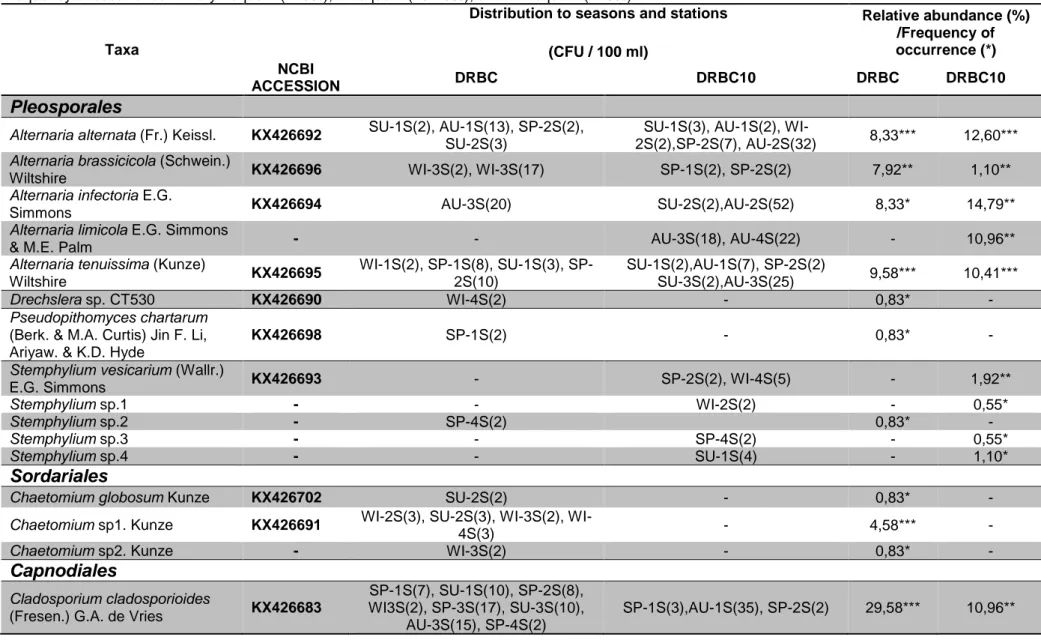
In this study, a total of 1280 fungal colonies, 620 of which are Dematiaceous fungi (48%), were counted and isolated from 16 water samples using DRBC and DRBC10 media. While 39% of these dematiaceous isolates was isolated from the DRBC plates, 61% of the isolates was isolated from the DRBC10 plates. According to the seasons, while the highest average number of dematiaceous fungi was determined in autumn in DRBC10 medium, no significant difference was observed among the seasons in DRBC medium (Figure 2),
suggesting that the seasonal conditions of autumn are growth-promoting for halotolerant / halophilic dematiaceous fungi.
The fungal isolates were identified depending on their morphological properties and the identification was confirmed using molecular sequencing of the ITS. Based on seasons and stations, the distribution of fungal isolates was organized by alphabetical order within taxa and presented in Table 2.
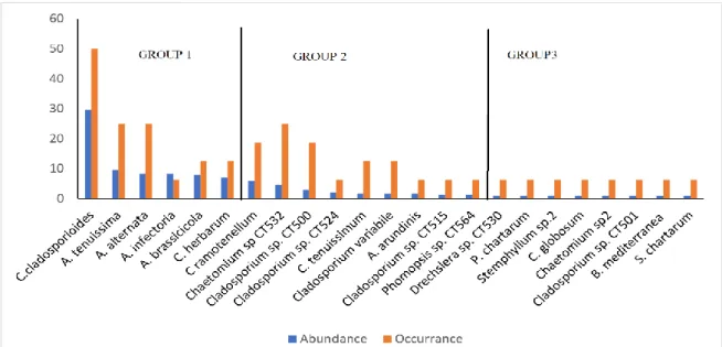
Thirty species of dematiaceous fungi belonging to 10 different genera were isolated from the water samples collected from four saltpans on İzmir Çamaltı Saltern, and identified. All identified fungal species belong to Ascomycota. The recovered species were distributed within Pleosporales (41.38%), Capnodiales (34.48%), Sordariales (10.34%), Diaporthales (3.45%), Xylariales (6.90%), and Hypocreales (3.45%). According to fungal taxa, the distribution of the species isolated from Çamaltı Saltern on DRBC and DRBC10 can be seen in Figure 3.
DRBC DRBC10
Figure 2. Distribution of the total colony numbers of Dematiaceae members according to the seasons and stations. Station 1 Station 2 Station 3 Station 4 0 20 40 60 80 100 WI SP SU AU CFU/100 ml Station1 Station 2 Station 3 Station 4 0 20 40 60 80 100 WI SP SU AU CFU /100 ml
33
Table 2. Seasonal and stational distribution of the fungal taxa isolated from four saltpans at Çamaltı Saltern (on two media DRBC and DRBC10). Frequency of occurrence ***very frequent (>20%), **frequent (10-20%), and *infrequent (<10%)
Taxa
Distribution to seasons and stations (CFU / 100 ml) Relative abundance (%) /Frequency of occurrence (*) NCBI ACCESSION DRBC DRBC10 DRBC DRBC10
Pleosporales
Alternaria alternata (Fr.) Keissl. KX426692 SU-1S(2), AU-1S(13), SP-2S(2), SU-2S(3)
SU-1S(3), AU-1S(2),
WI-2S(2),SP-2S(7), AU-2S(32) 8,33*** 12,60***
Alternaria brassicicola (Schwein.)
Wiltshire KX426696 WI-3S(2), WI-3S(17) SP-1S(2), SP-2S(2) 7,92** 1,10**
Alternaria infectoria E.G.
Simmons KX426694 AU-3S(20) SU-2S(2),AU-2S(52) 8,33* 14,79**
Alternaria limicola E.G. Simmons
& M.E. Palm - - AU-3S(18), AU-4S(22) - 10,96**
Alternaria tenuissima (Kunze)
Wiltshire KX426695 WI-1S(2), 1S(8), SU-1S(3), SP-2S(10) SU-1S(2),AU-1S(7), SP-2S(2) SU-3S(2),AU-3S(25) 9,58*** 10,41*** Drechslera sp. CT530 KX426690 WI-4S(2) - 0,83* - Pseudopithomyces chartarum
(Berk. & M.A. Curtis) Jin F. Li, Ariyaw. & K.D. Hyde
KX426698 SP-1S(2) - 0,83* -
Stemphylium vesicarium (Wallr.)
E.G. Simmons KX426693 - SP-2S(2), WI-4S(5) - 1,92**
Stemphylium sp.1 - - WI-2S(2) - 0,55*
Stemphylium sp.2 - SP-4S(2) 0,83* -
Stemphylium sp.3 - - SP-4S(2) - 0,55*
Stemphylium sp.4 - - SU-1S(4) - 1,10*
Sordariales
Chaetomium globosum Kunze KX426702 SU-2S(2) - 0,83* -
Chaetomium sp1. Kunze KX426691 2S(3), SU-2S(3), 3S(2),
WI-4S(3) - 4,58*** -
Chaetomium sp2. Kunze - WI-3S(2) - 0,83* -
Capnodiales
Cladosporium cladosporioides
(Fresen.) G.A. de Vries KX426683
SP-1S(7), SU-1S(10), SP-2S(8), WI3S(2), SP-3S(17), SU-3S(10),
AU-3S(15), SP-4S(2)
34
TaxaDistribution to seasons and stations (CFU / 100 ml) Relative abundance (%) /Frequency of occurrence (*) NCBI ACCESSION DRBC DRBC10 DRBC DRBC10
Cladosporium herbarum (Pers.)
Link KX426685 SP-1S(2), SP-3S(15) SP-1S(3),SU-1S(2), WI-2S(2),SP-2S(2), WI-3S(2),SU-3S(2), AU-4S(75) 7,08** 24,11*** Cladosporium ramotenellum K.
Schub., Zalar, Crous & U. Braun KX426686 SU-1S(10), SP-2S(2), SU-2S(2) - 5,83** -
Cladosporium sphaerospermum
Penz. KX426682 - WI-1S(5), AU-1S(30) - 9,59**
Cladosporium tenuissimum
Cooke KX426688 SU-1S(2), SP-4S(2) - 1,67** -
Cladosporium variabile (Cooke)
G.A. de Vries KX426684 SU-3S(2), SP-4S(2) - 1,67** -
Cladosporium sp1. Link KX426681 SP-1S(2) - 0,83* -
Cladosporium sp2.Link KX426689 SU-1S(5) SU-1S(5) 2,08* 1,37*
Cladosporium sp3. Link KX426680 SP-1S(2), SP-3S(3), SP-4S(2) - 2,92** -
Cladosporium sp4. Link KX426687 SU-2S(3) - 1,25* -
Diaporthales
-Phomopsis sp. KX426701 WI-4S(3) - 1,25*
Xylariales
Arthrinium arundinis (Corda)
Dyko & B. Sutton KX426697 WI-4S(4) - 1,67* -
Biscogniauxia mediterranea (De
Not.) Kuntze KX426700 WI-4S(2) - 0,83* -
Hypocreales
Stachybotrys chartarum (Ehrenb.)
S. Hughes KX426699 SU-3S(2) - 0,83* -
Unidentified isolates -
SU, Summer; AU, Autumn; WI, Winter; SP, Spring; 1S, Station 1; 2S, Station 2; 3S, Station 3; 4S, Station 4 The isolates with no NCBI accession number were identified by conventional methods.
35
The species diversity is as follows; Cladosporium (10 spp), Alternaria (5 spp), Stemphylium (5 spp),Chaetomium (3 spp), Arthrinium (1 sp), Biscogniauxia (1
sp), Drechslera (1 sp), Phomopsis (1 sp),
Pseudopithomyces (1 sp), and Stachybotrys (1 sp).
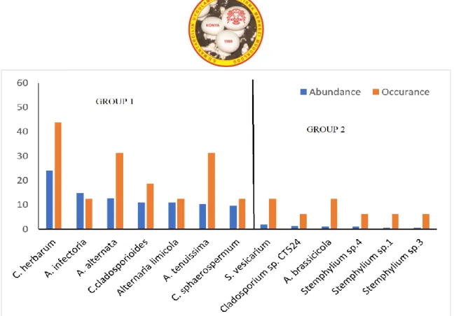
The relative abundances and frequency of occurrences of the isolated fungal species on DRBC and DRBC10 media are expected to be related to their growth, survival or transient status (contamination) in these environments. Based on the abundance on DRBC and DRBC10 media, the figures 4 and 5 can be divided into 3 groups and 2 groups, respectively. In both media, the first group included species possessing the highest abundance (7.08-29.58 for DRBC and 9.59-24.11 for
DRBC10) and thus the most actively developing in them. All species in DRBC group1 were also isolated in DRBC10 group 1. However, A. lumicola and C.
sphaerospermum were isolated only on DRBC10
medium. The relative abundance and frequency of occurrence of C. cladosporioides (29.58%; 50.00%) on DRBC and C. herbarum (24.11%; 43.75%) on DRBC10 media were the highest among all the isolated species, respectively. The second group contained the species with significantly lower abundance than that of the previous group of species (from 1.25 % to 5.83 % on DRBC and from 0.55 % to 1.92 % on DRBC10). The frequency of occurrence of these species were low with no significant difference (Figure 4 and 5).
DRBC DRBC10
Figure 3. The distribution of the species according to fungal taxa isolated from Çamaltı Saltern on DRBC and DRBC10.
Figure 4. The relative abundance (%) and frequency of occurrence (%) of the fungal species isolated on DRBC medium Pleosporales Sordariales Capnodiales Diaporthales Xylariales Hypocreales Pleosporales Capnodiales
36
Figure 5. The relative abundance (%) and frequency of occurrence (%) of the fungal species isolated on DRBC10 medium
Discussion
Hypersaline environments are characterized as thalassohaline and athalassohaline on the basis of their salt compositions (Oren, 2016). Since the brine in Çamaltı Saltern originate by evaporation of seawater, it reflects the ionic composition of the seawater and therefore it is known as thalassohaline which Na+ is the predominant cation. The salinity of the water samples collected from saltpans varied between 16% and 28% throughout the year (Table 1). The weather conditions such as temperature, precipitation and wind during the year cause fluctuations in the salinity levels of the stations. At the same time, the precipitation of different chemical salts occurs during the flow of salty water from the ponds. This situation causes the formation of salt layers in the bottom of the crystallization ponds and the water in the ponds to have a certain salt concentration despite the changing weather conditions. Seasonal sampling from different ponds was predicted to yield positive results in determining species diversity.
The fungal growth limitation of an isolation medium, such as DRBC, gives positive results in the isolation of melanized / dematiaceous fungi requiring longer duration of incubation. However, the use of a selective medium in which the fungi that survive in / adapted to hypersaline environments can be isolated was needed. For this reason, DRBC medium containing 10% salt (DRBC10)
was used as the second isolation medium. The water activities were 0.99 and 0.93 for DRBC and DRBC10, respectively. Although this water activity is a high value for xerophilic ones, it is particularly selective for halotolerant and moderate xerophilic ones.
It is noteworthy that Dematiaceae members had a high rate in summer and autumn seasons, where the temperature and salinity reach the highest values and the ponds are exposed to intense radiation. The fungi of the Dematiaceae family are dark-colored as they produce melanin pigment on the cell walls and are able to cope with stress conditions in their natural environment thanks to their adaptation mechanism. These fungi with spores and mycelia surrounded by dark cell walls can better tolerate dehydration and radiation than fungi belonging to the Moniliaceae family that do not contain melanin pigment in their cells (Gunde-Cimerman et al., 2004). This group of fungi are known to be natural members of hypersaline environments (Gunde-Cimerman et al., 2000).
Total colony number of Dematiaceae members varies according to the seasons and stations (Figure 2). There is a moderate fluctuation in the average number of colonies isolated from DRBC medium according to the seasons (7-27 CFU / 100 ml). In DRBC10 medium, Dematiaceae colonies showed similar fluctuations in winter, spring and summer seasons (5-9 CFU / 100 ml),
37
while a significant increase in autumn season (75 CFU / 100 ml). A similar situation was reported by Gunde Cimerman et al. (2000). They reported that they isolated a high number of Dematiaceae members when they used selective medium from autumn samples of a sea-linked salt on the Adriatic coast of Slovenia. They associate this situation with the fact that at the beginning of the autumn season when salt production is the highest, as a result of the decrease in water activity to 0.75 due to the increase of various ions in the water, only species that have adapted to these conditions can be found here. The fact that the salinity reached the highest value during the autumn season sampling (Table 1) supports the relatively high isolation of Dematiaceae members in selective medium.The most abundant and thus the most actively developing species, i.e. representatives of the first group of both media were A. alternata, A. brassicicola, A.
infectoria, A. limicola, A. tenuissima, C. cladosporioides, C. herbarum, C. sphaerospermum (Figure 3,4). These
species are present in various habitats including stressful ones. Several dematiaceous fungi identified in this study have been previously reported in various saline environments including A. alternata (Diguță et al., 2018),
C. cladosporioides (Fotedar et al., 2018), C. herbarum, C. sphaerospermum (Butinar et al., 2005).
Chaetomium is a large genus of Chaetomiacea
(Ascomycota) family with over 100 marine and soil members. They are known as remarkable cellulase producers but can also be isolated from water and soil samples (Steiman et al., 2004). It was reported that different types of Chaetomium species, especially Ch.
globosum, which is a marine endophytic species (Pitt and
Hocking, 2009), are isolated from the water and soil samples belonging to hypersaline environments (Cantrell et al., 2006; Kis-Papo et al., 2003). Kis-Papo et al. (2003), who pointed out that Chaetomium spores are highly resistant to extreme conditions, reported that Ch.
globosum spores remained alive even after 12 weeks of
standing in undiluted Dead Sea (Israel) water samples. In our study, Ch. globosum and two Chaetomium isolates
unidentified at species level were isolated at low number (3 CFU /100ml) on non-selective DRBC medium (Table 2). When the data obtained from different studies are evaluated together, even though the numbers are low, the representatives of this genus are always isolated from hypersaline environments and are thought that they have an adaptation that allows them to withstand high salinity concentrations for a long time.
Conclusion
In conclusion, this is the first comprehensive report concerning the composition of native dematiaceous fungal community in a saline environment Çamaltı Saltern in Turkey. Thirty dematiaceous fungal isolates were identified belonging to genera Alternaria, Artrinium,
Biscogniauxia, Chaetomium, Cladosporium, Drechslera, Stemphylium, Phomopsis, Pithomyces and Stachybotrys
using morphological and molecular approaches. Two of these, Alternaria and Cladosporium, were the most frequently isolated genera from Çamaltı Saltern hypersaline water. All identified fungal species are belonging to Ascomycota. The members of the Capnodiales and Pleosporales are the main inhabitants of the hypersaline environments.
Seasonal sampling from different ponds was predicted to yield positive results in determining species diversity. Furthermore, it has been revealed that the use of selective media with different salt concentration in fungal isolations is effective in detecting fungal diversity. Further studies will be focused on halotolerance test of the fungal isolates, as well as their biotechnological potential. This study will be expanded to the fungal communities of other salt environments distributed in Turkey.
Acknowledgement
This study was supported by Scientific Research Projects Commission of Eskişehir Osmangazi University with a Project number of 201319A101. We thank Assoc. Prof. Dr. R. Demirel for her help in identifying the samples.
References
Abdel-Hafez, S. (1981). Halophilic fungi of desert soils in Saudi Arabia. Mycopathol., 75(2), 75-80.
Altschul, S. F., Gish, W., Miller, W., Myers, E. W., and Lipman, D. J. (1990). Basic local alignment search tool. J. Molecular Biol., 215(3), 403-410.
Altschul, S. F., Madden, T. L., Schäffer, A. A., Zhang, J., Zhang, Z., Miller, W., & Lipman, D. J. (1997). Gapped BLAST and PSI-BLAST: a new generation of protein database search programs. Nucleic Acids Res., 25(17), 3389-3402. Barnett, H., and Hunter, B. (1999). Illustrated genera of imperfect fungi (4th ed.): St. Paul: APS.
38
Buchalo, A., Nevo, E., Wasser, S., Oren, A., and Molitoris, H. (1998). Fungal life in the extremely hypersaline water of the Dead Sea: first records. Proceedings of the Royal Society B: Biological Sciences, 265(1404), 1461-1465.
Butinar, L., Sonjak, S., Zalar, P., Plemenitaš, A., & Gunde-Cimerman, N. (2005). Melanized halophilic fungi are eukaryotic members of microbial communities in hypersaline waters of solar salterns. Botanica Marina, 48(1), 73-79.
Cantrell, S. A., Casillas-Martínez, L., and Molina, M. (2006). Characterization of fungi from hypersaline environments of solar salterns using morphological and molecular techniques. Mycol Res, 110(8), 962-970.
Cronin, E. A., and Post, F. J. (1977). Report of a dematiaceous hyphomycete from the Great Salt Lake, Utah. Mycologia, 69(4), 846-847.
Diguță, C. F., Proca, I. G., Jurcoane, Ș., and Matei, F. (2018). Molecular characterization by PCR-RFLP of indigenous fungal isolates from hypersaline stream water in România. Folia Microbiologica, 1-8.
Ellis, M. B. (1965). Dematiaceous hyphomycetes. Commonwealth Mycological Institute. Kew.
Fotedar, R., Kolecka, A., Boekhout, T., Fell, J. W., Al-Malki, A., Zeyara, A., & Al Marri, M. (2018). Fungal diversity of the hypersaline Inland Sea in Qatar. Botanica Marina, 61(6), 595-609.
Geer, L. Y., Marchler-Bauer, A., Geer, R. C., Han, L., He, J., He, S., Bryant, S. H. (2010). The NCBI BioSystems database. Nucleic Acids Res., 38(suppl_1), D492-D496.
Graham, G., Mayers, P., and Henry, R. (1994). A simplified method for the preparation of fungal genomic DNA for PCR and RAPD analysis. Biotechniques, 16(1), 48-50.
Gunde-Cimerman, N., and Zalar, P. (2014). Extremely halotolerant and halophilic fungi inhabit brine in solar salterns around the globe. Food Technol. Biotechnol. 52(2), 170-179.
Gunde-Cimerman, N., Zalar, P., de Hoog, S., and Plemenitaš, A. (2000). Hypersaline waters in salterns–natural ecological niches for halophilic black yeasts. FEMS Microbiol. Ecol., 32(3), 235-240.
King Jr, A., Hocking, A., and Pitt, J. (1979). Dichloran-rose bengal medium for enumeration and isolation of molds from foods. Appl. and Environ. Microbiol., 37(5), 959-964.
Kis-Papo, T., Oren, A., Wasser, S., and Nevo, E. (2003). Survival of filamentous fungi in hypersaline Dead Sea water. Microbial Ecol., 45(2), 183-190.
Koru, E., and Perçin, F. (2018). Characteristics of Biological Systems in Çamaltı Solar Saltworks (İzmir/Turkey). Qual. Stud. (NWSAQS), 13(3), 15-25.
Leong, W.F., Tan, TX. and E.B.G. Jones. (1991). Fungal colonization of submerged Bruguiera cylindrica and Rhizophora apiculata wood. Botanica Marina, 34, 69-76.
Maria, G., and Sridhar, K. (2003). Diversity of filamentous fungi on woody litter of five mangrove plant species from the southwest coast of India. Fungal Divers., 14, 109-126.
Mc Carthy. (1996). Chromas, (Version 1.45). School of Health science, Griffifth University, Gold Coast Campus: Queensland, Australia, .
Murray, M., and Thompson, W. (1980). Rapid isolation of high molecular weight plant DNA. Nucleic Acids Res., 8(19), 4321-4326.
Oren, A. (2016). Life in High-Salinity Environments Manual of Environmental Microbiology, Fourth Edition. American Society of Microbiology.
Pitt, J. I., and Hocking, A. A. D. (2009). Fungi and food spoilage. Springer.
Samson, R. A., Seifert, K. A., Kuijpers, A. F., Houbraken, J., and Frisvad, J. C. (2004). Phylogenetic analysis of Penicillium subgenus Penicillium using partial β-tubulin sequences. Stud Mycol., 49, 175-200.
Sarma, V., and Hyde, K. D. (2001). A review on frequently occurring fungi in mangroves. Fungal Divers., 8, 1-34.
Schubert, K., Groenewald, J. Z., Braun, U., Dijksterhuis, J., Starink, M., Hill, C., . . . Crous, P. W. (2007). Biodiversity in the
Cladosporium herbarum complex (Davidiellaceae, Capnodiales), with standardisation of methods for Cladosporium
taxonomy and diagnostics. Stud Mycol, 58(1), 105-156.
Selbmann, L., Zucconi, L., Isola, D., & Onofri, S. J. C. g. (2015). Rock black fungi: excellence in the extremes, from the Antarctic to space. Curr. Genet., 61(3), 335-345.
Simmons, E. G. (2007). Alternaria: An Identification Manual. American Society of Microbiology Washington D.C. USA. Steiman, R., Ford, L., Ducros, V., Lafond, J.-L., and Guiraud, P. (2004). First survey of fungi in hypersaline soil and water
39
Tamura, K., Peterson, D., Peterson, N., Stecher, G., Nei, M., and Kumar, S. (2011). MEGA5: molecular evolutionary genetics analysis using maximum likelihood, evolutionary distance, and maximum parsimony methods. Mol. Bio.l Evol., 28(10), 2731-2739.
White, T., Bruns, T., Lee, S., and Taylor, J. (1990). Amplification and direct sequencing of fungal ribosomal RNA genes for phylogenetics. In PCR protocols: a guide to methods and applications (pp. 315-322): Academic Press.
Yenice Gursu, B., Aytar, P., Ilhan, S., Kocabiyik, Y. E., Gedikli, S., and Cabuk, A. (2017). Diversity of microfungi in acid mine drainages. Biol. Divers. Conserv., 10(3), 184-192.