A Superficially Located Soft Tissue Mass in Upper Leg of a 7-Year-Old Boy
Tam metin
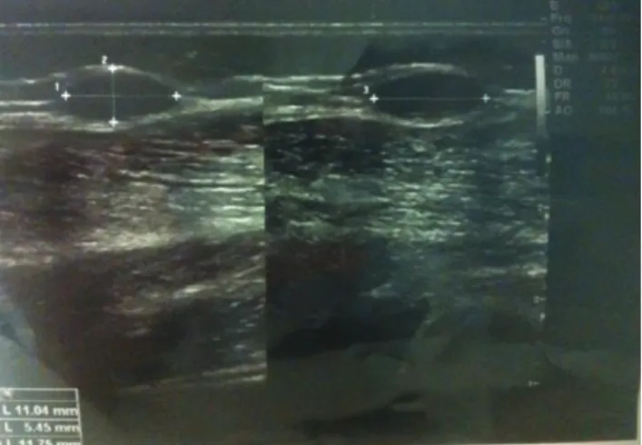
Şekil
Benzer Belgeler
Psoriazis hastasında ustekinumab ile ilişkili gelişen büllöz pemfigoid ve literatür derlemesi Ustekinumab associated bullous pemphigoid in a psoriasis patient and a review of
For example, the open space (well-court) of the temple in the northeast corner of the settlement of Hacilar IIA from the Chalcolithic Age [6] (Figure 2) and the open garden
In a magnetic particle imaging (MPI) scanner, utilizing a tunable gradiometer receive coil can aid in achieving greater degree of decoupling of direct feedthrough signal.. However,
With a large surplus of labor in agricultural and other primary services, and with informal economies of considerable size, premature deindustrialization and lack of
Hem on altıncı yüzyıl kadın giysisi kol kalıplarına yer verileceği hem de bu eski kalıplardan ve kol formlarından yola çıkarak son bölümde yeni
The significance of social influences of tourism expansion can not be overrated; all the agencies and sectors which are involved in the process of planning have to be
İlkçağ Anadolu’sunun Troya kentinde gerçekleşen ve Homeros’un “İlyada” destanına da konu olan “Troya savaşı”; rivayete göre, güzeller güzeli bir kadın olan
Hallaq’a göre oryantalistlerin İslam hukukunun teşekkül döneminde diğer kültürlerden ödünç alarak geliştiği iddiası, kolonyal güçlerin İslam ülkelerinde cari
![Ambrosius Aurelianus [called Emrys Wledig] (fl. 5th cent.), military leader](data:image/gif;base64,R0lGODlhAQABAIAAAP///wAAACH5BAEAAAAALAAAAAABAAEAAAICRAEAOw==)