Case Report
A Rare and Serious Syndrome That Requires Attention in
Emergency Service: Traumatic Asphyxia
Gultekin Gulbahar,
1Tevfik Kaplan,
2Ahmet Gokhan Gundogdu,
1Hatice Nurdan Baran,
3Burak Kazanci,
4Bulent Kocer,
5and Serdar Han
21Division of Thoracic Surgery, Dr. Nafiz Korez Sincan State Hospital, Ankara, Turkey 2Department of Thoracic Surgery, Ufuk University School of Medicine, Ankara, Turkey
3Division of Anesthesiology and Reanimation, Dr. Nafiz Korez Sincan State Hospital, Ankara, Turkey 4Department of Neurosurgery, Ufuk University School of Medicine, Ankara, Turkey
5Division of Thoracic Surgery, Ankara Numune Teaching and Research Hospital, Ankara, Turkey Correspondence should be addressed to Tevfik Kaplan; [email protected]
Received 27 February 2015; Accepted 5 May 2015 Academic Editor: Ching H. Loh
Copyright © 2015 Gultekin Gulbahar et al. This is an open access article distributed under the Creative Commons Attribution License, which permits unrestricted use, distribution, and reproduction in any medium, provided the original work is properly cited.
Traumatic asphyxia is a rare syndrome caused by blunt thoracoabdominal trauma and characterized by cyanosis, edema, and subconjunctival and petechial hemorrhage on the face, neck, upper extremities, and the upper parts of the thorax. Traumatic asphyxia is usually diagnosed by history and inspection; however, the patient should be monitored more closely due to probable complications of thoracoabdominal injuries. Treatment is conservative, but the prognosis depends on the severity of the associated injuries. Herein we present a traumatic asphyxia due to an elevator accident in a 32-year-old male patient and discuss the diagnosis, treatment, and prognosis by reviewing the relevant literature.
1. Introduction
Traumatic asphyxia is a rare syndrome resulting from sud-den, severe blunt trauma of the thorax and upper abdomen [1, 2]. It is characterized by cervicofacial cyanosis, edema, subconjunctival hemorrhage, and petechial eruptions on the face, neck, upper parts of the thoracic cage, and the upper extremities [3]. Commonly, thoracic injuries like pulmonary contusion, hemothorax, and pneumothorax accompany the situation [1,2].
2. Case
A 32-year-old male patient was admitted to the emergency service after an industrial accident, in which an elevator cabin fell on him. The patient was conscious and had stable hemo-dynamic state on his initial examination. He had dyspnea, back pain, facial cyanosis, subconjunctival hemorrhage, and petechial eruptions on the anterior surface of the thoracic
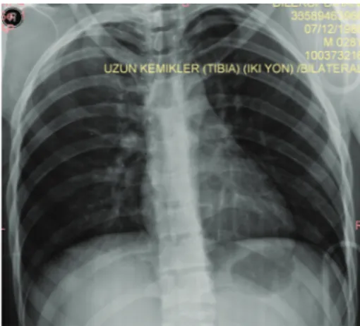
cage and on the left upper extremity (Figures1(a)and1(b)). Laboratory workup was consistent with the trauma. Bilateral pulmonary contusions, minimal pneumothorax, and fracture of the transverse processes of the first lumbar vertebrae were detected on computed axial tomography of the chest and lumbar vertebrae (Figures2(a)and 2(b)). Chest X-ray examination of the patient revealed bilateral heterogeneous density increases (Figure 3).
The patient was hospitalized in the intensive care unit for follow-up. He was monitored in a continuous fashion. The head of the bed was elevated to 30 degrees to help overcome the increased intracranial pressure. Uninterrupted oxygen therapy was administered. Hemodynamic state of the patient, arterial blood gas analysis, oxygen saturation levels, and hematologic and biochemical parameters were checked at certain intervals. He was taken to the ward since no complication was observed on the first day of the trauma. The symptoms regressed and no complication was observed; thus he was discharged on the fifth day of hospitalization. Volume 2015, Article ID 359814, 4 pages
2 Case Reports in Emergency Medicine
(a) (b)
Figure 1: (a) The patient had bilateral subconjunctival hemorrhage. (b) The patient had facial cyanosis, petechial eruptions on the anterior surface of the thoracic cage and on left upper extremity.
(a) (b)
Figure 2: (a) Minimal pneumothorax was detected on computed axial tomography of the chest. (b) Fracture of the transverse processes of the first lumbar vertebrae was detected on computed axial tomography of the lumbar vertebrae.
3. Discussion
Traumatic asphyxia is quite rare [3]. In 1937, when Oliver d’Angers first defined traumatic asphyxia, he used the term “ecchymotic mask” [4]. Traumatic cyanosis, compressive cyanosis, traumatic apnea, Oliver’s syndrome, and Perth’s syndrome have also been used to describe the situation [5,6]. Usually, it follows a sudden and severe thoracoabdominal trauma, but it may also be seen in asthmatic attacks, epileptic seizure episodes, excessive vomiting, and cough cases [6,7].
Sudden increase in the intrathoracic pressure is thought to be transmitted to the venous and capillary systems in head and neck, and these yields in stasis and rupture giving rise to the characteristic findings [3,8]. Other possible mechanism is the lack of the valve system in superior vena cava, innominate vein, and jugular veins. Deeply situated structures drain into the internal jugular veins whereas the superficial ones drain into the external jugular system. Since the valves of the external jugular system cannot prevent the backflow, the symptoms are more common in the superficial structures like
Figure 3: Chest X-ray of the patient.
the scalp compared to the deeper structures like brain and the respiratory tract [9]. The compressed IVC protects the lower part of the body from injury during the course of the trauma. The extent of the signs and symptoms depend on the duration and severity of the compression that thorax and upper abdomen are exposed to [10]. Many signs may accom-pany the characteristic findings. Confusion, amnesia, dis-orientation, uneasiness, agitation, hypoxia, cerebral edema, and hemorrhage are the most common ones [6]. Other than subconjunctival hemorrhage, decreased vision, blurred vision, papillary changes, optic nerve atrophy, diplopia, and exophthalmia are the most frequent ocular findings. Epistaxes due to capillary rupture, hearing loss due to the edema of the Eustachian tubes, or hemotympanum are the other probable findings [6, 8, 9]. Severe life-threatening conditions may coexist since the traumatic asphyxia develops as a result of major trauma to thorax, mediastinum, and upper abdomen. These are thoracic and extrathoracic injuries as pulmonary contusion, hemothorax, pneumothorax, flail chest, and hepatic laceration [1, 2, 9]. Signs of pulmonary injury like dyspnea, tachypnea, and hemoptysis may be observed. In rare cases, cardiac injury may be encountered [9]. An increase in the abdominal pressure may lead to organ injury, hematemesis, and hematuria. Apnea and hypoxemia associated with prolonged thoracic compression may be life threatening and give rise to increased mortality. The results may be such severe as shock, cerebral anoxia, neurological sequelae, and sudden death [7,9].
Although traumatic asphyxia can be diagnosed easily through short anamnesis and physical examination, other reasons like superior vena cava syndrome and skull base fractures leading to subconjunctival hemorrhage and peri-orbital ecchymosis must be evaluated [7]. Careful and thor-ough examination is necessary to detect the thoracic and extrathoracic injuries that need urgent intervention. No severe injury other than bilateral pulmonary contusion and minor pneumothorax was found in our case.
Traumatic asphyxia cases must be monitored after secur-ing the airway and fixsecur-ing the cervical spine. Oxygen inhala-tion therapy and intravenous fluid replacement need to be initiated and the patient shall be intubated and followed on
mechanical ventilation as needed [9]. The intubation may be difficult because of the airway edema, which is seen rarely [11]. Uncomplicated cases are treated conservatively. The head of the bed is preferred to be elevated at 30 degrees and oxygen administration is essential. This procedure decreases the intracranial pressure. Continuous monitorization of the patient, arterial blood gas, oxygen saturation level exami-nations at certain intervals, and supportive oxygen therapy are the key factors for the management throughout the hospitalization period [6,10].
Traumatic asphyxia has almost perfect prognosis. It heals spontaneously within weeks except the neurological and ocular signs. In cases without accompanying injuries death may occur due to prolonged compression, apnea, and hypoxia [6,10].
4. Conclusion
Traumatic asphyxia is a rare entity. Prognosis usually depends on the duration of the compression and the severity of the accompanying injuries. Quick and careful evaluation of the probable life-threatening conditions in the emergency service is crucial; however oxygen inhalation therapy, efficient ven-tilation, intravenous hydration, and measures to prevent the increase of intracranial pressure should be initiated as soon as possible.
Consent
Written consent form was taken from the patient for using his photographs in this scientific paper.
Conflict of Interests
The authors declare that there is no conflict of interests regarding the publication of this paper.
References
[1] Y. A. Karamustafaoglu, I. Yavasman, S. Tiryaki, and Y. Yoruk, “Traumatic asphyxia,” International Journal of Emergency
Medicine, vol. 3, no. 4, pp. 379–380, 2010.
[2] S. Kamali, S. Kesici, I. Gunduz, and U. Kesici, “A case of traumatic asphyxia due to motorcycle accident,” Case Reports in
Emergency Medicine, vol. 2013, Article ID 857131, 3 pages, 2013.
[3] C. Eken and ¨O. Yi˘git, “Traumatic asphyxia: a rare syndrome in
trauma patients,” International Journal of Emergency Medicine, vol. 2, no. 4, pp. 255–256, 2009.
[4] J. Dweek, “Ecchimotic mask,” The Journal of the International
College of Surgeons, vol. 9, pp. 257–265, 1946.
[5] N. S¸enoglu, H. Oksuz, B. Zıncırcı, M. Ezbercı, and A. Yasım, “Severe traumatic asphyxia: two case report and literature review,” Turkish Journal of Thoracic and Cardiovascular Surgery, vol. 14, pp. 78–81, 2006.
[6] T. R. Hurtado and D. A. Della-Giustina, “Traumatic asphyxia in a 6-year-old boy,” Pediatric Emergency Care, vol. 19, no. 3, pp. 167–168, 2003.
4 Case Reports in Emergency Medicine
[7] J. R. Dunne, G. Shaked, and M. Golocovsky, “Traumatic asphy-xia: an indicator of potentially severe injury in trauma,” Injury, vol. 27, no. 10, pp. 746–749, 1996.
[8] J. S. Williams, S. L. Minken, and J. T. Adams, “Traumatic asphyxia-reappraised,” Annals of Surgery, vol. 167, no. 3, pp. 384–392, 1968.
[9] E. Sertaridou, V. Papaioannou, G. Kouliatsis, V. Theodorou, and I. Pneumatikos, “Traumatic asphyxia due to blunt chest trauma: a case report and literature review,” Journal of Medical Case
Reports, vol. 6, article 257, 2012.
[10] I. Ertok, G. Kurtoglu C¸ elik, G. Ercan Haydar, O. Karakayali, M.
Yılmaz, and T. Ers¸en, “Review of traumatic asphyxia syndrome with a case presentation,” Journal of Academic Emergency
Medicine Case Reports, vol. 4, no. 2, pp. 58–61, 2013.
[11] P. Ibarra, L. M. Capan, S. Wahlander, and K. M. Sutin, “Difficult airway management in a patient with traumatic asphyxia,”
Submit your manuscripts at
http://www.hindawi.com
Stem Cells
International
Hindawi Publishing Corporationhttp://www.hindawi.com Volume 2014
Hindawi Publishing Corporation
http://www.hindawi.com Volume 2014
INFLAMMATION
Hindawi Publishing Corporation
http://www.hindawi.com Volume 2014
Behavioural
Neurology
Endocrinology
International Journal ofHindawi Publishing Corporation
http://www.hindawi.com Volume 2014 Hindawi Publishing Corporation
http://www.hindawi.com Volume 2014
Disease Markers
Hindawi Publishing Corporation
http://www.hindawi.com Volume 2014
BioMed
Research International
Oncology
Journal ofHindawi Publishing Corporation
http://www.hindawi.com Volume 2014
Hindawi Publishing Corporation
http://www.hindawi.com Volume 2014
Oxidative Medicine and Cellular Longevity
Hindawi Publishing Corporation
http://www.hindawi.com Volume 2014
PPAR Research
The Scientific
World Journal
Hindawi Publishing Corporation
http://www.hindawi.com Volume 2014
Immunology Research
Hindawi Publishing Corporation
http://www.hindawi.com Volume 2014
Journal of
Obesity
Journal ofHindawi Publishing Corporation
http://www.hindawi.com Volume 2014
Hindawi Publishing Corporation
http://www.hindawi.com Volume 2014
Computational and Mathematical Methods in Medicine
Ophthalmology
Journal ofHindawi Publishing Corporation
http://www.hindawi.com Volume 2014
Diabetes Research
Journal ofHindawi Publishing Corporation
http://www.hindawi.com Volume 2014
Hindawi Publishing Corporation
http://www.hindawi.com Volume 2014
Research and Treatment
AIDS
Hindawi Publishing Corporation
http://www.hindawi.com Volume 2014
Gastroenterology Research and Practice
Hindawi Publishing Corporation
http://www.hindawi.com Volume 2014