728
Case Report
Introduction
Acute aortic syndromes (AAS) are life-threatening condi-tions, and despite advances in imaging techniques, their diag-nosis and treatment remain challenging. While intramural hema-toma is much less common than acute aortic dissection (AD), which is the most common form of AAS, they have similar mor-tality rates (1). On the other hand, aortitis may mimic intramural hematoma (IMH) due to radiologic similarity and, thus, may lead to misdiagnosis and treatment.
Here, we describe an IgG4-related aortitis case mimicking intramural hematoma (IMH) due to radiologic image and present-ing with chest pain and severe aortic regurgitation.
Case Report
A 49-year-old male patient with new-onset retrosternal chest pain was admitted to our clinic, and unstable angina was sus-pected. He had a history of coronary artery by-pass grafting surgery 7 years ago and stenting for right coronary artery 1 year ago. He was under statin treatment for hyperlipidemia. On physi-cal examination, his heart rate was 110/min, blood pressure was 104/45 mm Hg, and an aortic diastolic murmur was auscultated. Electrocardiography was free from signs of new ischemia. Echo-cardiographic examination revealed normal left ventricle systolic function, ascending aortic aneurysm with maximal diameter of 4.7 cm, and severe aortic regurgitation, which were not present in previous exams. Aortic wall thickness was visually noted to be increased in parasternal long-axis (Fig. 1a). A preliminary diagno-sis of AAS was established using computed tomography (CT). CT images were consistent with a circular IMH image with a thick-ness of 12 mm and extending from aortic root to aortic arch and also containing orifices of major branches of aorta (Fig. 1b, c). No entry for flap or dissection was seen. CT coronary angiography showed normal circumflex artery, patent LIMA-AD, and IMH con-taining ostia of both left main coronary artery and right coronary artery RCA. Moreover, external pressure by IMH caused LMCA stenosis >50%. Heart team recommended surgery to the patient with ongoing chest pain. Re-sternotomy was performed through an old incision line. Because of unexpectedly severe adhesive and firm character of tissues in the surgical area, the ascending aorta, branches of aortic arch, and thoracic aorta could not be clearly distinguished during surgery. Aortic lumen was reached
by cutting the stiff tissue around the aortic outer surface, and aortic wall thickness was measured as 11 mm. A 23-mm St. Ju-de’s prosthetic valve was implanted with Ti-Cron suture. After 2 days, during follow-up in the coronary care unit, the patient died. Pathological examination of the aorta revealed IgG4-related aor-titis and chronic inflammation containing medial, adventitial, and periadventitial lymphocytes and plasma cells (Fig. 2a–c).
Discussion
IgG4-related aortitis is defined as an inflammatory and non-infectious type of vasculitis. The exact mechanism underlying this pathology remains to be elucidated; however, genetic profile and autoimmune mechanism triggered by bacterial infections
IgG4-related aortitis mimicking
intramural hematoma
Gamze Babur Güler, Emir Cantürk*, Ekrem Güler, Gülbin Oran**, Gültekin Günhan Demir, Atıf Akçevin*, İrfan Barutçu
Departments of Cardiology, *Cardiovascular Surgery, **Pathology, Faculty of Medicine, İstanbul Medipol University; İstanbul-Turkey
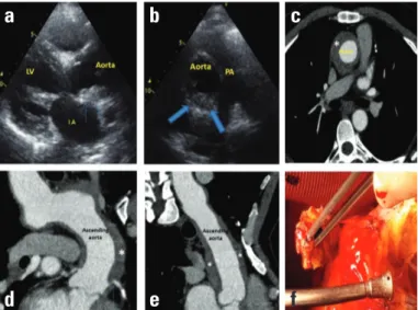
Figure 1. (a,b) Parasternal long and short-axis images showing thick-ened aortic wall (blue arrows) starting just above aortic valve (c,d,e) CT image showing circular thickened aortic Wall (*) extending from aortic valve level to aortic arch and mimicking intramural hematoma image, and the relationship of coronary ostium with wall thickness (blue arrow), (f) macroscobic thickened aortic wall image obtained from intraoperative specimens
a
d
b
e
c
f
Figure 2. Lymphoplasmocytic infiltration in biopsy material obtained from aorta (a) Immunohistochemical IgG staining (original magnifica-tion x20), (b) Stain acquisimagnifica-tion with IgG4 in 50 % of the cells stained with immunohistochemical IgG (x20), (c) Hematoxylin and eosin stain-ing showstain-ing inflammation includstain-ing lymphocytes and plasma cells but no granuloma formation (lymphoplasmocytic pattern ) (x40)
have been implicated. Estimated prevalence of IgG4-related tho-racic aortitis is 9–22% (2). It is associated with increased eryth-rocyte sedimentation rate, C-reactive protein, and Ig-G4 levels in plasma (2).
Long-term mortality of IMH is similar to that of aortic dissec-tion; however, it differs from dissection with bleeding into the aortic wall and absence of false lumen and intimal tear. It con-stitutes 10–25% of AAS cases. IgG4-related aortitis is a relatively uncommon form of aortitis, which may mimic IMH and usually affects individuals older than 50 years. Moreover, it can cause pancreatitis, sialadenitis, lymphadenopathy, sclerosing cholan-gitis, nephritis, and retroperitoneal fibrosis via systemic inflam-matory response. According to 2015 dated consensus statement of Society for Cardiovascular Pathology, the requirements for the diagnosis of IgG4-related aortitis/periaortitis were increased levels of serum IgG4, histopathologically confirmed fibrosis, an IgG4/IgG ratio greater than 50%, and more than 50 IgG4+ plasma cells per 400x HPF (3).
IgG4-related aortitis can be confused with IMH because of increased aortic wall thickness, and in this context, various cas-es have been reported. Tay et al. (4) made a post-operative di-agnosis of IG4-related aortitis in a patient with hoarseness who was previously diagnosed as IMH on CT and had undergone sur-gery for IMH. Moreover, regression in simultaneously increased abdominal aortic wall thickness was noted after prednisolone treatment for 1 month. Rituximab was shown to be effective in regression of refractory or recurrent disease despite steroid therapy. Holmes et al. (5) reported an IgG4-related aortitis case presenting with sudden cardiac death associated with aortic dissection extending into LMCA. Similarly, our patient suffered new-onset chest pain and severe aortic regurgitation due to complications of progressive IgG4-related aortitis. The reasons responsible for greater difficulty during surgery than anticipated were; a) involvement of a wide segment of ascending aorta, b) involvement of coronary arteries thus leading severe aortic re-gurgitation, c) re-sternotomy, d) lack of being on medical therapy for aortitis. Although early surgical intervention is recommend-ed for Type A IMH, investigation for aortitis mimicking IMH will
change the course of management. Unless patients diagnosed with IMH is unstable, examination of levels of plasma IgG and IgG4 should help establishment of accurate diagnosis.
Conclusion
Our ability to detect intramural hematoma has increased with the advancement of diagnostic technology and better clas-sification of acute aortic syndromes. Not only morphological but also pathological properties such as inflammatory reaction that develop during these syndromes are very similar to each other. Keeping this fact in mind is critical when deciding on diagnostic and therapeutic strategies.
References
1. Harris KM, Braverman AC, Eagle KA, Woznicki EM, Pyeritz RE, Myr-mel T, et al. Acute aortic intramural hematoma: an analysis from the International Registry of Acute Aortic Dissection. Circulation 2012; 126: S91-6.
2. Kasashima S, Zen Y, Kawashima A, Endo M, Matsumoto Y, Kasashi-ma F, et al. A clinicopathologic study of immunoglobulin G4-related sclerosing disease of the thoracic aorta. J Vasc Surg 2010; 52: 1587-95.
3. Stone JR, Bruneval P, Angelini A, Bartoloni G, Basso C, Batoroeva L, et al. Consensus statement on surgical pathology of the aorta from the Society for Cardiovascular Pathology and the Association for European Cardiovascular Pathology: I. Inflammatory diseases. Cardiovasc Pathol 2015; 24: 267-78.
4. Tay DZ, Goh PY, Teo TK, Boey ML, Chachlani N, Wong PS. Immu-noglobulin G4-related aortitis mimicking an intramural hematoma. Asian Cardiovasc Thorac Ann 2015; 23: 1083-6.
5. Holmes BJ, Delev NG, Pasternack GR, Halushka MK. Novel cause of sudden cardiac death: IgG4-related disease. Circulation 2012; 125: 2956-7.
Address for Correspondence: Dr. Gamze Babur Güler Tem Avrupa Otoyolu Göztepe çıkış No: 1 Bağcılar 34214 İstanbul-Türkiye E-mail: [email protected]
©Copyright 2016 by Turkish Society of Cardiology - Available online at www.anatoljcardiol.com
DOI:10.14744/AnatolJCardiol.2016.7185
Case Report