CORRESPONDENCE
Double snaring of superior vena cava
Levent Çetin1&Erkan Kuralay1Received: 9 November 2018 / Revised: 12 November 2018 / Accepted: 19 November 2018 / Published online: 7 December 2018 # Indian Association of Cardiovascular-Thoracic Surgeons 2018
Introduction
Superior septal approach provides the best exposure of the mitral valve. It is the preferred approach for all mitral operations [1]. When the superior vena cava (SVC) is shorter than usual, cannulation of SVC becomes difficult. Air leaking and air blocking of pump lines may develop necessitating the surgeon to divert his attention to the cannulation system. Deairing of tubing set becomes nec-essary for effective pump perfusion in these situations, which besides being tedious and cumbersome, wastes time, and may compromise the safety. Double throw of SVC tape avoids this complication.
Technique
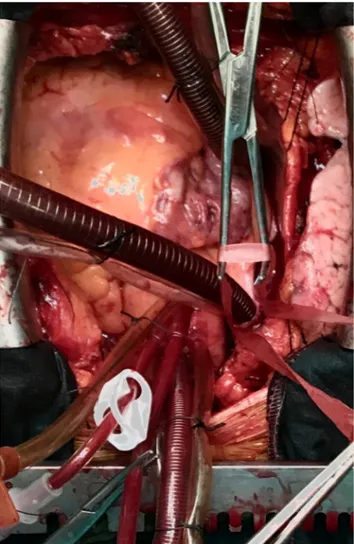
Pericardial reflections over the SVC are dissected and purse string stitch placed to SVC. Both SVC and inferior vena cava (IVC) are cannulated. Cardiopulmonary perfu-sion is instituted and the heart is decompressed. IVC snared. Then, SVC taped double times with same tape and the snare squeezed firmly so that air does not enter into the pump lines (Fig.1).
Discussion
Air blocking of venous system is much more common when superior septal approach is used. Excessive air may impede pump perfusion due to air blocking of
venous line of pump. The most common cause of air invasion is loosening of SVC snare during surgical in-tervention. We have taped/snared SVC double times as a routine to avoid air leaking into pump line in all patients since 2008. We think that double snaring tech-nique should be in surgical armamantarium of valve surgeons.
Fig. 1 SVC taped double times with same tape and the snare s queezed firmly
* Erkan Kuralay
1 Cardiovascular Surgery Department, Lokman Hekim Hospital,
Citipiti Sokak no:6 Angora Evleri Çayyolu, Çankaya, Ankara, Turkey
https://doi.org/10.1007/s12055-018-0774-4
Compliance with ethical standards
Conflict of interest The authors declare that they have no conflict of interest.
Ethical approval Lokman hekim univercity has approved our technique since 2012.
Ap number: 2012/23-16
Informed consent Informed consent was obtained from all individual participants included in the study.
Publisher’s Note Springer Nature remains neutral with regard to jurisdic-tional claims in published maps and institujurisdic-tional affiliations.
Reference
1. Utley JR, Leyland SA, Nguyenduy T. Comparison of outcomes with three atrial incisions for mitral valve operations. Right lateral, supe-rior septal, and transseptal. J Thorac Cardiovasc Surg. 1995;109: 582–7.