http://journals.tubitak.gov.tr/medical/ © TÜBİTAK
doi:10.3906/sag-1712-80
Ankaferd Blood Stopper with antibiofilm potential successfully inhibits the extracellular
matrix degradation enzymes and promotes wound healing of 3T3 fibroblasts in vitro
Rukiye BORAN1, Tuba BAYGAR2,*, Nurdan SARAÇ3, Aysel UĞUR4
1Medical Laboratory Program, Department of Medical Services and Techniques, Vocational School of Health Services,
Aksaray University, Aksaray, Turkey
2Material Research Laboratory, Research Laboratories Center, Muğla Sıtkı Koçman University, Muğla, Turkey 3Department of Biology, Faculty of Science, Muğla Sıtkı Koçman University, Muğla, Turkey
4Section of Medical Microbiology, Department of Basic Sciences, Faculty of Dentistry, Gazi University, Ankara, Turkey
1. Introduction
Ankaferd Blood Stopper (ABS) is an herbal agent composed of Thymus vulgaris, Urtica dioica, Alpinia
officinarum, Glycyrrhiza glabra, and Vitis vinifera (1),
and it contains no inorganic or synthetic additives (2). Each of the plants used in ABS has some effects on blood cells, endothelium, cell proliferation, vascular dynamics, angiogenesis, apoptosis, inflammation, or cell mediators (3,4). This product has been licensed for use for external, dental, and postsurgical major or minor hemorrhages (5). The basic mechanism of action for ABS is the formation of an encapsulated protein network, representing focal points for vital erythrocyte aggregation (6).
ABS can be used against bleeding, such as in adenoidectomy/tonsillectomy, gastrointestinal hemorrhages, anterior epistaxis, and urological surgery (5–9). Dental use of ABS has also been reported (10–12). In addition, several scientific papers have been published representing the potential use of ABS as an anticancer (13), antiangiogenesis
(14), antifungal (15), antimicrobial (16,17), antioxidant, antimutagenic (18), and wound healing agent (19–21).
Wound healing is a highly complex biological process, which requires the coordinated behavior of some factors such as blood cells, connective tissue and epithelial cells, inflammatory cells, molecular and humoral components, and many soluble factors (22). Among the important factors involved in the control of the wound healing process are the extracellular matrix (ECM) macromolecules including collagens, elastins, glycosaminoglycans, glycoproteins, proteoglycans, and polysaccharides (23).
Collagen plays an essential role in all phases of the wound healing process, including hemostasis, inflammation, proliferation, and remodeling. Collagen creates an appropriate healing environment by facilitating wound healing with the organization and accumulation of newly formed fibers and granulation tissue in the wound bed (24). Deficiency in specific collagens is closely linked to Background/aim: The potential inhibitory effects of Ankaferd Blood Stopper (ABS) against biofilm formation of oral microorganisms and its capacity for collagenase, hyaluronidase, and elastase inhibitions that have important roles in wound healing have been investigated. Materials and methods: The wound healing potential was determined by its inhibition ability on collagenase, hyaluronidase, and elastase enzyme activities and was evaluated via scratch wound healing assay on murine 3T3 fibroblasts. The antibiofilm activity was tested against eight oral microorganisms using the crystal violet staining method.
Results: At 10% ABS successfully inhibited the biofilm formation of the tested microorganisms. Enzyme inhibition analysis revealed that 3% ABS significantly inhibited all three enzymes related to wound healing. The scratch assay showed that wound closure was faster than that of the control for the 3% ABS/plate.
Conclusion: The findings of the present study indicated that ABS has effective wound healing potential with its strong antibiofilm activity against oral cavity microorganisms.
Key words: Ankaferd Blood Stopper, enzyme inhibition, antibiofilm, wound healing
Received: 13.12.2017 Accepted/Published Online: 07.04.2018 Final Version: 14.06.2018 Research Article
abnormal wound closure, so collagen-based formulations have played roles in providing wound healing (25), dural closure, reinforcement of compromised tissue, and guided tissue regeneration (24). Elastin is an insoluble ECM protein that provides flexibility and elasticity to skin arteries and lungs. Because of its highly cross-linked nature, elastin is not soluble and is difficult to process into new biomaterials (26). Glycosaminoglycan chains are very important players in the wound healing process. The most important is hyaluronic acid, which is present in large quantities in the skin and provides viscoelasticity and hydrophilicity of the tissue. Hyaluronic acid interacts with cell surface receptors. Interaction of hyaluronic acid with its receptor causes considerable events in the wound repair process, such as collagen secretion, modulation of inflammation, cell migration, chemotaxis, and angiogenesis (27,28).
However, the inflammatory response accelerates the synthesis of dermal enzymes, (matrix metalloproteinase, elastase, hyaluronidase, etc.), leading to the degradation of ECM. Collagenase can break down molecules such as collagen, elastin, fibronectin, aggrecan, gelatin, and laminin (29). Elastase has a broad substrate portfolio, such as elastin, fibronectin, collagen, and other ECM proteins (30). Hyaluronidases play an important role in the degradation of cellular hyaluronan. The scratch wound healing assay is used for testing the effects of crude extracts of medicinal plants, isolated compounds, and pharmaceutical preparations on fibroblast migration and proliferation into artificially wounded monolayers (31).
Shortly after an injury, microbial colonization begins to occur in the wound, and at about the same time, there is confusion between the innate immune response, pathogens, and microflora. However, microorganisms can cause infection, which can impair the wound healing process and result in chronic wounds (32). The human oral cavity includes different living areas such as the teeth, gums, cheeks, tongue, palate, and tonsils, which are colonized by bacteria. The oral cavity is a propitious environment for bacteria with different tissue tropisms to colonize and grow due to its diverse surfaces. Reports have shown that almost 800 species of microorganisms inhabit this environment and a number of these are associated with oral diseases (33). Microorganisms from the oral cavity have been shown to cause oral infectious diseases such as periodontitis, caries, endodontic infections, tonsillitis, and alveolar osteitis. Various evidence emerged that links oral bacteria to some systemic diseases, including pneumonia, cardiovascular disease, preterm birth, stroke, and diabetes (34). The use of antimicrobial agents to prevent contamination of oral wounds is highly needed for healing, particularly during periodontal and oral and maxillofacial surgery, specifically in the scope of implantodontics. In various cases in dentistry, infections of oral wounds can be stopped with different antimicrobials and antiseptics (35).
The presence of microbial infection may prevent the wound healing process (36). Microbial colonization may occur in all chronic or acute injuries (37) and the effects on the healing process of microbial wound colonization are of great importance because infection destroys the wound repair process, slowing epithelization (38) and delaying healing (39). Deterioration in the wound healing process causes morbidity and mortality and significant economic and social impacts in a large proportion of the population (40). Therefore, a higher level of interest and research is needed to assess new therapeutic agents that can speed wound healing and reduce the incidence of infected wounds (41).
Therefore, in this study, the wound healing effect of ABS was determined by assessing its potential inhibitory activity on collagenase, elastase, and hyaluronidase enzymes, all of which have essential roles in the wound healing process. Thereafter, the wound healing properties of ABS were screened on 3T3 murine fibroblasts with an in vitro scratch assay and its antibiofilm activity was analyzed against oral cavity microorganisms.
2. Materials and methods
ABS is a patented product (Turkish Patent No. 2007-0-114485) comprising a standardized mixture of the plants
G. glabra (90 µg/mL), V. vinifera (80 µg/mL), U. dioica
(60 µg/mL), T. vulgaris (50 µg/mL), and A. officinarum (70 µg/mL). A 2-mL vial of ABS was obtained from the manufacturer (Trend Teknoloji İlaç AŞ, Ankara, Turkey). Throughout the study, a 10% (v/v) final concentration of ABS was tested for antibiofilm activity; 3% and 0.3% (v/v) ABS were tested for enzyme inhibition assay and 3% (v/v) ABS was tested for in vitro scratch assay.
2.1. Microorganisms and culture conditions
Candida albicans (ATCC 10239), Staphylococcus aureus
(ATCC 25923), and Streptococcus mutans (ATCC 25175) were purchased from the American Type Culture Collection (ATCC). Streptococcus mitis (DSMZ 12643),
Streptococcus oralis (DSMZ 20395), Streptococcus sobrinus
(DSMZ 20742), Streptococcus sanguinis (DSMZ 20068), and
Streptococcus parasanguinis (DSMZ 6778) were purchased
from the German Collections of Microorganisms and Cell Cultures (DSMZ). S. aureus was cultured in nutrient broth at 37 °C. S. mutans, S. sanguinis, S. mitis, S. oralis, S.
sobrinus, and S. parasanguinis were cultured in brain heart
infusion broth in a humidified atmosphere of 5% CO2 at 37 °C. C. albicans was cultured in Sabouraud dextrose broth at 30 °C.
2.2. Antibiofilm activity
Antibiofilm activities of ABS were determined against the abovementioned strains using crystal violet staining. Inocula containing 5 × 10⁵ cfu/mL cells and fresh broth medium containing 5% sucrose and 10% ABS (final
concentration in each tube) were inoculated into tubes and were incubated under appropriate conditions for each microorganism for 72 h. After the incubation period, tubes were rinsed 3 times with phosphate buffer solution (PBS) to remove planktonic cells and fixed by drying at 37 °C for 2 h. Dried tubes were stained with 0.1% (w/v) crystal violet for 10 min and were rewashed with water before being left to dry. The bound dye was extracted from the adherent cells using 33% acetic acid and the optical densities of the solutions were measured at 550 nm (Multiskan GO UV/Vis Microplate Spectrophotometer, Thermo Fisher Scientific, USA). The experiments were performed in duplicate. 2.3. Scanning electron microscopy (SEM)
Biofilm inhibition of the ABS was also observed by SEM using glass coverslips (42). Sterile circle glass coverslips (20 × 20 mm) were placed in biofilm assay tubes, which were prepared as described above. After appropriate incubation periods for each microorganism, the coverslips were gently rinsed with PBS (pH 7.4) and fixed with 2.5% glutaraldehyde at 4 °C for 2 h. After glutaraldehyde fixation, the coverslips were washed again with PBS for 1 h and dehydrated by increasing concentrations of ethanol. Specimens were air-dried and coated by gold (Emmitech K550, UK) before examination by SEM (JSM-7600F, JEOL Ltd., Tokyo, Japan).
2.4. Collagenase inhibitory activity
Collagenase (EC 3.4.24.3) (0.8 U/mL) was mixed with the same volume of tricine buffer (pH 7.5) (prepared with 400 mM NaCl and 10 mM CaCl2) and ABS (0.3% and 3%). The mixture was incubated at 37 °C for 20 min before 50 µL of N-(3-[2-furyl]-acryloyl)-Leu-Gly-Pro-Ala (1.6 mM) was added. Epigallocatechin gallate (EGCG) and deionized water were used as the reference and negative control, respectively. To measure the collagenase inhibitory activity of ABS, absorbances were determined at 335 nm quickly and at 2-min intervals for 20 min (43).
2.5. Hyaluronidase inhibitory activity
One hundred microliters of ABS (0.3% and 3%) and 50 µL of bovine hyaluronidase (7900 U/mL) were stirred and incubated at 37 °C for 20 min. Then 100 µL of CaCl2 (12.5 mM) was added and again incubated under the same conditions. After incubation, 250 µL of sodium hyaluronate (1.2 mg/mL) was added and incubated at 37 °C for 40 min. The solution was blended with 100 µL of 0.2 M sodium borate and 50 µL of 0.2 M NaOH and incubated in a bath of boiled water for 3 min. After cooling to room temperature, a p-dimethylaminobenzaldehyde solution was added to the mixture and incubated under the same conditions (44). Tannic acid and deionized water were used as the reference and negative control, respectively. To measure the hyaluronidase inhibitory activity of ABS, absorbances were determined at 585 nm.
2.6. Elastase inhibitory activity
Fifty microliters of Tris-HCl buffer (200 mM, pH 8.0), 25 µL of elastase (EC 3.4.21.36), and 50 µL of ABS (0.3% and 3%) were stirred and incubated at 25 °C for 20 min. Then 125 µL of N-succinyl-Ala-Ala-Ala-p-nitroanilide solution was added and incubated under same conditions. EGCG and deionized water were used as the reference and negative control, respectively. The absorbance was monitored at 410 nm for 20 min (44). For enzyme inhibition analysis, the following formula was used to determine the percentage of inhibition of collagenase, hyaluronidase, and elastase enzymes:
Inhibition (%) = [(A – B) – (T – D)] / (A – B) × 100 Here, A is the absorbance without the test sample, B is the absorbance without the test sample and enzyme, T is the absorbance with the test sample, and D is the absorbance with the test sample without the enzyme. 2.7. Determination of in vitro scratch wound healing assay
2.7.1. Cell culture
The mouse embryonic fibroblast cell line NIH 3T3 (3T3), obtained from the ATCC (Manassas, VA, USA), was grown in Dulbecco’s minimal essential medium supplemented with 10% heat-inactivated fetal calf serum, L-glutamine (2 mM), penicillin (100 IU/mL), and streptomycin (100 µg/mL). Cells were maintained at 37 °C in a humidified atmosphere of 5% CO2 and subcultured after being detached from culture flasks with 0.05% trypsin and 0.002% EDTA solution. Exponentially growing cells were seeded onto a cell culture dish (60 × 10 mm) at a density of 75 × 104/cm2 and were maintained in culture for 24 h prior to treatments.
2.7.2. Scratch wound healing assay
The spreading and migration capabilities of 3T3 fibroblasts were assessed using a scratch wound assay, which measures the expansion of a cell population on surfaces (45). Before deciding the amount of the ABS, a pilot study was employed at different concentrations of ABS and 3% ABS/plate was found to be the most effective concentration to exhibit the wound healing property. Briefly, a linear wound was generated in the monolayer with a sterile 100-µL plastic pipette tip. Any cellular debris was removed by washing the plate with Dulbecco’s phosphate-buffered saline. Thereafter, cells were treated with 3% ABS/plate and maintained in culture for a period of 48 h. Controls consisted of cells cultured in basal medium (untreated controls). Representative images from each cell culture dish of the scratched areas were photographed using a Leica DM IL microscope (Leica Microsystems, Wetzlar, Germany) to estimate the relative migration of cells. The experiments were performed in duplicate.
3. Result
The Table shows the inhibition of collagenase, hyaluronidase, and elastase of ABS. In vitro collagenase, hyaluronidase, and elastase inhibitory activity assay results showed that ABS displayed a significant inhibitory activity on all three enzymes at 10 µg/mL concentrations. The inhibition values of ABS on collagenase, hyaluronidase, and elastase were 88 ± 0.31%, 96.8 ± 0.47%, and 66 ± 0.07%, respectively (Table). ABS exhibited enzyme inhibitory activity in a dose-dependent manner. ABS showed 10.5 ± 0.1% collagenase inhibitory activity, 45 ± 0.3% hyaluronidase inhibitory activity, and 12 ± 0.01% elastase inhibitory activity at the dose of 0.3%. In addition, ABS showed very strong enzyme inhibitory activity at 3% as compared to standard epigallocatechin gallate (36 ± 0.31% inhibition for collagenase and 47 ± 0.7% inhibition for elastase at 100 µg/mL) and tannic acid (61.6 ± 0.81% inhibition for hyaluronidase at 1 mg/mL concentration).
For in vitro determination of biofilm formation, crystal violet is commonly used and its density is measured spectrophotometrically. Crystal violet dye not only displays stains but also displays a small quantity of molecules that change the formation of biofilm. ABS inhibited the growth of the biofilm layer of S. aureus, S. sanguinis, S. mitis, S.
sobrinus, and S. parasanguinis by 94.48 ± 0.96%, 87.00 ±
0.92%, 86.57 ± 0.97%, 82.38 ± 0.88%, and 80.17 ± 1.12%, respectively. However, ABS at the same concentration inhibited S. oralis, C. albicans, and S. mutans biofilm growth by 62.13 ± 0.04%, 38.80 ± 0.57%, and 7.89 ± 0.57%, respectively (Figure 1).
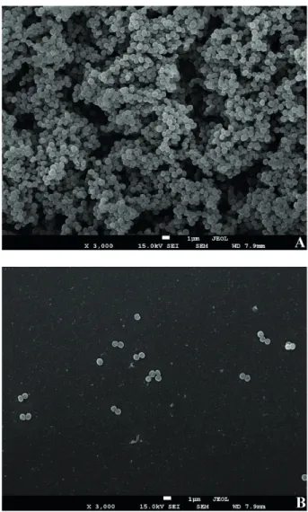
The highest antibiofilm activity of ABS of 94.48 ± 0.96% was also monitored by SEM against S. aureus biofilm formation (Figure 2). After ABS treatment, planktonic S. aureus cells were observable, but there was no biofilm formation on the coverslips. Similar to all other microorganisms, the control group of S. aureus without ABS exhibited typical biofilm structure (Figure 2).
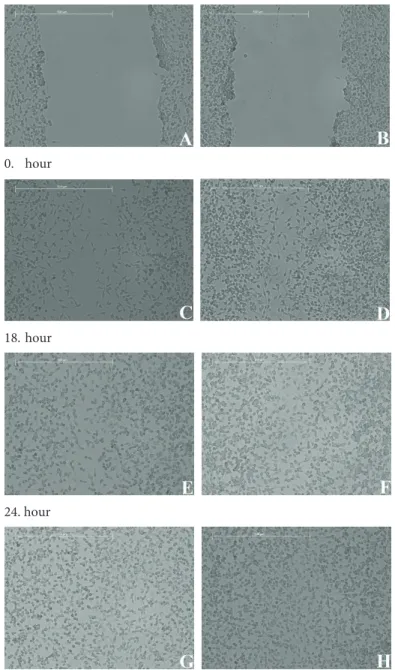
To investigate the effect of ABS on fibroblast migration and proliferation, the scratch wound healing assay was applied to 3T3 fibroblasts and images were taken at regular intervals. After administration of 1 µL of ABS to cell culture plates, fibroblasts were stimulated for cell migration. After 48 h, the scar had completely healed on both control group and ABS-treated plates (Figure 3).
4. Discussion
ABS is a medicinal plant extract that is produced by a registered Turkish company. ABS has been used as a local hemostatic agent in surgery, but it was also shown to promote healing. There are some studies that have been performed to investigate the efficacy of ABS on soft-tissue healing in animal models.
Akbal et al. (19) showed that oral ABS application prevented inflammation, scar formation, weight loss, and mortality in caustic esophageal injury in the study of mucosal wound healing in a rat model. In another study based on a rat skin defect model, Akalin et al. (20) reported higher collagen deposition and fibroblast proliferation scores and lower inflammatory scores in the ABS group. Similarly, according to Yüce et al. (21), collagen deposits of the tissues increased until day 10 in an experimental skin incision model. However, Aydın et al. (46) did not obtain positive results after histological administration to mice in tendon healing.
In wound healing, there are three consecutive steps of inflammation, tissue formation, and tissue remodeling, and it is a complex process involving cells, intermediates, and ECM components. Traditional wound healing treatments are expected to contribute to the wound healing process by influencing one or more of these stages (47). Therefore, in this study, the inhibition of enzymes that break down the ECM molecules of ABS was investigated. It is now recognized that the ECM is not only an architectural support for tissues but also plays an important role in cell regulation. It is now Table. Anticollagenase, antihyaluronidase, and antielastase activities of the ABS.
Inhibition % ± SEMa
Material Concentration Collagenase Hyaluronidase Elastase ABS 3%0.3% 88 ± 0.3110.5 ± 0.1 96.8 ± 0.4745 ± 0.3 66 ± 0.0712 ± 0.01 Epigallocatechin gallate (reference) 100 µg/mL 36 ± 0.31 NTc 47 ± 0.7
Tannic acid (reference) 1 mg/mL100 µg/mL NTNTcc 61.6 ± 0.81–b NT c
NTc a Standard error of the mean, b no activity, c not tested.
clear that significant ECM changes during wound healing have made it a very important player in this process (23).
Effects of the individual plant extracts found in ABS on enzyme inhibitions are reported. Extracts of T. vulgaris showed collagenase and elastase inhibition of almost 25% and 17%, respectively, while the hyaluronidase inhibition rate was 100% (48). In another study, ursolic acid extracted from U. dioica exhibited elastase and collagenase inhibition activity of 24.51% and 16.23%, respectively (49).
Similarly, Carini et al. (50) reported that procyanidins from V. vinifera seeds inhibited elastase enzyme (IC50 = 5.4 µM). In another study, A. officinarum rhizome extract showed very low elastase inhibition at 100 µg/mL (1%) and 1000 µg/mL (8%) concentrations (44). The results of the present study show that ABS has higher enzyme inhibition activity than the plants used in its formulation. This is probably due to the synergistic activity of the extracts found in ABS. The cytotoxic activity of ABS was also evaluated for cancer cell lines. Turk et al. (51) determined the effect of ABS on the viability of melanoma cells; cells treated with ABS showed a significant decrease in cell viability compared to control groups.
Antimicrobial resistance levels within a biofilm can reach 1000-fold higher than in planktonic cells. An estimated 65%–80% of all infections are thought to be biofilm-related; therefore, this increase in antibiotic resistance presents a serious challenge (33). The wound healing process can also affect the microbial flora. It has been found that the total absence of microflora affects the wound healing process positively. However, colonization occurs immediately after injury and microbial colonization is inevitable in clinical practice. When there is injury, initially, the host’s immune system controls microbial proliferation but gradually develops microorganisms of the wound area, developing biofilms and resistance to host immunity (32,52). In previous works, the antimicrobial activity of ABS was tested against clinical isolates (16,53), but, as far as we know, there is no study in the literature that examines the effects of ABS
on biofilm development. The results of the present study showed that ABS can effectively inhibit biofilm formation of oral microorganisms.
There are a few studies about the in vivo wound healing potential of ABS. İşler et al. (54) observed that the application of ABS decreased the occurrence of inflammation and necrosis while increasing new bone formation in the early bone healing period for rats. Akalin et al. (20) investigated the efficacy of ABS on the healing of dermal wounds in a rat model and concluded that the ABS-treated group was superior to the control group in terms of inflammatory scoring, type I/type III collagen ratio, and wound contraction rates. Kaya et al. (55) investigated the effects of ABS on burn healing using a rat burn model and wound diameter and inflammation were found to be significantly decreased while fibrosis was significantly increased in the ABS group
38.80 94.48 7.89 86.57 62.13 82.38 87.00 80.17 0.00 20.00 40.00 60.00 80.00 100.00 120.00 % inhibition
Antibiofilm activity of ABS
Figure 1. Antibiofilm activity of the ABS against oral cavity microorganisms. Error bars represent standard deviations.
Figure 2. Scanning electron micrographs of ABS against biofilm development of S. aureus: A) the untreated group, B) ABS-treated group. Scale bars are shown within the figures.
on the 14th day. To our knowledge, this is the first study on the in vitro wound healing potential of ABS.
ABS has been used as a hemostatic agent for different types of bleeding. In this study, the antibiofilm activity of ABS against oral streptococci has been revealed with in vitro analysis and also visualized by SEM. ECM components are required at each stage of wound healing and it is known that recovery does not continue without these ECM components. In this study, the collagenase, hyaluronidase,
and elastase enzyme inhibitory activities of ABS were investigated for the first time. In vitro experiments have shown that ABS can enhance the wound healing process by providing inhibition of ECM-degrading enzymes during wound repair. Our findings also demonstrated that ABS enhanced the stimulated migration of 3T3 fibroblasts to an artificial wounded area. Besides its hemostatic activity, ABS can be used effectively as an antibiofilm and wound healing agent for oral cavity wounds.
0. hour 18. hour 24. hour 48. hour
Figure 3. Images of scratch wound healing assay for 0, 18, 24, and 48 h after creating the scratch. Images on the left side represent the control group.
References
1. Haznedaroglu BZ, Beyazit Y, Walker SL, Haznedaroglu IC. Pleiotropic cellular, hemostatic, and biological actions of Ankaferd hemostat. Crit Rev Oncol Hematol 2012; 83: 21-34. 2. Orhan I, Dogan R, Soylu E, Aksoy F, Veyseller B, Ozturan
O, Esrefoglu M, Aydın MS. Histopathological evaluation of Ankaferd blood stopper use in the rabbit septoplasty model. Int J Pediatr Otorhinolaryngol 2015; 79: 305-309.
3. Goker H, Haznedaroglu IC, Ercetin S, Kirazli S, Akman U, Ozturk Y, Firat HC. Haemostatic actions of the folkloric medicinal plant extract Ankaferd Blood Stopper. J Int Med Res 2008; 36: 163-170.
4. Mumcuoglu M, Akin DF, Ezer U, Akar N. Ankaferd Blood Stopper induces apoptosis and regulates PAR1 and EPCR expression in human leukemia cells. Egyptian Journal of Medical Human Genetics 2015; 16: 19-27.
5. Huri E, Akgül T, Ayyildiz A, Germiyanoğlu C. Hemostasis in retropubic radical prostatectomy with Ankaferd BloodStopper: a case report. Kaohsiung J Med Sci 2009 25: 445-447.
6. Iynen I, Bozkus F, San I, Alatas N. The hemostatic efficacy of Ankaferd Blood Stopper in adenoidectomy. Int J Pediatr Otorhi 2011; 75: 1292-1295.
7. Teker AM, Korkut AY, Gedikli O, Kahya V. Prospective, controlled clinical trial of Ankaferd Blood Stopper in children undergoing tonsillectomy. Int J Pediatr Otorhinolaryngol 2009; 73: 1742-1745.
8. Kurt M, Akdogan M, Ibis M, Haznedaroglu IC. Ankaferd blood stopper for gastrointestinal bleeding. J Invest Surg 2010; 23: 239.
9. Teker AM, Korkut AY, Kahya V, Gedikli O. Prospective, randomized, controlled clinical trial of Ankaferd Blood Stopper in patients with acute anterior epistaxis. Eur Arch Otorhinolaryngol 2010; 267: 1377-1381.
10. Ercetin S, Haznedaroglu IC, Kurt M, Onal IK, Aktaş A, Kurt OK, Göker H, Ozdemir O, Kirazli S, Firat HC. Safety and efficacy of Ankaferd Blood Stopper in dental surgery. International Journal of Hematology and Oncology 2010; 27: 1-5.
11. Yaman E, Görken F, Erdem AP, Sepet E, Aytepe Z. Effects of folk medicinal plant extract Ankaferd Blood Stopper® in vital primary molar pulpotomy. Eur Arch Paediatr Dent 2012; 13: 197-202.
12. Çakarer S, Eyüpoğlu E, Günes ÇÖ, Küseoğlu BG, Berberoğlu HK, Keskin C. Evaluation of the hemostatic effects of Ankaferd blood stopper during dental extractions in patients on antithrombotic therapy. Clin Appl Thromb Hemost 2013; 19: 96-99.
13. Goker H, Cetinkaya D, Kilic E, Haznedaroglu IC, Kirazli S, Firat H. Anti-cancer activity of Ankaferd Blood Stopper on osteosarcom (SAOS-2) cell lines in vitro. In: Haznedaroglu IC, Goker H, Ozdemir O, Kosar A, Firat H, editors. Ankaferd: Scientific Perspectives and Basic-Clinical Data. İstanbul, Turkey: Naviga Publications; 2008. p. 109.
14. Gülşen MR, Uzunay NS, Fermanlı O, Çoban ZD, Öztürk D, Hamidi M, Avcu F, Güran Ş. Anti-angiogenic role of Ankaferd on chick chorioallontoic membrane model. Gülhane Tıp Dergisi 2015; 57: 274-279.
15. Ciftci S, Keskin F, Keceli Ozcan S, Erdem MA, Cankaya B, Bingol R, Kasapoglu C. In vitro antifungal activity of Ankaferd Blood Stopper against Candida albicans. Curr Ther Res Clin Exp 2011; 72: 120-126.
16. Tasdelen Fisgin N, Tanriverdi Cayci Y, Coban AY, Ozatli D, Tanyel E, Durupinar B, Tulek N. Antimicrobial activity of plant extract Ankaferd Blood Stopper. Fitoterapia 2009; 80: 48-50. 17. Çinar Ç, Odabaş ME, Akca G, Işik B. Antibacterial effect of
a new haemostatic agent on oral microorganisms. J Clin Exp Dent 2012; 4: 151-155.
18. Uğur A, Saraç N, Çankal DA, Özle M. The antioxidant and antimutagenic activities of Ankaferd blood stopper, a natural hemostatic agent used in dentistry. Turk J Med Sci 2016; 46: 657-663.
19. Akbal E, Köklü S, Karaca G, Astarci HM, Koçak E, Taş A, Beyazit Y, Topcu G, Haznedaroğlu IC. Beneficial effects of Ankaferd Blood Stopper on caustic esophageal injuries: an experimental model. Dis Esophagus 2012; 25: 188-194. 20. Akalin C, Kuru S, Barlas AM, Kismet K, Kaptanoglu B, Demir
A, Astarci HM, Ustun H, Ertas E. Beneficial effects of Ankaferd Blood Stopper on dermal wound healing: an experimental study. Int Wound J 2014; 11: 64-68.
21. Yüce S, Çandırlı C, Yenidünya S, Muslu B. New hemostatic agent: the effect of Ankaferd Blood Stopper on healing wounds in experimental skin incision model. Turk J Med Sci 2014; 44: 288-294.
22. Reinkes JM, Sorg H. Wound repair and regeneration. Eur Surg Res 2012; 49: 35-43.
23. Maquart FX, Monboisse JC. Extracellular matrix and wound healing. Pathol Biol 2014; 62: 91-95.
24. Lee CH, Lee Y. Collagen-based formulations
for wound healing applications. In: Ågren MS, editor. Wound Healing Biomaterials. Volume 2: Functional Biomaterials. Amsterdam, the Netherlands: Elsevier; 2016. pp. 135-149.
25. Nyström A. Collagens in wound healing. In: Ågren MS, editor. Wound Healing Biomaterials. Volume 2: Functional Biomaterials. Amsterdam, the Netherlands: Elsevier; 2016. pp. 171-201.
26. Vasconcelos A, Gomes AC, Cavaco-Paulo A. Novel silk fibroin/ elastin wound dressings. Acta Biomater 2012; 8: 3049-3060. 27. Prodicimi M, Bevilacqua C. Exogenous hyaluronic acid and
wound healing: an up-dated vision. Panminerva Med 2012; 54: 129-135.
28. Frenkel JS. The role of hyaluronan in wound healing. Int Wound J 2014; 11: 159-163.
29. Raffetto JD, Khalil RA. Matrix metalloproteinases and their inhibitors in vascular remodeling and vascular disease. Biochem Pharmacol 2008; 15: 346-359.
30. Thring TSA, Hili P, Naughton DP. Anti-collagenase, anti-elastase and anti-oxidant activities of extracts from 21 plants. BMC Complement Altern Med 2009; 9: 1-27.
31. Fronza M, Heinzmann B, Hamburger M, Laufer S, Merfort I. Determination of the wound healing effect of Calendula extracts using the scratch assay with 3T3 fibroblasts. J Ethnopharmacol 2009; 126: 463-467.
32. Misic AM, Gardner SE, Grice EA. The wound microbiome: modern approaches to examining the role of microorganisms in impaired chronic wound healing. Adv Wound Care 2014; 3: 502-510.
33. Costa EM, Silva S, Veiga M, Tavaria FK, Pintado MM. A review of chitosan’s effect on oral biofilms: perspectives from the tube to the mouth. Journal of Oral Biosciences 2017; 59: 205-210. 34. Dewhirst FE, Chen T, Izard J, Paster BJ, Tanner AC, Yu
WH, Lakshmanan A, Wade WG. The human oral microbiome. J Bacteriol 2010; 192: 5002-5017.
35. Mariano RC, Oliveira MR, Silva LC, Ferreira S, Júnior IRG, de Carvalho Silva. Effect of topical application of chlorhexidine and metronidazole on the tissue repair of palatal wounds of rats: a clinical and histomorphometric study. Oral Surg Oral Med Oral Pathol Oral Radiol 2015; 119: 505-513.
36. Mensah AY, Sampson J, Houghton PJ, Hylands PJ, Westbrook J, Dunn M, Hughes MA, Cherry GW. Effects of Buddleja
globosa leaf and its constituents relevant to wound healing. J
Ethnopharmacol 2001; 77: 219-226.
37. Schrezenmeir J, de Vrese M. Probiotics, prebiotics, and synbiotics—approaching a definition. Am J Clin Nutr 2001; 73: 361-364.
38. Koslovsky A, Artzi Z, Israeli-Tobias C, Hirshberg A. Effect of local antimicrobial agents on excisional palatal wound healing: a clinical and histomorphometric study in rats. J Clin Periodontol 2007; 34: 164-171.
39. Salami AA, Imosemi IO, Owoeye OO. A comparison of the effect of chlorhexidine, tap water and normal saline on healing wounds. Int J Morphol 2006; 24: 673-676.
40. Järbrink K, Ni G, Sönnergren H, Schmidtchen A, Pang C, Bajpai R, Car J. The humanistic and economic burden of chronic wounds: a protocol for a systematic review. Syst Rev 2017; 6: 15-22.
41. Tsiouris CG, Tsiouri MG. Human microflora, probiotics and wound healing. Wound Medicine 2017; 19: 33-38.
42. Baygar T, Ugur A. In vitro evaluation of antimicrobial and antibiofilm potentials of silver nanoparticles biosynthesized by
Streptomyces griseorubens. IET Nanobiotechnol 2017; 11:
667-681.
43. Barrantes E, Guinea M. Inhibition of collagenase and metalloproteinases by aloins and aloe gel. Life Sci 2003; 72: 843-850.
44. Lee KK, Kim JH, Cho JJ, Choi JD. Inhibitory effects of 150 plant extracts on anti-elastase activity and their anti-inflammatory effects. Int J Cosmet Sci 1999; 21: 71-82.
45. Liang CC, Park AY, Guan JL. In vitro scratch assay: a convenient and inexpensive method for analysis of cell migration in vitro. Nat Protoc 2007; 2: 329-333.
46. Aydın BK, Altan E, Acar MA, Erkoçak ÖF, Ugraş S. Effect of Ankaferd blood stopper® on tendon healing: an experimental study in a rat model of Achilles tendon injury. Joint Diseases and Related Surgery 2015; 26: 31-37.
47. Süntar I, Küpeli Akkol E, Keles H, Yesilada E, Sarker SD. Exploration of the wound healing potential of Helichrysum
graveolens (Bieb.) Sweet: isolation of apigenin as an active
component. J Ethnopharmacol 2013; 26: 103-110.
48. Duque L, Bravo K, Osorio E. A holistic anti-aging approach applied in selected cultivated medicinal plants: a view of photoprotection of the skin by different mechanisms. Ind Crop Prod 2017; 97: 431-439.
49. Bourgeois C, Leclerc EA, Corbin C, Doussot J, Serrano V, Vanier JR, Seigneuret JM, Auguin D, Pichon C, Lainé E et. al. Nettle (Urtica dioica L.) as a source of antioxidant and anti-aging phytochemicals for cosmetic applications. CR Chim 2016; 19: 1090-1100.
50. Carini M, Stefani R, Aldini G, Ozioli M, Facino RM. Procyanidins from Vitis vinifera seeds inhibit the respiratory burst of activated human neutrophils and lysosomal enzyme release. Planta Med 2001; 67: 714-717.
51. Turk S, Malkan UY, Ghasemi M, Hocaoglu H, Mutlu D, Gunes G, Aksu S, Haznedaroglu IC. Growth inhibitory activity of Ankaferd hemostat on primary melanoma cells and cell lines. SAGE Open Medicine 2017; 5: 1-7.
52. Percival SL, McCarty SM, Lipsky B. Biofilms and wounds: an overview of the evidence. Adv Wound Care 2015; 4: 373-381. 53. Bilgili H, Captug O, Kosar A, Kurt M, Kekilli M, Shorbagi
A, Kurt OK, Ozdemir O, Goker H, Haznedaroglu IC. Oral systemic administration of Ankaferd blood stopper has no short-term toxicity in an in vivo rabbit experimental model. Clin Appl Thromb Hemost 2010; 16: 533-536.
54. İşler SC, Demircan S, Çakarer S, Çebi Z, Keskin C, Soluk M, Yüzbaşıoğlu E. Effects of folk medicinal plant extract Ankaferd Blood Stopper® on early bone healing. J Appl Oral Sci 2010; 18: 409-414.
55. Kaya H, Gokdemir MT, Sogut O, Demir T, Kocarslan S. Effects of folk medicinal plant extract Ankaferd blood stopper on burn wound healing. Acta Medica Mediterr 2013; 29: 497-502.