A Multidisciplinary Approach To Localized Gingival
Recession: A Case Report
Burcu KARADUMAN
1Cenker KOYUNCUOĞLU
2Sevda ATALAY
3Ece ÇALIŞKAN
4Nurcan TEZCİ
5Sabri Hasan MERİÇ
6Abstract
Background: Many techniques are available for the treatment of gingival recessions. Platelet rich fibrin (PRF) is being used for root coverage and periodontal regeneration with better wound healing.
Objective: The purpose of this case report is to present the treatment stages and outcomes of a female patient who referred to our clinic with the complaint of gingival recession.
Case Description: A 29-year-old patient with porcelain fused to metal (PFM) restorations on the maxillary incisors, had Miller Class I recession on her left lateral incisor. Initially, Phase I periodontal therapy was performed. One month later, the exposed root surface was covered with PRF and a coronally advanced flap was done. Postoperative healing was uneventful and 2 mm of root coverage was gained. Maxillary incisors were restored with zirconia crown prostheses. During 6 months follow-up no further exposure occured.
Practical Implications: PRF is a preferable method enhancing success rates of the operation for recession coverage.
Keywords: Gingival recession, Platelet rich fibrin, Zirconia
1 (DDS, PhD)İstanbul Aydin University, Faculty of Dentistry, Department of Periodontology, Corresponding Author: [email protected] 2 (DDS, PhD)İstanbul Aydin University, Faculty of Dentistry, Department of Periodontology
Lokalize Dişeti Çekilmesine Multidisipliner Yaklaşım: Bir Olgu Sunumu Özet
Giriş: Günümüzde dişeti çekilmeleri için farklı tedavi seçenekleri gündeme gelmiştir. Kök yüzeyi kapatılması ve periodontal rejenerasyonun yanı sıra yara iyileşmesinin de hızlanması amacıyla trombositten zengin plazma (TZF) kullanılarak tedavi başarısı arttırılmaya çalışılmaktadır.
Amaç: Bu olgu sunumunun amacı dişeti çekilmesi şikâyetiyle kliniğimize başvuran kadın hastanın operasyon süreci, endodontik ve protetik tedavi aşamaları ile tedavi sonucunun sunulmasıdır. Olgu Sunumu: Sol üst lateral dişinde Miller Sınıf I dişeti çekilmesi ve üst kesici dişlerinde metal destekli porselen restorasyonları var olan 29 yaşındaki kadın hastaya ilk aşamada Faz I periodontal tedavi uygulandı. 1 ay sonra kök yüzeyine TZF yerleştirilerek koronale kaydırılan flep yöntemiyle örtüldü. Postoperatif iyileşme sorunsuz olarak gerçekleşti ve 2 mm kök yüzeyi kapanması sağlandı. Üst kesici dişler zirkonya seramik kuron protezleriyle restore edildi. Olgunun 6 aylık takibi sonrasında dişeti çekilmesi gözlenmedi.
Klinik Bağlantı: TZF, dişeti çekilmelerinin kapatılmasında operasyon başarısını arttıran alternatif tedavi yöntemleri arasında tercih edilen bir uygulamadır ve daha fazla kullanım alanı bulmaktadır. Anahtar Kelimeler: Dişeti çekilmesi, Trombositten zengin fibrin, Zirkonya
Introduction
Gingival recession is a common problem resulting from periodontal diseases. The main causes are trauma, inflammation, occlusal forces, surgical interventions or orthodontic therapy. Gingival recession usually results in hypersensitivity and caries problems. However, when the defects occur in the anterior area, esthetic concerns should be involved in treatment strategy (1). Recession defects are classified by Miller based on the periodontal tissue support and the mucogingival line. Miller Class I defects are recessions that do not extend to the mucogingival junction where hard/ soft tissue loss is absent in the interdental area. Complete root coverage is obtainable in this type of defect (2).
Treatment approach includes many techniques mainly combined with coronally advanced flaps (3). Connective tissue graft combined with a coronally positioned flap is considered as a gold standard in root coverage (1). Due to the well-known obvious disadvantages of connective tissue grafts such as creating a second wound site, many attempts have been made to prevent the patient from suffering this situation. Xenografts or allografts are substitutes obtained from external sources and may have different results related to foreign body reactions. However PRF membrane is obtained from the patients’ blood and has no known side effects. It is easy to prepare and use by clinicians. Studies including PRF membrane with the coronally advanced flap procedure show promising results with good esthetic outcomes and better wound healing (4, 5).
Conventional metal-ceramic restorations have been the treatment of choice for decades because of their superior mechanical properties, adequate marginal and internal fit and acceptable esthetic appearance. However, patients’ growing esthetic demand for natural looking, tooth coloured restorations has led the development of various all ceramic materials with different optical and mechanical properties (6). The selection criteria for dental materials include strength, esthetics and fit. The main goal of utilizing all ceramic materials is to fabricate restorations with significant esthetic and biocompatible properties and favourable mechanical behaviour (7). Zirconia has superior machanical properties and causes less inflammatory reaction in gingival tissues than other restorative materials. In addition to its colour that is similar to natural teeth, opaque zirconia core makes it possible to cover a dischromic restoration ot tooth (8). These favourable features of zirconia allow clinicians to utilize it as a ceramic restorative material in most cases. Therefore, the purpose of this case report is to present the multidisciplinary treatment procedure and the treatment outcomes of a female patient who referred to our clinic with the complaint of gingival recession on her left maxillary lateral tooth.
Case Report
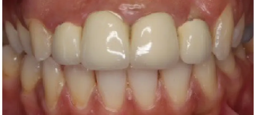
In this case report, periodontal operation procedure, endodontic and prosthodontic treatment and maintenance of a 29-year-old patient with the chief complaint of root exposure on her left maxillary lateral incisor were presented. The patient was systemically healthy with no ongoing medication. Intraoral examination revealed Miller Class I recession defect on tooth #22 (Fig 1). Recession depth and width was 3 mm and 4 mm, respectively. Slight plaque accumulation, gingival erythema and edema were present. She had previous porcelain fused to metal (PFM) restorations on the maxillary incisors. Radiographical findings revealed a periapical pathology; well-defined radiolucent area near the left maxillary lateral tooth apex. The tooth #22 had no mobility, no response to vertical or horizontal percussion tests. It was devital according to the results of the electric pulp test. Plaque associated gingivitis and chronic apical periodontitis were diagnosed based on the clinical and radiographical examination. Following the removal of PFM restorations, Phase I periodontal treatment consisting of oral hygiene instructions, scaling and root planing, was performed by an experienced periodontist under local anesthesia. Mechanical periodontal therapy was not accompanied by any medications. The tooth #22 was endodontically treated in two sessions with rotary instrumentation.
Periodontal operation included a coronally advanced flap with the use of a PRF membrane. The
PRF was prepared according to the protocol developed by Choukroun et al.(9) Prior to surgery, 10
ml of intravenous blood of the patient has been collected in test tubes without adding anticoagulant and immediately centrifuged at 2700 revolutions/min for 12 min. (10). The fibrin clot was easily separated from the RBC base (preserving a small part of the RBC layer) using sterile tweezers and scissors, and PRF is prepared by using a PRF box.
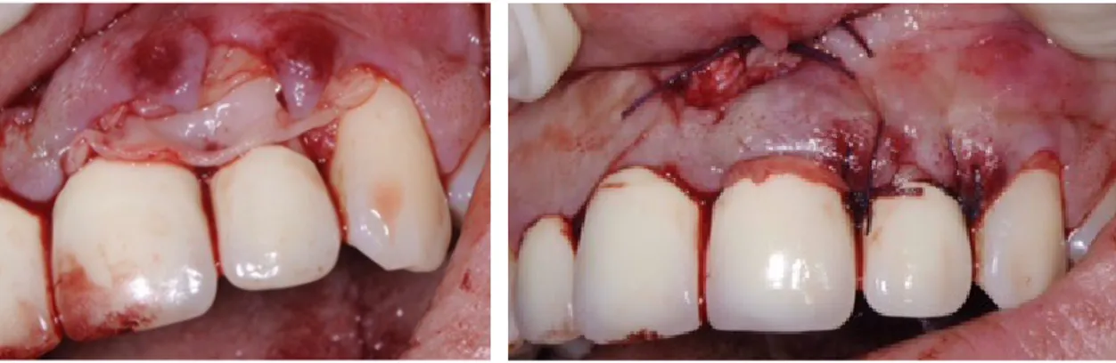
Following administration of local anesthesia, sulcular incisions through each recession area were given with a number 15 blade. Tunnel technique was designed first and the tip of the interdental papilla was not included in the incision. A split thickness mucoperiosteal flap was reflected, extending beyond the mucogingival junction in order to reduce the tension on the flap and facilitate coronal displacement of the tissue. However, the flap ruptured near the papilla area during the incisions. The design of the flap was then changed with a modified envelope flap. The PRF membrane was covered on the exposed root surface with the most part of it placing on the connective tissue bed, in order to gain adequate blood supply (Fig 2). Interdental sutures were placed stabilizing both the membrane and the flap (Fig 3). The suturing was made with 5/0 polyglycolide-co-lactide braided absorbable
suture material (Dogsan Pegelak® 5/0, 16 mm 3/8, cutting suture, Trabzon, Turkey).Following
surgical operation, naproxen sodium 2 × 1 for 5 days (Apranax Fort® 550 mg 10 film tablets, Abdi İbrahim, İstanbul, Turkey) and chlorhexidine gluconate rinse 2 x 1 for 5 days (Andorex® 120 ml Delta Vital, İstanbul, Turkey) were prescribed to the patient. Clinical healing was achieved without
any postoperative complication such as pain, hemorrhage, swelling or recurrence. 2 mm of root
coverage has been gained. Smile design was performed with gingivoplasty approach on maxillary incisors and right canine due to uneven margins.
Figure 2. PRF membrane on the exposed root surface - Figure 3. Interdental sutures stabilizing the membrane and the flap
Maxillary incisors were decided to restore with zirconia ceramic crowns due to the esthetic and functional demands of the patient. The tooth preparation was completed using medium-grit diamond burs and a chamfer finish line was placed 0,5 mm subgingivally. Final impression was made with a combination of putty and light-body vinyl polysiloxane (VPS) elastomeric impression material (Elite HD+, Zhermack, Italy) for definitive restorations. Zirconia ceramic crowns were fabricated with a core (DC Zircon, DCS Dental AG, Allschwil, Switzerland) and veneering porcelain. Internal and marginal of the restorations were checked, proximal and occlusal contacts
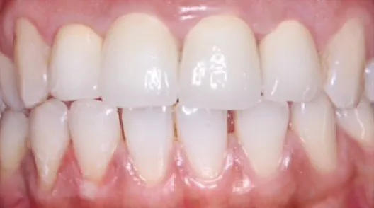
were adjusted gently with low-speed rotary instruments under irrigation. The crowns were then luted via self-adhesive, dual-cure resin cement (Panavia SA Cement, Kuraray, Japan). During 6 months follow-up no further recession or inflammation was detected (Fig 4).
Conclusion
PRF membrane is a preferable material in the management of gingival recessionss. It might be suggested as an alternative to the subepithelial connective tissue technique, especially for situations where harvesting from the palate is challenging or impossible. When needed, small ‘touch-ups’ could be performed by means of gingivoplasty techniques for the adjacent teeth. Zirconium dioxide ceramic crowns give clinicians the opportunity to finalize the treatment with maximum esthetic and functional outcomes.
Figure 4. Zirconia ceramic crown restorations after 6 months
Conflict of Interest
There is no disclosure that supported the work (no grant funds, commercial sources or funds from a contributors’ institution) and authors report no conflict of interest related to this study.
REFERENCES
[1] Dominiak M, Gedrange T. New perspectives in the diagnostic of gingival recession. Adv
Clin Exp Med. 2014;23:857-63.
[2] Miller PD Jr: A classification of marginal tissue recession. Int J Periodontics Restorative Dent. 1985;5:8–13.
[3] Sanz M, Simion M. Working Group 3 of the European Workshop on Periodontology.
Surgical techniques on periodontal plastic surgery and soft tissue regeneration: consensus report of Group 3 of the 10th European Workshop on Periodontology. J Clin Periodontol.
[4] Aleksić Z, Janković S, Dimitrijević B, Divnić-Resnik T, Milinković I, Leković V. The use of platelet-rich fibrin membrane in gingival recession treatment. Srp Arh Celok Lek.
2010;138:11-8.
[5] Jankovic S, Aleksic Z, Klokkevold P, Lekovic V, Dimitrijevic B, Kenney EB, Camargo
P. Use of platelet-rich fibrin membrane following treatment of gingival recession: a randomized clinical trial. Int J Periodontics Restorative Dent. 2012;32:41-50.
[6] Zarone F, Russo S, Sorrentino R. From porcelain-fused-to-metal to zirconia: Clinical and experimental considerations. Dent Mater. 2011;27:83–96.
[7] Amir Rad FA, Succaria FG, Morgano SM. Fracture resistance of porcelain veneered
zirconia crowns with exposed lingual zirconia for anterior teeth after thermal cycling: An in vitro study. Saudi Dent J. 2015;27: 63–9.
[8] Manicone PF, Iommetti PR, Raffaelli L. An overview of zirconia ceramics: Basic properties and clinical applications. J Dent. 2007;35:819–26.
[9] Choukroun J, Adda F, Schoeffler C, Vervelle A. An opportunity in perio-implantology: The PRF (in French) Implantodontie. 2001;42:55–62.
[10] Ghanaati S, Booms P, Orlowska A, Kubesch A, Lorenz J, Rutkowski J, Landes C, Sader R, Kirkpatrick C, Choukroun J. Advanced platelet-rich fibrin: a new concept for cell-based tissue engineering by means of inflammatory cells. J Oral Implantol. 2014;40:679-89.