Selçuk Matyar
1, A, B, D–F, Esmeray Acartürk
2, A, D, E, Gülen Attila
1, A, B, D,
İlker Ünal
3, C, Levent Soyer
2, B, Onur Akpınar
2, B, E, FGene-Gene Interaction of ACE I/D, Endothelial Nitric
Oxide Synthase 4 a/b and ApoE does not Affect Coronary
Artery Disease Severity
1 Department of Biochemistry, Çukurova University, Adana, Turkey 2 Department of Cardiology, Çukurova University, Adana, Turkey 3 Department of Biostatistics, Çukurova University, Adana, Turkey
A – research concept and design; B – collection and/or assembly of data; C – data analysis and interpretation; D – writing the article; E – critical revision of the article; F – final approval of article; G – other
Abstract
Objectives. Previous studies have shown the impact of angiotensin converting enzyme (ACE) insertion/deletion
(I/D), endothelial nitric oxide synthase (eNOS) polymorphisms and ApoE genotypes on coronary artery disease (CAD). The aim of this study is to investigate the relationship between the genetic polymorphisms and the severity of CAD and to evaluate their potential interactions.
Material and Methods. All patients underwent coronary angiography; coronary score (CS) and severity score (SS)
were calculated for them. ACE I/D, eNOS and ApoE polymorphisms were detected by polymerase chain reaction (PCR).
Results. Neither CS nor SS showed a direct relationship with eNOS and ApoE genotypes. CS and SS were found
to be high in patients carrying the ACE DD allele (p = 0.034 and p = 0.009). In the gene interactions, there was an increase in the SS only in patients with coexisting eNOS b/b genotype and ACE DD allele (p = 0.043).
Conclusions. The interactions of the gene polymorphisms investigated don’t play an important role in
determin-ing an individual’s risk for the severity of CAD (Adv Clin Exp Med 2014, 23, 2, 215–223).
Key words: ACE gene, ApoE gene, eNOS gene, gene–gene interaction, severity of coronary artery disease.
Adv Clin Exp Med 2014, 23, 2, 215–223 ISSN 1899-5276
ORIGINAL PAPERS
© Copyright by Wroclaw Medical University
Coronary artery disease (CAD) is the most common cause of death in the world. It is a com-plex disease with both environmental and genetic determinants. Genetics, in addition to other well-known major risk factors, is an important mecha-nism in the development of CAD [1–7]. Numerous studies have indicated an association of CAD with gene polymorphisms [1–5].
Angiotensin converting enzyme (ACE) is a member of the renin-angiotensin-aldosterone system, which converts angiotensin I to angio-tensin II. The studies on the association of ACE gene insertion/deletion (I/D) polymorphism with CAD have shown that the DD genotype is asso-ciated with increased risk of developing CAD [3– 5]. In addition, nitric oxide (NO) derived from the
endothelium is considered as an important media-tor, and synthesized from L-arginine by the enzyme endothelial nitric oxide synthase (eNOS). Numer-ous studies have analyzed the association between eNOS polymorphisms and CAD [1, 8]. Another molecule, Apolipoprotein E (ApoE), plays a major role in lipoprotein metabolism and the ApoE gene is polymorphic with three common alleles (ε2, ε3 and ε4), which produce three homozygous (ε2∕2, ε3∕3 and ε4∕4) and three heterozygous (ε2∕3, ε2∕4 and ε3∕4) genotypes [2, 9]
We previously evaluated the impact of ACE I/D polymorphism, eNOS intron 4 a/b variable number of tandem repeats (VNTR) polymorphism and ApoE genotypes on CAD1–3. This retrospective
polymorphisms and the severity of CAD and eval-uates their potential interactions.
Material and Methods
Patients
The study population consisted of 239 patients (161 male and 78 female, with a mean age of 54.1 ± 10.3 years) who were admitted for diagnostic coronary angiography for the assessment of a sus-pected or confirmed clinical diagnosis of CAD. As a routine procedure, an informed written consent was obtained from all patients before angiography. The study was also approved by the local Ethics Committee.
Risk Factor Assessment
The risk factors were considered as hyper-tension (HT), hyperlipidemia, diabetes melli-tus (DM), cigarette smoking and family history of CAD. A sustained blood pressure greater than 140 mm Hg systolic and 90 mmHg diastolic or us-ing an antihypertensive medication was defined as HT [10]. DM was defined as hyperglycemia, re-quiring antidiabetic drugs or testing blood sug-ar over 6.7 mmol/L [11]. Patients reporting ciga-rette use during the year prior to examination were considered as smokers. Hyperlipidemia was de-fined as plasma low-density lipoprotein cholester-ol (LDL-C) > 3.37 mmcholester-ol/L or using lipid-lowering drugs at the time of investigation, hypertriglyceri-demia was defined as triglyceride (TG) level > 1.70 mmol/L and high-density lipoprotein cholesterol (HDL-C) was considered present if the concentra-tion of HDL-C was < 1.04 mmol/L according to The Third Report of The National Cholesterol Ed-ucation Program (NCEP ATP III) guidelines [12]. NonHDL-C was defined as the difference between total cholesterol (TC) and HDL-C.
Coronary Angiography
Coronary angiography was performed accord-ing to the Judkins technique and images of the coronary tree were obtained in routine, standard-ized projections. The angiograms were assessed by at least two cardiologists. The coronary tree was divided into 15 segments and each segment was graded on a 4-level scale as conventionally used [13, 14]. Patients without angiographic lesions were considered as patients without CAD. Coro-nary artery diameter narrowing was determined according to the previously described method as 0, no visible wall irregularity or stenosis ≤ 25%; 1,
stenosis ≤ 50%; 2, stenosis ≤ 75%; 3, stenosis ≥ 75 [13]. CS was defined as the number of coronary arteries (0–3) with a stenosis ≥ 75 % [15]. Severi-ty score was calculated as the average grade of the coronary segments graded 1 or more [14].
Laboratory Analysis
Blood Chemistry
Venous blood samples were collected after a 12-h fasting period. Plasma was separated with-in 4 h and stored at – 20oC. Subsequent analysis
of plasma TC and TG was performed using en-zymatic and colorimetric assays (CHOD-PAP and GPO-PAP methods, respectively). Plasma HDL-C was measured using an enzymatic and colorimetric method (CHOD-PAP) without sam-ple pretreatment. Plasma LDL-C was calculated using the Friedewald formula when the TG levels were < 4.5 mmol/L. In case of high levels of TG (> 4.5 mmol/L), a direct enzymatic and colorimet-ric method (CHOD-PAP) without sample pretreat-ment was used. Non-HDL cholesterol was defined as the difference between TC and HDL-C [12].
Genetic Analysis
Venous blood samples were collected into EDTA. Genomic DNA from leukocytes was puri-fied according to the method of Miller [16].
Determination
of ACE I/D Polymorphism
A 287 bp I/D polymorphism in intron 16 of the ACE gene was examined with PCR by using the primer sequences of the targeted region of the genome (17q23) [17].
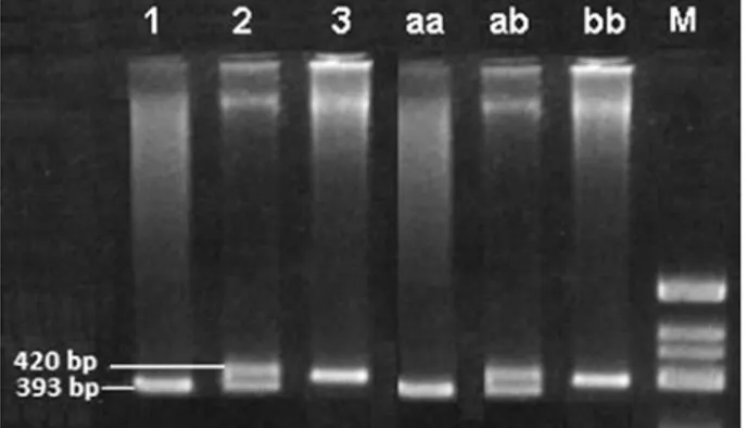
The template DNA (0.4 mg per sample) was am-plified by the following primers: (forward) 5’-CTG GAG ACC ACT CCC ATC CTT TCT 3’, and (re-verse) 5’-GAT GTG GCC ATC ACA TTC GTC AGA T 3‘. These primers (10 pmol of each) were add-ed to a mixture containing 5 µL of 10X Cetus buffer (pH 8.3), 0.5 mM dNTP (dATP, dCTP, dGTP, dTTP) and 1.0 units of Taq DNA polymerase (Perkin Elmer Cetus). A PCR program (Perkin Elmer 9600 Thermal Cycler) was initiated in a final total of 50 mL volume with thirty cycles of denaturation for 1 min at 94°C, annealing for 1 min at 58°C and primer extension for 1 min at 72°C was applied for amplification. PCR products of ACE gene locus were examined by aga-rose gel electrophoresis (3% agaaga-rose) at 150 V for 60 min and visualized at room temperature under UV after ethidium bromide staining (Fig. 1).
Determination of eNOS
4 a/b VNTR Polymorphism
The eNOS gene locus, located on chromosome 7q 35 to 36, which comprises 26 exons spanning 21 kilobases, shows a 27-bp repeat polymorphism in intron 4 of the eNOS gene [intron 4VNTR]. The eNOS gene has two common alleles containing 4 repeats (a) and 5 repeats (b) which produce two homozygous (aa and bb) and one heterozygous (ab) genotype [1].
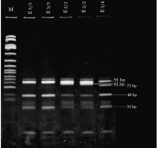
eNOS gene intron 4, 27 bp. VNTR polymor-phism was detected by PCR according to the meth-od described by Wang et al. [18]. The template DNA (0.5 µg per sample) was amplified using the following primers: (forward) 5’-’AGG CCC TAT GGT AGT GCC TTT-3’ and 5’-TCT CTT AGT GCT GTG CTC AC-3’ (reverse). These primers (25 pmol of each) were added to a mixture containing 0.2 µmol/L each of the dATP, dCTP, dGTP, dTTP, 5 µL of 10x Cetus buffer (pH 8.3), 5 µL of DM-SO (100%) and 0.5 units of Taq DNA polymerase (Perkin Elmer Cetus) in a final volume of 50 µL. The PCR was initiated with a denaturation by first heating the samples for 5 min at 94°C. Thirty five cycles of denaturation for 1 min at 94°C, annealing for 1 min at 56°C, primer extension for 2 min at 72°C and last extension for 5 min at 72°C was ap-plied for amplification. PCR products of NO gene locus were examined by gel electrophoresis (2 % NuSieve agarose-agarose) at 150 V for 30 min and visualized at room temperature under UV after ethidium bromide staining (Fig. 2).
Determination
of ApoE Genotypes
The ApoE gene locus located on chromosome 19 shows a polymorphism with three common al-leles (ε2, ε3 and ε4), which produce three homozy-gous (ε2∕2, ε3∕3 and ε4∕4) and three heterozyhomozy-gous (ε2∕3, ε2∕4 and ε3∕4) genotypes[2, 19].
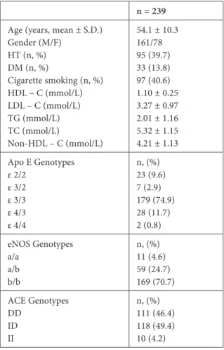
ApoE gene polymorphism was detected by PCR according to the method described by Wenham et al. [19]. The template DNA (0.4 µg per sample) was amplified using the following primers: (for-ward) 5’-TCCAAGGAGCTGCAGCTGCAGGCG-GCGCA-3’ and (reverse) 5’-ACAGAATTCGC-CCCGGC-3’. These primers (25 pmoL of each) were added to a mixture containing 0.2 µmoL/each of the dATP, dCTP, dGTP, DTTP, 5 µL of 10X Cetus buf-fer (pH 8.3), 10 µL of DMSO (50%) and 1.5 units of Taq DNA polymerase (Perkin Elmer Cetus) in a fi-nal volume of 50 µL. The PCR was initiated with a denaturation by first heating the samples for 5 min at 95°C. Forty cycles of denaturation for 0.5 min at 94°C, annealing for 0.5 min at 65°C and primer ex-tension for 1.5 min at 70°C were applied for ampli-fication. PCR products of ApoE locus were digest-ed with a CfoI restriction enzyme. Then the samples were examined by gel electrophoresis (3% agarose) at 150 V for 30 min after incubation at 37°C over-night and visualized at room temperature under UV after ethidium bromide staining (Fig. 3).
Statistical Analysis
All statistical analyses were performed using the SPSS v15.0 package program. Categorical da-ta such as sex, hypertension, etc. were presented as
Fig. 1. Polymorphism in intron 16 of the ACE gene.
The PCR products were examined by agarose gel elec-trophoresis (3% agarose) at 150 V for 60 min and visu-alized at room temperature under UV after ethidium bromide staining 100 bp DNA ID DD ID II Ladder 490 bp 190 bp
Fig. 2. Polymorphism in intron 4 of the eNOS gene.
The PCR products were examined by gel electropho-resis (2%NuSieve agarose–agarose) at 150 V for 30 minutes and visualized at room temperature under UV after ethidium bromide staining, 1 – aa control, 2 – ab control, 3 – bb control, M – Marker (x174 DNAIHinf I)
percents and, for continuous data, mean and stan-dard deviation were used. To evaluate gene-gene interaction, all possible subgroups were created. A Chi square test was used to evaluate the trend in severity score and CS. ANOVA (or Kruskal Wallis Test) was applied to compare age and lipid mea-surements in severity score and CS groups. In all analyses, p values less than 0.05 were considered statistically significant.
Results
Demographic patterns and the gene distribu-tions of the patients are shown in Table 1.
Coronary and Severity
Scores and Risk Factors
CS is significantly higher in male patients (p < 0.001), patients with DM (p = 0.003), and smokers (p = 0.006). It is also higher in patients with HT, although the difference is not signifi-cant (p = 0.052) (Table 2). SS is also higher in males (p < 0.001), diabetics (p = 0.006) and smok-ers (p < 0.001) (Table 3). There was no relation-ship between CS and SS and lipid profile (Tables 2 and 3, respectively).
Fig. 3. The apoE gene
locus located on chro-mosome 19 shows a polymorphism with three common alleles (ε2, ε3 and ε4). PCR products of Apo E locus were digested with CfoI restriction enzyme. Then, the samples were examined by gel electro-phoresis (3% agarose) at 150 V for 30 min after the incubation at 37°C overnight and visualized at room temperature under UV after ethidium bromide staining, M – Marker, ε2∕2 – 91bp–81bp, ε2∕3 – 91bp–81bp– 48bp–33bp, ε2∕4 – 91bp– 81bp–72bp–48bp–33bp, ε3∕3 – 91bp–48bp–33bp, ε3∕4 – 91bp–72bp–48bp– 33bp, ε4∕4 – 72bp–48bp– 33bp.
Risk Factors and Genes
In patients with HT, the coexistence of ACE DD allele increases the SS (p = 0.024). This is not observed in patients without HT (p = 0.323).
Coronary and Severity Scores
and Genes
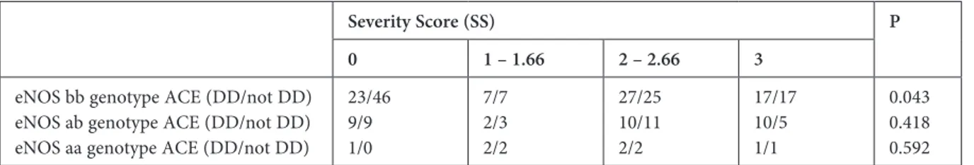
Gene distributions according to the CS and the SS are shown in Tables 2 and 3. Coronary and se-verity scores did not show a direct relationship with eNOS and ApoE genotypes. There is also no rela-tionship between ACE and CS (p = 0.095) (Table 2). However, in patients carrying the ACE DD allele, the severity score was higher than the patients with other ACE alleles (p = 0.031) (Table 3). When gene interactions were investigated, SS were higher only in patients with coexisting eNOS b/b genotype and ACE DD allele (p = 0.043) (Table 4).
Discussion
The present study investigated the effects of gene-gene interaction on CAD severity and showed that only ACE I/D polymorphism was associat-ed with CAD severity, whereas eNOS 4a/b VNTR
polymorphism and ApoE genotypes were not as-sociated with severity of CAD. We previously found that ApoE polymorphism (presence of ε4 allele), I/D polymorphism of the ACE gene (carry-ing D allele) and eNOS intron a/b polymorphism (presence of a allele) were the risk factors for CAD in Southern Turkey [1–3]. These findings were consistent with other studies [4, 5, 20–23]. We did not evaluate whether these individual genes have an effect on the severity of CAD.
It is known that serum lipid levels are strong-ly correlated with atherosclerosis [24]. Lipid accu-mulation may lead to a cascade of events resulting in serious obstructive atherosclerotic lesions. It is hard to determine the hemodynamic consequences of obstructions with angiography, since quantita-tive angiography is not sufficient for 3-dimensional sensitive evaluation of coronary lesions. However, clinical symptoms in atherosclerosis usually evolve due to degeneration and rupture of the atheroscle-rotic plaque, rather than its stable development.
This may be an explanation as to why the severity of coronary artery obstruction might not be asso-ciated with clinical disease severity.
In addition to conventional risk factors, genet-ics also contributes to CAD development. A great number of genetic polymorphisms may be involved in the development of CAD. On the other hand, in-teractions of multiple genes may also play a role in the development of CAD. Genetic polymorphisms and interactions of different genes on CAD have been subject to many studies in medicine [23, 25]. The association of ApoE and ACE genotypes with healthy aging have been investigated, and a signif-icantly higher frequency of ApoE/ε2 was observed in men between 60–90 years of age. No relation-ship has been observed in ApoE and ACE gene polimorphisms[25]. Genetic polymorphisms have also been studied in other cardiovascular diseas-es. XuGung et al. [26] found unfavorable geno-type combinations, which act synergistically in the development of ischemic stroke. In a study, ACE I/D polymorphism was found to be positively as-sociated with type 2 DM and synergistic effects of DD-33 and ID-23 were also shown. No association was found with ApoE polymorphism [27].
The CORGENE study [14] investigated the re-lationship between renin-angiotensin system ge-netic polymorphisms and the severity and/or ex-tent of coronary atherosclerosis, evaluated their potential interactions and no significant associa-tions were found.
There is conflicting data on gene-gene interac-tions. In some studies, significant interactions be-tween ApoE and ACE alleles have been found in patients with stroke and Alzheimer’s disease [28, 29]. A significant interaction was found between PPARγ CT and ApoE/ε4 genotypes in patients with CAD [30]. eNOS and ACE gene polymor-phisms were examined in patients with CAD and eNOS gene polymorphism was found as a frequent risk factor for vascular abnormalities in CAD [20]. Ji et al. [21] found an association between ACE and eNOS gene polymorphisms and CAD risk. How-ever, in this study, the diagnosis of CAD was not based on coronary angiography. Another study showed that ACE gene polymorphism was a sig-nificant predictor for CAD, but was not a mark-er for the sevmark-erity of coronary athmark-erosclmark-erosis [22]. On the other hand Qiu et al. [31] showed that ACE (DD genotype) and angiotensin II type 1 receptor gene (C allele) polymorphism increased the risk of CAD.
In our study, coronary and severity scores did not show a relationship with eNOS and ApoE genotypes. There was also no relationship be-tween ACE and CS (p = 0.095) (Table 2). How-ever, in patients carrying the ACE DD allele, the
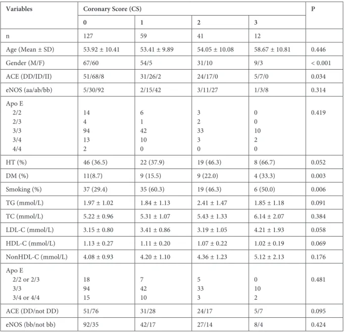
Table 1. Demographic patterns and the gene distributions
of the patients
n = 239
Age (years, mean ± S.D.) Gender (M/F) HT (n, %) DM (n, %) Cigarette smoking (n, %) HDL – C (mmol/L) LDL – C (mmol/L) TG (mmol/L) TC (mmol/L) Non-HDL – C (mmol/L) 54.1 ± 10.3 161/78 95 (39.7) 33 (13.8) 97 (40.6) 1.10 ± 0.25 3.27 ± 0.97 2.01 ± 1.16 5.32 ± 1.15 4.21 ± 1.13 Apo E Genotypes ε 2/2 ε 3/2 ε 3/3 ε 4/3 ε 4/4 n, (%) 23 (9.6) 7 (2.9) 179 (74.9) 28 (11.7) 2 (0.8) eNOS Genotypes a/a a/b b/b n, (%) 11 (4.6) 59 (24.7) 169 (70.7) ACE Genotypes DD ID II n, (%) 111 (46.4) 118 (49.4) 10 (4.2)
ACE – Angiotensin converting enzyme, DM – diabetes mellitus, eNOS – Endothelial nitric oxide synthase, F – female, HDL-C – high density lipoprotein cholesterol, HT – hypertension, M–male, LDL-C – low-density lipo-protein cholesterol, TC – total cholesterol, TG – triglyc-eride.
severity score was higher than patients with oth-er ACE alleles (p = 0.031) (Table 3). When gene interactions were investigated, there was an in-crease in the severity score of patients with co-existing eNOS b/b genotype and ACE DD allele (p = 0.043) (Table 4). Variability of the results of the different studies could be partially explained by differences in ethnicities of the studied pop-ulations, environmental factors, studied genes, methods used to determine CAD and also statis-tical methods.
In conclusion, the interactions of investigat-ed gene polymorphisms do not play any impor-tant role in determining an individual’s risk for the severity of CAD. Thus, genotype screening is not a useful method in the evaluation of the severity of coronary atherosclerosis in clinical practice. Using conventional risk factors is easier than using com-plex genetic factors for CAD risk determination in daily clinical practice.
Table 2. Gene distributions according to the coronary score
Variables Coronary Score (CS) P
0 1 2 3 n 127 59 41 12 Age (Mean ± SD) 53.92 ± 10.41 53.41 ± 9.89 54.05 ± 10.08 58.67 ± 10.81 0.446 Gender (M/F) 67/60 54/5 31/10 9/3 < 0.001 ACE (DD/ID/II) 51/68/8 31/26/2 24/17/0 5/7/0 0.034 eNOS (aa/ab/bb) 5/30/92 2/15/42 3/11/27 1/3/8 0.314 Apo E 2/2 2/3 3/3 3/4 4/4 14 4 94 13 2 6 1 42 10 0 3 2 33 3 0 0 0 10 2 0 0.419 HT (%) 46 (36.5) 22 (37.9) 19 (46.3) 8 (66.7) 0.052 DM (%) 11(8.7) 9 (15.5) 9 (22.0) 4 (33.3) 0.003 Smoking (%) 37 (29.4) 35 (60.3) 19 (46.3) 6 (50.0) 0.006 TG (mmol/L) 1.97 ± 1.02 1.84 ± 1.13 2.41 ± 1.47 1.85 ± 1.18 0.091 TC (mmol/L) 5.22 ± 0.96 5.31 ± 1.07 5.43 ± 1.33 6.14 ± 2.07 0.384 LDL-C (mmol/L) 3.15 ± 0.80 3.41 ± 0.86 3.19 ± 1.05 4.21 ± 1.93 0.058 HDL-C (mmol/L) 1.13 ± 0.27 1.11 ± 0.20 1.07 ± 0.22 1.02 ± 0.19 0.069 NonHDL-C (mmol/L) 4.08 ± 0.93 4.20 ± 1.10 4.36 ± 1.23 5.12 ± 2.13 0.176 Apo E 2/2 or 2/3 3/3 3/4 or 4/4 18 94 15 7 42 10 5 33 3 0 10 2 0.481 ACE (DD/not DD) 51/76 31/28 24/17 5/7 0.095 eNOS (bb/not bb) 92/35 42/17 27/14 8/4 0.424
ACE – Angiotensin converting enzyme, DM– diabetes mellitus, eNOS – Endothelial nitric oxide synthase, F – female, HDL-C – high density lipoprotein cholesterol, HT – hypertension, M – male, LDL-C – low-density lipoprotein cholesterol, TC – total cholesterol, TG – triglyceride.
Table 3. Gene distributions according to the severity score
Variables Severity Score (SS) P
0 1 – 1.66 2 – 2.66 3 n 88 23 77 51 Age (Mean ± SD) 53.45±10.02 55.13 ± 10.92 54.03 ± 10.11 54.67±10.76 0.866 Gender (M/F) 45/43 13/10 63/14 40/11 < 0.001 ACE (DD/ID/II) 33/48/7 11/11/1 39/37/1 28/22/1 0.009 eNOS (aa/ab/bb) 1/18/69 4/5/14 4/21/52 2/15/34 0.117 Apo E 2/2 2/3 3/3 3/4 4/4 12 2 65 8 1 2 0 17 4 0 8 3 54 11 1 1 2 43 5 0 0.144 HT (%) 32 (36.4) 9 (39.1) 29 (37.7) 25 (49.0) 0.227 DM (%) 5 (5.7) 5 (21.7) 11 (14.3) 12 (23.5) 0.006 Smoking (%) 22 (25.0) 8 (34.8) 36 (46.8) 31 (60.8) < 0.001 TG (mmol/L) 1.96 ± 1.01 1.94 ± 1.07 2.00 ± 1.24 2.13 ± 1.30 0.912 TC (mmol/L) 5.15 ± 1.01 5.38 ± 0.85 5.40 ± 1.00 5.47 ± 1.59 0.387 LDL-C (mmol/L) 3.14 ± 0.87 3.05 ± 0.59 3.31 ± 0.83 3.53 ± 1.34 0.180 HDL-C (mmol/L) 1.13 ± 0.13 1.17 ± 0.19 1.09 ± 0.17 1.08 ± 0.24 0.091 NonHDL-C (mmol/L) 4.02 ± 0.97 4.21 ± 0.87 4.31 ± 0.96 4.39 ± 1.60 0.243 Apo E 2/2 or 2/3 3/3 3/4 or 4/4 14 65 9 2 17 4 11 54 12 3 43 5 0.276 ACE (DD/not DD) 33/55 11/12 39/38 28/23 0.031 eNOS (bb/not bb) 69/19 14/9 52/25 34/17 0.109
ACE – Angiotensin converting enzyme, DM – diabetes mellitus, eNOS – endothelial nitric oxide synthase, F – female, HDL-C – high density lipoprotein cholesterol, HT–hypertension, M – male, LDL-C – low-density lipoprotein cholesterol, TC – total cholesterol, TG – triglyceride.
Table 4. Gene – gene and gene – risk factors interaction according to the severity score
Severity Score (SS) P
0 1 – 1.66 2 – 2.66 3
eNOS bb genotype ACE (DD/not DD) eNOS ab genotype ACE (DD/not DD) eNOS aa genotype ACE (DD/not DD)
23/46 9/9 1/0 7/7 2/3 2/2 27/25 10/11 2/2 17/17 10/5 1/1 0.043 0.418 0.592 ACE – Anjiotensin convertig enzym, DM – diabetes mellitus, eNOS – endothelial nitric oxide synthase, HT – hypertension.
References
Matyar S, Attila G, Acartürk E, Akpinar O, Inal T:
[1] eNOS gene intron 4 a/b VNTR polymorphism is a risk factor for coronary artery disease in Southern Turkey. Clin Chim Acta 2005, 354, 153–158.
Attila G, Acartürk E, Eskandari G, Akpinar O, Tuli A, Kanadaşı M, Kayrin L:
[2] Effects of apolipoprotein E
geno-types and other risk factors on the development of coronary artery disease in Southern Turkey. Clin Chim Acta 2001, 312, 191–196.
Acarturk E, Attila G, Bozkurt A, Akpinar O, Matyar S, Seydaoglu G:
[3] Insertion/deletion polymorphism of the angiotensin converting enzyme gene in coronary artery disease in southern Turkey. J Biochem Mol Biol 2005, 38, 486–490.
Cambien F, Poirier O, Lecerf L, Evans A, Cambou JP, Arveiler D, Luc G, Bard JM, Bara L, Ricard S:
[4] Deletion
polymorphism of the angiotensin-converting –enzyme is a potent risk factor for myocardial infaction. Nature 1992, 359, 641–644.
Wang XL, McCredie RM, Wilcken DE:
[5] Genotype distribution of ACE polymorphism in Australian healthy and coronary populations and relevance to myocardial infarction and coronary artery disease. Arterioscler Tromb Vasc Biol 1996, 16, 115–119.
Sabatino L, Botto N, Borghini A, Turchi S, Andreassi MG:
[6] Development of a new multiplex quantitative real-time PCR assay for the detection of the mtDNA (4977) deletion in coronary artery disease patients: a link with telomere shortening. Environ Mol Mutagen 2013, 54, 299–307.
van’t Hof FN, Ruigrok YM, Baas AF, Kiemeney LA, Vermeulen SH, Uitterlinden AG, Hofman A, Rivadeneira F, [7]
Rinkel GJ, de Bakker PI: Impact of inherited genetic variants associated with lipid profile, hypertension, and
coronary artery disease on the risk of intracranial and abdominal aortic aneurysms. Circ Cardiovasc Genet 2013, 6, 264–270.
Casas JP, Cavalleri GL, Bautista LE, Smeeth L, Humphries SE, Hingorani AD:
[8] Endothelial nitric oxide synthase
gene poly morphisms and cardiovascular disease: a HuGE review. Am J Epidemiol 164, 921–935, 2006.
Li SS, Yang J, Li LS, Wang HC:
[9] Apolipoprotein E polymorphism and the characteristics of diseased vessels in male Chinese patients with angiographic coronary artery disease: a case-case study. Clin Cardiol 2010 Jun, 33, E30-4. World Health Organization – International Society of Hypertension Guidelines for the Management of
[10]
Hypertension. J Hypertens 1999, 17, 151–183.
American Diabetes Association, the Expert Committee on the diagnosis and classification of diabetes mellitus.
[11]
Diabetes Care 1999, 22, Suppl 1, 5–19.
Executive Summary of the Third Report of the National Cholesterol Education Program (NCEP) Expert Panel on
[12]
Detection, Evaluation and Treatment of High Cholesterol in Adults (Adult Treatment Panel III). JAMA 2001, 285, 2486–2497.
Miller NE, Hammett F, Saltissi S, Rao S, van Zeller H, Coltart J, Lewis B:
[13] Relation of angiographically defined
coronary artery disease to plasma lipoprotein subfractions and apolipoproteins. Br Med J 1981, 282, 1741–1744.
Jeunemaitre X, Ledru F, Battaglia S, Guillanneuf MT, Courbon D, Dumont C, Darmon O, Guize L, Guermonprez JL, [14]
Diebold B, Ducimetière P: Genetic polymorphisms of the rennin-angiotensin system and angiographic extent and
severity of coronary artery disease: the CORGENE study. Hum Genet 1997, 99, 66–73.
CASS Principal Investigators. The National Heart, Lung, Blood Institute Coronary Artery Surgery Study. Circulation
[15]
1981, 63, II-1 – II-81.
Miller SA, Dykes DD, Polesky HF:
[16] A simple salting out procedure for extracting DNA from human nucleated cells. Nucleic Acids Res 1988, 16, 1215.
Zee RY, Lou YK, Griffiths LR, Morris BJ:
[17] Association of a polymorphism of the angiotensin I-converting enzyme gene with essential hypertension. Biochem Biophys Res Commun 1992, 184, 9–15.
Wang XL, Mahaney MC, Sim AS, Wang J, Wang J, Blangero J, Almasy L, Badenhop RB, Wilcken DE:
[18] Genetic
contribution of the endothelial constitutive nitric oxide synthase gene to plasma nitric oxide levels. Arterioscler Thromb Vasc Biol 1997, 17, 3147–3153.
Wenham PR, Price WH, Blubdell G:
[19] Apolipoprotein genotyping by one stage PCR. Lancet 1991, 337, 1158–1159.
Heltianu C, Costache G, Gafencu A, Diaconu M, Bodeanu M, Cristea C, Azibi K, Poenaru L, Simionescu M: [20]
Relationship of eNOS gene variants to diseases that have in common an endothelial cell dysfunction. J Cell Mol Med 2005, 9, 135–142.
Ji XW, Zhang AY, Guan LX:
[21] Association between angiotensin-converting enzyme and endothelial nitric oxide synthase gene polymorphism and risk of coronary artery disease. Zhonghua Xin Xue Guan Bing Za Zhi 2007, 35, 1024–1028.
Shi YP, Meng WH, Shan J, Fu GS, Xu G:
[22] Polymorphism of angiotensin converting enzyme in Han populations and its relevance to the severity of coronary atherosclerosis. Zhejiang Da Xue Xue Bao Yi Xue Ban 2006, 35, 287–291.
Freitas AI, Mendonça I, Brion M, Sequeira MM, Reis RP, Carracedo A, Brehm A:
[23] RAS gene polymorphisms,
classical risk factors and the advent of coronary artery disease in the Portuguese population. BMC Cardiovasc Disord 2008, 8, 15.
Castelli WP, Garrison RJ, Wilson PWF, Abbott RD, Kalousdian S, Kannel WB:
[24] Incidence of coronary heart
Seripa D, Franceschi M, Matera MG, Panza F, Kehoe PG, Gravina C, Orsitto G, Solfrizzi V, Di Minno G, [25]
Dallapiccola B, Pilotto A: Sex differences in the association of apolipoprotein E and angiotensin-converting
enzyme gene polymorphisms with healthy aging and longevity: a population-based study from Southern Italy. J Gerontol A Biol Sci Med Sci 2006, 61, 918–923.
XuGuang G, Yang H, ZhiPing T:
[26] Association studies of genetic polymorphism, environmental factors and their interaction in ischemic stroke. Neurosci Lett 2006, 398, 172–177.
Singh PP, Naz I, Gilmour A, Singh M, Mastana S:
[27] Association of Apo E (Hha 1) and ACE (I/D) gene polymor-phisms with type 2 diabetes mellitus in North West India. Diabetes Res Clin Pract 2006, 74, 95–102.
Szolnoki Z:
[28] Evaluation of the interactions of common genetic mutations in stroke. Methods Mol Med 2005, 104, 241–250.
Isbir T, Agachan B, Yılmaz H, Aydin M, Kara I, Eker D, Eker E:
[29] Interaction between apolipoprotein-E and angiotensin-converting enzyme genotype in Alzheimer’s disease. Am J Alzheimers Dis Other Demen 2001, 16, 205–210.
Peng DQ, Zhao ZP, Nie S, Li J:
[30] Gene-gene interaction of PPARgamma and Apo E affects coronary heart disease risk. Int J Cardiol 2003, 92, 257–263.
Qiu C, Han Z, Lu W, Zhang C:
[31] Association of polymorphisms in angiotensin-converting enzyme and type 1 angio-tensin II receptor genes with coronary heart disease and the severity of coronary artery stenosis. J Huazhong Univ Sci Technolog Med Sci 2007, 27, 660–663.
Address for correspondence
Onur Akpınar
Department of Cardiology, Çukurova University 01330 Adana
Turkey
Tel.: +90 322 338 69 33
E-mail: [email protected] Conflict of interest: None declared Received: 19.11.2012
Revised: 28.08.2013 Accepted: 7.04.2014