Ankara Üniv Vet Fak Derg, 65, 439-442, 2018
Case Report / Olgu Sunumu
Microtia and anotia in a dog
Murat KİBAR
Artvin University, Artvin Vocational School, Hunting and Wild Life Programme, Artvin, Turkey.
Summary: The aim of this case presentation is to report microtia and anotia in a 3‑year‑old female dog. Owner complained about hearing loss and absence of auricles. There were no auricles in both control ears in clinical examination. Degree of the hearing problem was nearly deafness. A fistula was determined in left lateral cranial side. Grade III microtia and grade IV anotia were diagnosed in the ear canal of the left and right sides, respectively. Under general anesthesia, the dog underwent auricular reconstructive surgery for microtia on the left side. Two weeks after operation, the owner evaluated the patient’s hearing as moderate. This finding was clinically confirmed through performance of the Pyror’s reflex. In conclusion, grade III microtia can be treated surgically to improve the conductive hearing of the patient.
Keywords: Anotia, dog, microtia, surgery.
Bir köpekte mikrotia ve anotia
Özet: Bu olgu sunumunun amacı 3 yaşındaki dişi bir köpekteki mikrotia ve anotiayı rapor etmektir. Hasta sahibinin şikayeti işitme kaybı ve kulakların olmamasıydı. Klinik muayenede kontrol edilen her iki kulakta, kulak kepçeleri yoktu. İşitme problem derecesi sağırlığa yakındı. Sol tarafta fistül belirlendi. Kulak kanalında III. derece mikrotia ve IV. derece anotia sol ve sağ tarafta belirtilen sıra ile belirlendi. Hastada genel anestezi ile sol aurikulada rekonstruktif cerrahi planlandı. Operasyondan 2 hafta sonra hasta sahibi işitme düzeyinin orta seviye olduğunu değerlendirdi. Bu bulgu, klinik olarak yapılan Pyror’s testi ile teyit edildi. Sonuç olarak, III. derece mikrotia hastada işitme kaybını iyileştirmek için cerrahi olarak düzeltilebilir.
Anahtar sözcükler: Anotia, cerrahi, köpek, mikrotia.
The external ear composes of the pinna and the external auditory canal. Microtia is a comprehensive term that summarizes various sequences of visually abnormal auricles (6, 17). Conductive hearing is sacrificed on the affected site for more than 90% of patients with microtia (6, 7). Various grading systems have been described for microtia, with the most frequently used system being the Marx classification. Various grading systems were described for microtia, by most frequently used Marx classification. Individuals with grade I microtia demonstrate all the normal ear components but have a smaller auricle. Grade II is determined by absence of normal properties of the external ear. Grade III, in which only a rudiment of soft tissue is present, is the most common form. The extreme form is grade IV (anotia), which is characterized by the absence of an external ear or auditory canal (8).
There are a few reports about the microtia in the veterinary medicine (1, 2, 5, 11). To the best of our knowledge, there is no report on microtia and anotia in
dogs. The aim of this case presentation is to report microtia and anotia in a dog.
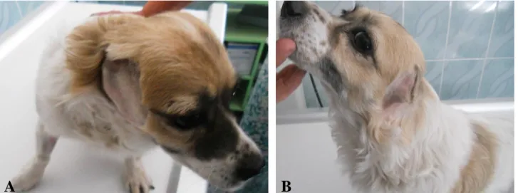
A 3-year-old, female, Spaniel Cocker mixed breed dog was referred to small animal surgery clinics for conductive hearing loss because of the absence of both auricles (Figure 1). Her body weight was 13 kg. The patient’s chest radiograph and preoperative laboratory findings were all normal. X-ray of the patient’s skull showed a radiolucent external ear canal on left side, but not on right side. It is applied on ventro-dorsal position. Soft tissue opacity was noted on the right side. Bulla tympanicas were determined bilaterally as non-radiolucent. Grade III microtia was diagnosed in the ear canal on the left side, and anotia was classified as grade IV on the right side. There were no auricles in both sides. A small fistulized area was determined on the left side and brown color fluid was seen on hair. The external auditory canal (EAC) was measured on the horizontal plane on radiogram, parallel to the tympanic membrane, along the medial- aspect of the external auditory canal. It was 4.3
Murat Kibar 440
mm long (Figure 2). No ear canal was determined on the right side for anotia. The size of the middle ear was compared with the contralateral side and subjectively measured as normal (16.3 × 13.5 mm on the right side, 15.7 × 11.5 mm on the left side).
There was no information concerning past life history of the dog including her birth since the owner had found her on the street. The dog had profound hearing loss
(information given by owner). Hearing was assessed by Pyror’s reflex behind the dog at a distance of 30 cm, 1 and 3 m. The dog was diagnosed with severe conductive hearing loss after evaluation with the Pyror’s test. The neurological examination was normal. No mental retardation was detected during the examination. The patient owner allowed for surgery. We informed the patient owner about surgical technique.
Figure 1. View of the auricular region from right side (A) and left side (B). Şekil 1. Kulak bölgesinin sağ (A) ve sol taraftan (B) görünümü.
Figure 2. Ventro-dorsal radiogram of skull; it is noted that the radiolucent ear canal (white arrow) and measurement method for the EAC diameter (white line) on the horizontal plane on the left side.
Şekil 2. Başın ventrodorsal radyogramı. Radyolusent görülen kulak kanalı (beyaz ok) ve sol tarafta horizontal kesitte çapının ölçümüne (beyaz çizgi) dikkat ediniz.
Ankara Üniv Vet Fak Derg, 65, 2018 441
Figure 3. View of atresic cartilage (arrow) brought up with Allice forceps (A), inclined incision of atresic ear canal to obtain a wider canal entrance (arrow) (B).
Şekil 3. Atrezik olan kıkırdak (ok) Allice pensi ile yukarı kaldırıldı (A), geniş bir kanal ağzı (ok) sağlamak için kulak kanalı eğimli kesildi.
Dog was premedicated with xylazine HCl (2 mg/kg, IM) and atropine sulphate (0.04 mg/kg SC), and general anesthesia was induced with ketamine HCl (10 mg/kg IM). Though many other surgical techniques are being used for microtia, the following surgical technique was performed a single stage-procedure started with fish-mouth incision around the atresic external ear canal followed by the below-mentioned stages: 1: Soft tissues were dissected and separated between the skin and cartilage. 2: The atresic cartilage was brought up with Allice forceps. 3: It was cut at an oblique angle with scissors in order to obtain a wider canal entrance (Figure 3). 4: The inner ear canal was cleaned and then checked by the otoscope. 5: The canal wall was separately sutured by 4/0 non-absorbable suture. Appropriate cefezoline (20 mg/kg IM, Sefazol, Mustafa Nevzat, Turkey) and carprofen (4.4 mg/kg PO, Rimadyl, Pfizer, Germany) were provided. Postoperatively, a disposable 5 ml syringe was used to frequently irrigate the ear canal with an antiseptic 2% iodine solution.
At each follow-up day, the sutures were checked. During each visit, the owner was given a questionnaire concerning their dog’s conductive hearing and their degree of satisfaction concerning the surgery. The questionnaire contained three items including good, moderate, and no satisfaction. At second week post operatively, the owner evaluated the hearing as moderate. This assessment was confirmed clinically with the aid of Pyror’s test.
Approximately 80% of microtia cases are unilateral in human (3, 4, 15) and the male to female ratio is 2:1 (19). To the best of our knowledge, there are few reports of unilateral microtia in veterinary medicine (2, 5, 11, 14, 16, 18). This is the first case to be reported bilaterally in a dog. Therefore, more research should be done on the ear as
congenital anomalies occur in animals and the etiology can differ depending on the species.
Congenital external auditory canal stenosis is diagnosed as an external auditory canal with a diameter of less than 4 mm by using computed tomography (9). From the radiogram results, grade III microtia was diagnosed on the left side, as there was a very small auricular hole and an auditory canal; anotia was diagnosed as grade IV on the right side due to soft tissue opacifications.
Microtia has heterogeneous natures in both pathology and etiology. Researches demonstrated the involvement of the genetic and environmental factors, as well as their combined influences on the disease (10). Epidemiological investigations of microtia showed many environmental risk factors, such as maternal medication usage, lower birth weight, and older maternal age or parity (10, 20). There was no information concerning birth because the owner had found her on the street.
Congenital conductive hearing loss can belong to an isolated anomaly of the middle ear without anomalies of the external canal or ear (12, 15). The majority of cases (80-90%) have moderate-severe to severe grade conductive hearing loss on the effected side. Patients with unilateral aural atresia usually have normal hearing on the unaffected side, unless an underlying syndrome is associated (10). The owner complained of severe deafness; thus, the case was referred to our clinic. Due to microtia and anotia, the dog had loss of conductive hearing.
Every patient with aural atresia should have an audiological evaluation to assess the type and the degree of hearing loss (10). The Pyror’s test was performed far from the dog, at 1 and 3 m behind the dog; she could not hear. This was then repeated at 30 cm behind the dog. She
Murat Kibar 442
reacted slightly, and it was understood that she could slightly hear the sounds.
It was reported that patients with congenital external auditory canal stenosis have greater postoperative hearing improvement and fewer postoperative complications than patients with a congenital atresic auricle (12, 20). Two week after operation, conductive hearing was determined to be moderate considering clinical examination and owner observation. No complication was encountered in the postoperative period.
The significant limitations of the study are debated below. First, Schmidt et al. (16) noticed the external auditory canal atresia occurring probable congenital origin based on the absence of bacteria from culture, the lack of signs of previous trauma, the absence of previous clinical signs, and abnormal accumulation of sterile ceruminous material in the auditory canal. It was not any auditory canal on the right side, and the bacterial culture was not checked on the left side. Based on the same rationale, this case was hypothesized to be congenital in origin, although a culture from the ear canal would have been ideal. Second, a computed tomography scan is the modality of choice for evaluation of ear diseases in dogs and cats, although positive-contrast ear canalography and magnetic resonance imaging can also be beneficial (2). In our patient, direct radiography was essential for evaluation of the ear canal’s anatomy to precisely diagnose the type of atresia. Third and last, brainstem auditory evoked response is an objective, reliable, easy to perform and noninvasive method to assess auditory function in dogs (2). It would have been ideal to help investigating the sensorineural part. This technique can’t be used because of technical usefulness in our clinic.
The microtia and anotia are rare abnormalities that can be seen in dogs. In this study, we reported a case of a dog with microtia and anotia, which had not been previously reported in veterinary medicine. In conclusion, grade III microtia can be treated surgically to improve the conductive hearing of the patient.
References
1. Bähr C, Distl O (2004): Bilaterale Anophthalmie in
Verbindung Mit Weiteren Missbildungen Im Kopfbereich Bei Deutschen Holsteinkälbern. Dtsch Tierarztl Wochenschr, 110, 454-456.
2. Béraud R (2012): Surgical management of 2 different
presentations of ear canal atresia in dogs. Can Vet J, 53,
412–418.
3. Brown KK, Viana LM, Helwig CC, et al. (2013):
Haploinsufficiency in Dominant Bilateral Microtia and Hearing Loss. Hum Mutat, 34, 1347-1351.
4. Canfield MA, Langlois PH, Nguyen LM, et al. (2008):
Epidemiologic features and clinical subgroups of anotia/microtia in Texas. Birth Defects Res A Clin Mol
Teratol, 85, 905-913.
5. Coomer AR, Bacon N (2009): Primary anastomosis of
segmental external auditory canal atresia in a cat. J Felin
Med Surg, 11, 864-868.
6. Cox TC, Camci ED, Vora S, et al. (2014): The genetics of
auricular development and malformation: New findings in model systems driving future directions for microtia research. Europ J Med Gen, 57, 394-401.
7. Ishimoto S, Ito K, Karino S, et al. (2007): Hearing levels
in patients with microtia: correlation with temporal bone malformation. Laryngoscope, 117, 461-465.
8. Jacob R, Gupta S, Isaacson B, et al. (2015):
High-Resolution CT Findings in Children with a Normal Pinna or Grade I Microtia and Unilateral Mild Stenosis of the External Auditory Canal. Am J Neuroradiol, 36, 176-180.
9. Jahrsdoerfer RA, Yeakley JW, Aguilar EA, et al. (1992):
Grading system for the selection of patients with congenital aural atresia. Am J Otol, 13, 6-12.
10. Lo JFW, Tsang WSS, Yu JYK, et al. (2014):
Contemporary Hearing Rehabilitation Options in Patients with Aural Atresia. BioMed Res Intern Article, 2014,
Article ID 761579.
11. Messas NB, Cipriano BDL, Andreussi PAT, et al. (2016):
Microtia and congenital external auditory canal atresia in dog: case report. Arq Bras Med Vet Zootec, 68, 683-686.
12. Park K, Choung YH (2009). Isolated congenital ossicular
anomalies. Acta Otolaryngol, 129, 419-422.
13. Qin F, Zhang T, Dai P, et al. (2015): Anatomic Variants
on Computed Tomography in Congenital Aural Atresia and Stenosis. Clin Exp Otorhinolaryng, 8, 320-328.
14. Rezaei M, Mahmoudi T, Ebrahimi M, et al. (2015): First
report of microtia in dog. Comp Clin Pathol, 24, 699–702.
15. Ruimin Q, Yuyong H, Bo P, et al. (2015): Understanding
the molecular mechanisms of human microtia via a pig model of HOXA1 syndrome. Dis Mod Mechan, 8, 611-622.
16. Schmidt K, Piaia T, Bertolini G, et al. (2007): External
auditory canal atresia of probable congenital origin in a dog. J Small Anim Pract, 48, 233–236.
17. Suutarla S, Rautio J, Ritvanen A, et al. (2007): Microtia
in Finland: comparison of characteristics in different populations. Int J Pediatr Otorhinolaryngol, 71, 1211-1217.
18. Unny NM, Ajithkumar S (2016): A rare case of aural
atresia and microtia in a goat. IOSR J Agri Vet Sci
(IOSR-JAVS), 9, 23-24.
19. Verma R, Jana M, Bhalla AS, et al. (2016): Diagnosis of
osteopetrosis in bilateral congenital aural atresia: Turning point in treatment strategy. World J Clin Pediatr, 5,
228-233.
20. Zhang TY, Li CL, Fu YY, et al. (2012): Management of
congenital meatal stenosis and atresia: impact factors on long-term results and surgical strategy. Chin J Otol, 10,
15-18.
Geliş tarihi: 21.04.2017 / Kabul tarihi: 27.12.2017
Adress for correspondence:
Prof. Dr. Murat Kibar
Artvin University, Artvin Vocational School, Hunting and Wild Life Programme, Artvin, Turkey. Gsm: +905427474447